. 2024 Dec 23;15(1):10718.
doi: 10.1038/s41467-024-55493-3.

VLDLR mediates Semliki Forest virus neuroinvasion through the blood-cerebrospinal fluid barrier

Affiliations

VLDLR mediates Semliki Forest virus neuroinvasion through the blood-cerebrospinal fluid barrier

Miika Martikainen et al. Nat Commun. .

Abstract

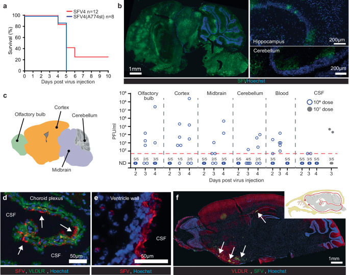
Semliki Forest virus (SFV) is a neuropathogenic alphavirus which is of interest both as a model neurotropic alphavirus and as an oncolytic virus with proven potency in preclinical cancer models. In laboratory mice, peripherally administered SFV infiltrates the central nervous system (CNS) and causes encephalitis of varying severity. The route of SFV CNS entrance is poorly understood but has been considered to occur through the blood-brain barrier. Here we show that neuroinvasion of intravenously administered SFV is strictly dependent on very-low-density-lipoprotein receptor (VLDLR) which acts as an entry receptor for SFV. Moreover, SFV primarily enters the CNS through the blood-cerebrospinal fluid (B-CSF) barrier via infecting choroid plexus epithelial cells which show distinctly high expression of VLDLR. This is the first indication of neurotropic alphavirus utilizing choroid plexus for CNS entry, and VLDLR playing a specific and crucial role for mediating SFV entry through this pathway.


Conflict of interest statement

Competing interests: The authors declare no competing interests. Ethical approval: The Swedish Work Environment Authority has approved the work with genetic modification of SFV (ID no. 202100-2932 v66a14 [laboratory] and v67a10 [mice]). All experiments regarding modified SFV were conducted under biosafety level 2. The local Animal Ethics Committee in Stockholm (13414/2020 and 04399/2023) approved the animal studies.

Figures

Fig. 1
Fig. 1. A genome-wide CRISPR/Cas9 screen identifies VLDLR as entry receptor for SFV4.
a Schematic presentation of the CRISPR/Cas9 screen experiment. b Results of the MAGeCK analysis. Statistically significant hits indicated with bigger dots. c Schematic illustration of the SFV virion. d Schematic illustration of the used SFV constructs. e Incubating HOS cells with VLDLR-blocking antibody reduces SFV4 infection, but not A774 or SFV4(A774st) (structural A774 proteins) infection. Cell viability of infected (MOI = 0.1) HOS cells pre-incubated with the LDLR class A-specific mAb 1H5, or isotype control, in an attempt to block SFV entry. Cell viability measured with MTT-assay 48 h after infection. Data plotted as mean (n = 3) ± SD. f Titration (PFU/ml) from cell culture medium collected 48 h after infection of K562 cells and K562 cells engineered to express VLDLR (K562-VLDLR) with SFV4, A774 or SFV4(A774st) (MOI = 0.01). Data presented in Log2 scale for statistical analysis. Data plotted as mean (n = 6, for K562 infected with A774 n = 5) ± SD. g Immunofluorescence staining showing VLDLR expression in K562-VLDLR. Each datapoint in (e, f) represents the result from an independent biological replicate. Statistical analysis is done using One-way ANOVA with Tukey’s multiple comparisons test. Source data and exact P values are provided as a Source Data file. PFU plaque forming units, MOI multiplicity of infection.
Fig. 2
Fig. 2. VLDLR is expressed in choroid plexus epithelial cells.
Staining for VLDLR and basement membrane (Type IV collagen, COL4) in the choroid plexus in C57BL/6 J mice (a, b) show clear expression of VLDLR in epithelial cells. b Magnified insert from (a). Epithelial cell nuclei indicated with white asterisks. c staining for choroid plexus marker aquaporin-1 (AQP1). d single cell RNAseq data (reproduced from) showing expression of Vldlr and Lrp8 (gene encoding ApoER2) in different cell types in 2–3 month old C57BL/6 mouse brain. Choroid plexus epithelial cells (red) show Vldlrhigh/Lrp8low expression profile. e immunostaining in choroid plexus shows apical ApoER2 polarization in epithelial cells (indicated with arrows). f Immunostaining for VLDLR and ApoER2 in ventricle wall. CSF: cerebrospinal fluid. g single cell RNAseq violin plot (reproduced from ref. ) of Vldlr and Lrp8 in choroid plexus endothelial cells (EC) and epithelial cells (EpiC) in adult mice. Maximum (max), median (med) and third quartile (Q3) values are provided below the graphs.
Fig. 3
Fig. 3. Early SFV neuroinvasion in C57BL/6J mice occurs through choroid plexus epithelial cells.
a Kaplan–Meier analysis of mice infected intravenously (IV) with 1 × 106 PFU SFV4 or SFV4(A774st). Statistical analysis done with log-rank test. Data from two independent experiments. b Representative immunostaining for SFV proteins in mouse brain at end point (day 5) after SFV4(A774st) injection. Magnified regions are shown from hippocampus and cerebellum. c Analysis of virus in different brain regions (as indicated), blood and CSF with plaque titration (PFU/ml). Samples from 5 mice collected at d2, 3 and 4 after 1 × 106 PFU (blue circles) IV injection of SFV4(A774st). Additional CSF samples analyzed at d3 after 1 × 107 PFU injection (gray dots). ND: not detected. Limit of detection: 50 PFU/ml. Each data point represents the result from an individual mouse. d Representative immunostaining of choroid plexus 2 days after 1 × 106 PFU IV SFV4(A774st) injection. SFV protein staining can be seen in the outer surface of VLDLR-positive epithelial cells (indicated with arrows). e Representative immunostaining of SFV protein in ventricle wall 2 days after 1 × 106 PFU IV SFV4(A774st) injection. f Representative immunostaining for SFV proteins (green) in WT mouse brain at day 3 after 1 × 106 PFU IV SFV4(A774st) injection show expression of SFV proteins in ventral regions. Top right corner shows schematic picture of CSF fluid flow in the mouse brain. Created in BioRender. Martikainen, M. (2024) https://BioRender.com/o42e737. All experiments are done with 7–9-week-old female C57BL/6J mice. Source data for (a, c) is provided as a Source Data file.
Fig. 4
Fig. 4. VLDLR is crucial for SFV neuroinvasion from the circulation.
Immunofluorescence staining for VLDLR in the choroid plexus of Vldlr KO (a) and B6129SF2/J (Vldlr wild-type control mouse) mice (b). No VLDLR can be detected in Vldlr KO sample. CSF: cerebrospinal fluid. c Kaplan–Meier analysis of Vldlr KO mice infected intravenously (IV) with 1 × 106 PFU SFV4 or SFV4(A774st) and B6129SF2/J infected intravenously (IV) with 1 × 106 PFU SFV4(A774st). Statistical analysis done with log-rank test. Data from two independent experiments. Representative immunostaining for SFV proteins in Vldlr KO (d) and B6129SF2/J (e) mouse brain at day 4 after 1 × 106 PFU IV SFV4(A774st) injection. f, g Analysis of virus in different brain regions, blood and CSF with plaque titration (PFU/ml). Samples from 5 Vldlr KO mice collected at d2, 3, and 4 after 1 × 106 PFU (red circles) IV injection of SFV4(A774st). Additional CSF samples analyzed at d3 after 1 × 107 PFU injection (gray dots). ND: not detected. g plaque titration of brain samples collected from B6129SF2/J mice at d4 after 1 × 107 PFU SFV4(A774st) injection. Limit of detection: 50 PFU/ml. Each data point represents the result from an individual mouse. h Kaplan–Meier analysis of Vldlr KO mice infected intracranially (IC) or intranasally (IN) SFV4(A774st). All mice succumb to neurological symptoms. Data from two independent experiments. i Representative immunostaining of Vldlr KO mouse olfactory epithelium (OE) 2 days after IN SFV4(A774st) injection. SFV staining can be seen at the surface layer of epithelium (indicated with arrow) but also in deeper tissues. j Representative immunostaining of Vldlr KO mouse brain (transverse plane) 4 days after IN SFV4(A774st) injection. SFV staining can be seen in olfactory bulb but also in deeper in the brain tissues. Experiments done with 7–10 week-old female mice. Source data for (c, f, g, h) and P values for (c) are provided as a Source Data file.
Fig. 5
Fig. 5. Refined model for SFV neuroinvasion through the blood-CSF barrier.
a Schematic presentation of the choroid plexus and the blood-CSF barrier. b Schematic presentation of VLDLR and ApoER2 expression in endothelial and epithelial cell layers and the effect knocking out Vldlr has on generalized SFV entry. Entry through the epithelial layer is solely mediated by VLDLR on the basolateral side, making Vldlr KO mice completely resistant to SFV neuroinvasion. Due to the strictly apical expression pattern, ApoER2 does not affect SFV passage through the epithelial cell layer from the basolateral side. Abbreviations: EC: endothelial cell, EpiC: epithelial cell, SFV: Semliki Forest virus, CSF: cerebrospinal fluid, WT: wild-type, KO: knock-out. Created in BioRender. Martikainen, M. (2024) https://BioRender.com/z30c241.

References

    1. Martikainen, M. et al. MicroRNA-Attenuated clone of virulent Semliki forest virus overcomes Antiviral Type I interferon in resistant Mouse CT-2A Glioma. J. Virol.89, 10637–10647 (2015). - PMC - PubMed
    1. Martikainen, M. et al. IFN-I-tolerant oncolytic Semliki Forest virus in combination with anti-PD1 enhances T cell response against mouse glioma. Mol. Ther. Oncolytics21, 37–46 (2021). - PMC - PubMed
    1. Ramachandran, M. et al. Safe and effective treatment of experimental neuroblastoma and glioblastoma using systemically delivered triple microRNA-detargeted oncolytic Semliki Forest Virus. Clin. Cancer Res.23, 1519–1530 (2017). - PubMed
    1. Fragkoudis, R. et al. Neurons and oligodendrocytes in the mouse brain differ in their ability to replicate Semliki Forest virus. J. Neurovirol.15, 57–70 (2009). - PubMed
    1. Fazakerley, J. K., Pathak, S., Scallan, M., Amor, S. & Dyson, H. Replication of the A7(74) strain of Semliki Forest virus is restricted in neurons. Virology195, 627–637 (1993). - PubMed

Publication types

Associated data