Review
. 2024 Dec 23;9(1):368.
doi: 10.1038/s41392-024-02069-8.

The molecular mechanisms of cardiac development and related diseases

Affiliations
Review

The molecular mechanisms of cardiac development and related diseases

Yingrui Li et al. Signal Transduct Target Ther. .

Abstract

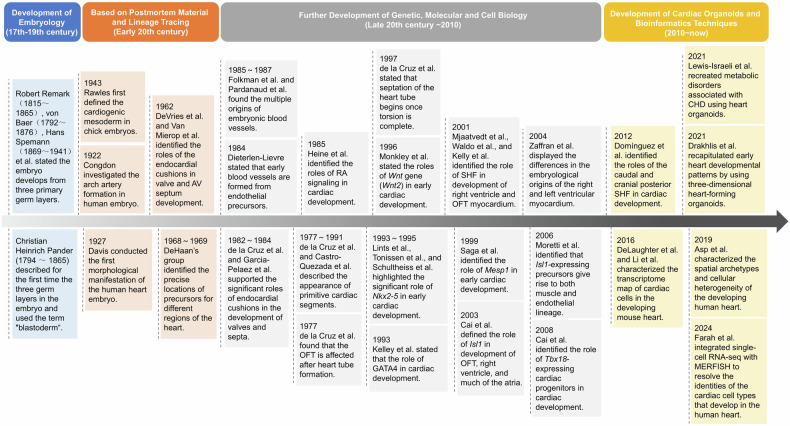
Cardiac development is a complex and intricate process involving numerous molecular signals and pathways. Researchers have explored cardiac development through a long journey, starting with early studies observing morphological changes and progressing to the exploration of molecular mechanisms using various molecular biology methods. Currently, advancements in stem cell technology and sequencing technology, such as the generation of human pluripotent stem cells and cardiac organoids, multi-omics sequencing, and artificial intelligence (AI) technology, have enabled researchers to understand the molecular mechanisms of cardiac development better. Many molecular signals regulate cardiac development, including various growth and transcription factors and signaling pathways, such as WNT signaling, retinoic acid signaling, and Notch signaling pathways. In addition, cilia, the extracellular matrix, epigenetic modifications, and hypoxia conditions also play important roles in cardiac development. These factors play crucial roles at one or even multiple stages of cardiac development. Recent studies have also identified roles for autophagy, metabolic transition, and macrophages in cardiac development. Deficiencies or abnormal expression of these factors can lead to various types of cardiac development abnormalities. Nowadays, congenital heart disease (CHD) management requires lifelong care, primarily involving surgical and pharmacological treatments. Advances in surgical techniques and the development of clinical genetic testing have enabled earlier diagnosis and treatment of CHD. However, these technologies still have significant limitations. The development of new technologies, such as sequencing and AI technologies, will help us better understand the molecular mechanisms of cardiac development and promote earlier prevention and treatment of CHD in the future.


Conflict of interest statement

Competing interests: The authors declare no competing interests.

Figures

Fig. 1
Fig. 1
Timeline of milestone events in cardiac development research history. Each significant breakthrough in cardiac development research has been accompanied by advancements in related disciplines and technologies. The research of cardiac development starting with early studies observing morphological changes to exploration of molecular mechanisms using various molecular biology methods. Nowadays, advancements in stem cell technology and sequencing technology, such as the generation of cardiac organoids and multi-omics sequencing, have enabled researchers to understand the molecular mechanisms of cardiac development better. AV atrioventricular; AVC atrioventricular canal, CHD congenital heart disease, GATA4 GATA binding protein 4, hPSCs human pluripotent stem cells, Isl1 islet 1, MERFISH multiplexed error-robust fluorescence in situ hybridization, Mesp1 mesoderm posterior 1, Nkx2-5 NK2 homeobox 5, OFT outflow tract, PE proepicardium, RA retinoic acid, SHF second heart field, Tbx18 T-box transcription factor 18. This figure was created using Microsoft Powerpoint
Fig. 2
Fig. 2
The molecular mechanisms of cardiac development were explored using pluripotent stem cell technology and multi-omics analysis. Pluripotent stem cells are generated by reprogramming adult somatic cells from CHD patients or animal models into a pluripotent state. These cells can differentiate into various cardiac cells, such as cardiomyocytes, smooth muscle cells, endothelial cells, and epicardial cells, through different differentiation protocols. Pluripotent stem cells can also form cardiac organoids through self-organization and specific differentiation methods, thereby creating an in vitro model of cardiac development. Multiomics analysis encompasses genomics, transcriptomics, proteomics, epigenomics, and metabolomics. By analyzing heart tissues obtained from CHD patients or animal models or cells derived from pluripotent stem cells, multi-omics analysis can be used to investigate the molecular mechanisms involved in the regulation of cardiac development at multiple levels. CHD congenital heart disease. This figure was created using Adobe Illustrator
Fig. 3
Fig. 3
Human cardiac development and its regulatory mechanisms. Cardiac development involves five distinct stages. The first stage, mesoderm cardiac induction (15-17 days), begins with cardiac mesoderm progenitors migrating from the primitive streak to the heart-forming region, an anterior lateral area relative to the streak. During migration, these mesodermal cells acquire the ability to differentiate into the cardiac lineage and express markers such as Brachyury and MESP1. The key signaling pathways involved include the Nodal/Activin, BMP, and WNT signaling pathways. In the second stage (17–19 days), the cardiac crescent forms, and MESP1-derived cardiac mesodermal cells differentiate into FHF and SHF progenitors. Molecular signals, including WNT signaling, BMP signaling, and various TFs, are critical. FHF progenitor cells, positioned more anteriorly and laterally in the crescent, readily respond to molecular cues and begin differentiation. SHF progenitors remain proliferative and undifferentiated until they reach later stages when they contribute to the heart tube. The third to fifth stages include heart tube formation (19–22 days), cardiac looping (22–35 days), and the formation of the four-chambered fetal heart (35–60 days). During these stages, the cardiac crescent fuses at the midline and folds into a Y-shaped FHF-derived linear heart tube. SHF cells proliferate rapidly and contribute to the arterial and venous poles of the heart tube. Subsequently, the heart tube undergoes rapid growth and rightward looping, forming distinct chambers by approximately day 32 in humans that are fully septated and connected to the pulmonary trunk and aorta by week 7 of fetal development. Events such as the migration of CNCCs, myocardial trabeculation, and the development of endocardial cushions and the epicardium are crucial during these stages. Signaling pathways, including FGF signaling, Notch signaling, and Shh signaling, are involved in the migration of CNCCs, promoting the development of the aortic arch and outflow tract. FGF signaling, Notch signaling, BMP signaling, and others contribute to myocardial trabeculation. Furthermore, FGF signaling, Notch signaling, TBX5, HAND1/2, and related signals aid in the development of endocardial cushions, facilitating subsequent septum and valve development. Finally, FGF signaling, TBX18, WT1, Notch signaling, and others contribute to epicardial development, promoting coronary artery formation and subsequent proliferation of myocardial cells. BRY brachyury, BMP bone morphogenetic protein, CNCCs cardiac neural crest cells, FGF fibroblast growth factor, FHF first heart field, GATA GATA binding protein, HAND heart and neural crest derivatives expressed, HFR heart-forming region, ISL1 islet 1, MEF myocyte enhancer factor, MESP1 mesoderm posterior 1, NKX2-5 NK2 Homeobox 5, RA retinoic acid, Shh sonic hedgehog, SHF second heart field, TBX T-box transcription factor, TGF transforming growth factor, TFs transcription factors, WT1 Wilms tumor 1. This figure was created using Adobe Illustrator
Fig. 4
Fig. 4
The role of hypoxia and metabolic transition during cardiac development. Significant changes in metabolic pathways accompany the maturation of cardiac development. During embryonic cardiac development, glucose and lactate serve as the primary sources of ATP. Hypoxic conditions activate HIF-1α during embryonic development, coupled with mitochondrial immaturity, which promotes a reliance on glycolysis for ATP production in the fetal heart, leading to increased lactate production. As cardiac development progresses, there is a simultaneous decrease in glycolysis and an increase in fatty acid β-oxidation during myocardial maturation. After birth, cardiac metabolism primarily relies on fatty acid oxidation, facilitated by an oxygen-rich environment. In this environment, the HIF-1α subunit is hydroxylated and targeted for degradation by VHL, thereby inhibiting its promotion of glycolysis. α-KG α-ketoglutarate, ATP adenosine triphosphate, CoA coenzyme A, HIF-1α hypoxia-inducible factor 1-alpha, OH hydroxide, O2 oxygen, TCA tricarboxylic acid, VHL von Hippel Lindau. This figure was created using Adobe Illustrator
Fig. 5
Fig. 5
The role of macrophages during cardiac development. Macrophages first appear during mouse embryo development around E10 near the outflow tract, sinus node, and endocardial cushions. As the heart matures, they are distributed beneath the epicardium, within the myocardial layer, and in the endocardial layer throughout the entire heart. Various macrophage subpopulations have been identified at different times and locations, with significant research focused on subepicardial space subpopulations (CX3CR1+ CD68+ F4/80+ Lyve-1+/-; CX3CR1high CD64+ Lyve-1+ CCR2-). These macrophages originate from the yolk sac and migrate to the epicardium under the influence of the transcription factor WT1, subsequently entering the subepicardial space. In the subepicardial space, these macrophages adhere to the endothelial cells of blood vessels, promoting the normal development of coronary arteries. Simultaneously, they adhere to the endothelial cells of lymphatic vessels and facilitate normal lymphatic development through direct interactions involving hyaluronic acid. Macrophages within endocardial cushions (Nfatc1+ CD45+ CD68+ F4/80+ CD206+), derived from endothelial cells, likely contribute to valve development through phagocytic activities. CCR2 C-C chemokine receptor type 2, CX3CR1 chemokine (C-X3-C motif) receptor 1, Lyve-1 lymphatic vessel endothelial hyaluronan receptor 1, Nfatc nuclear factor of activated T cells, cytoplasmic, WT1 Wilms tumor 1. This figure was created using Adobe Illustrator
Fig. 6
Fig. 6
Common congenital heart defects and pathogenic mechanisms. Ventricular septal defect is a defect in the ventricular septum involving the loss of functions of various TFs, GFs, Notch signaling, RA signaling, etc.; atrial septal defect is a defect in the atrial septum, involving the loss of functions of various TFs, GFs, WNT signaling, RA signaling, etc; bicuspid aortic valve is a pathological condition where the aortic valve has only two leaflets instead of the normal three, involving the loss of functions of TGF-β signaling, Notch signaling, various TFs, etc.; mitral valve prolapse is a pathological condition where the mitral valve does not close properly, involving the loss of functions of TGF-β signaling, ECM signaling, FBN1, etc.; patent ductus arteriosus is a pathological condition in which the ductus arteriosus fails to close after birth, allowing a portion of oxygenated blood from the left heart to flow back to the lungs through the aorta, involving the loss of functions of TFAP2B, prostaglandin signaling, Notch signaling, etc.; tetralogy of Fallot includes ventricular septal defects, overriding of the aorta, right ventricular outflow obstruction and right ventricular hypertrophy, involving the loss of functions of various TFs, Notch signaling, VEGF signaling, etc.; double-outlet right ventricle is the defect that both the pulmonary artery and the aorta arise from the right ventricle instead of their normal positions, involving the loss of functions of various TFs, RA signaling, Notch signaling, etc.; transposition of the great arteries is a pathological condition where the pulmonary artery and aorta are switched in position, causing oxygen-rich blood from the lungs is pumped back to the lungs instead of being circulated to the rest of the body, and oxygen-poor blood from the body is pumped back to the body instead of being sent to the lungs to pick up oxygen, involving the loss of functions of cilia, various TFs, Nodal signaling, etc.; persistent truncus arteriosus is a pathological condition characterized by single large vessel arising from the heart that supplies blood to both the systemic and pulmonary circulations, involving the loss of functions of various TFs, RA signaling, Nodal signaling, etc.; hypoplastic left heart syndrome presents with different degrees of stenosis or atresia of the aortic and mitral valve along with hypoplasia of the left ventricle and ascending aorta involving the loss of functions of various TFs, Notch signaling, ECM signaling, etc. ECM extracellular matrix, FBN1 fibrillin 1, FLNA filamin A, FOG2 friend of GATA 2, GATA GATA binding protein, GFs Growth factors, HAND1 heart and neural crest derivatives expressed 1, HOXA1 homeobox A1, ISL1 islet 1, NKX2-5 NK2 homeobox 5, PITX2 paired-like homeodomain transcription factor 2, RA retinoic acid, TBX T-box transcription factor, TFs transcription factors, TFAP2B transcription factor AP-2 beta, TGF-β transforming growth factor beta, VEGF vascular endothelial growth factor, ZIC3 Zic family member 3. This figure was created using Adobe Photoshop
Fig. 7
Fig. 7
Current and future advanced treatment strategies for CHD. Currently, CHD is considered a lifelong condition and is primarily treated through early surgical interventions, multiple interventional catheterizations, and cell therapies. However, postsurgery CHD patients often face challenges such as decreased exercise tolerance, multiple cardiovascular and cerebrovascular complications, increased risk of sudden cardiac death, and decreased quality of life. Therefore, lifelong management, including long-term medication and care, is necessary for CHD patients. Advances in technology and a deeper understanding of cardiac development mechanisms aim to achieve earlier diagnosis and prevention of CHD, minimizing its impact and improving patient quality of life and longevity. Early intervention strategies, such as FCI in select CHD patients, are already underway, although further research into related drug therapies and gene treatments is needed. The development of cardiac imaging combined with AI technology enhances CHD diagnosis accuracy, while genetic testing aids in detecting pathogenic variants associated with CHD, facilitating early diagnosis. PGT offers the possibility for early prevention of CHD by identifying genetic defects or chromosomal abnormalities in embryos early in pregnancy; thus, selecting embryos most likely to result in successful pregnancies and healthy offspring is highly desirable. Utilizing AI to integrate multi-omics and clinical data for constructing risk prediction models will enable the identification of more precise genetic or environmental risk factors, further promoting early prevention strategies for CHD in the future. AI artificial intelligence, CHD congenital heart disease, FCI fetal cardiac intervention, PGT preimplantation genetic testing. This figure was created using Adobe Illustrator

References

    1. Brade, T., Pane, L. S., Moretti, A., Chien, K. R. & Laugwitz, K. L. Embryonic heart progenitors and cardiogenesis. Cold Spring Harb. Perspect. Med.3, a013847 (2013). - PMC - PubMed
    1. Meilhac, S. M. & Buckingham, M. E. The deployment of cell lineages that form the mammalian heart. Nat. Rev. Cardiol.15, 705–724 (2018). - PubMed
    1. Buijtendijk, M. F. J., Barnett, P. & van den Hoff, M. J. B. Development of the human heart. Am. J. Med. Genet. C Semin. Med. Genet.184, 7–22 (2020). - PMC - PubMed
    1. Moorman, A. F. & Christoffels, V. M. Cardiac chamber formation: development, genes, and evolution. Physiol. Rev.83, 1223–1267 (2003). - PubMed
    1. Abu-Issa, R. & Kirby, M. L. Heart field: from mesoderm to heart tube. Annu. Rev. Cell Dev. Biol.23, 45–68 (2007). - PubMed

Publication types

MeSH terms

LinkOut - more resources