

Application of apparent diffusion coefficient of extraocular muscles from diffusion tensor imaging scanning in the assessment of disease activity of thyroid eye disease
- PMID: 39716188
- PMCID: PMC11668034
- DOI: 10.1186/s12902-024-01818-8
Application of apparent diffusion coefficient of extraocular muscles from diffusion tensor imaging scanning in the assessment of disease activity of thyroid eye disease
Abstract
Purpose: To evaluate the utility of apparent diffusion coefficient (ADC) values of extraocular muscles (EOMs) in differentiating activity of thyroid eye disease (TED).
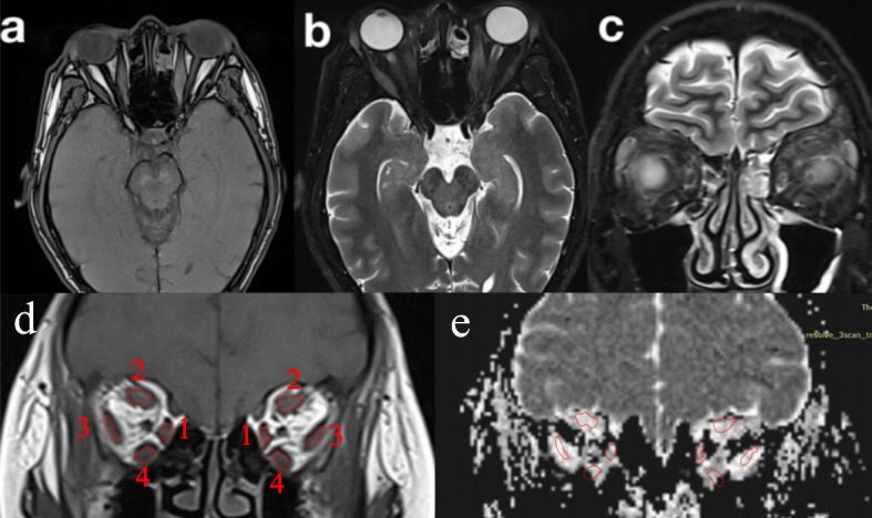
Method: Forty-two TED patients who underwent diffusion tensor imaging(DTI) were retrospectively enrolled in this study, including 29 patients in analysis group and 13 patients in validation group. The mean, maximum and minimum ADC value of each EOM were regarded as ADCmean, ADCmax and ADCmin. The difference between ADCmax and ADCmin was regarded as △ADC. The correlations between ADCmean or △ADC of each EOM and clinical activity score (CAS) were assessed.
Results: In analysis group, ADCmean differed between active and inactive eyes and positively correlated with CAS in IR (P < 0.05), not in SR,LR and MR(all p > 0.05). While △ADC differed between two groups and negatively correlated with CAS in all EOMs (all P < 0.05). ADCmean predicted active disease at cut-off value of 1259.3 × 10-6mm2s-1 with sensitivity of 66.7% and specificity of 71.4% in IR[area under curve = 0.667, P < 0.05]. △ADC predicted disease activity in all EOMs [area under curve 0.658-0.746,all P < 0.05]. The cut-off values of △ADC were 382, 823,520 and 572 × 10-6mm2s-1 with sensitivity of 80.0%, 50.0%, 43.3%, 83.3% and specificity of 67.9%, 85.7%, 89.3%, 60.7% in SR, IR, MR, and LR respectively. There were no significant differences in the predictive efficacy among all cut-off values.
Conclusions: Our results showed that DTI is a valuable tool in the assessment of disease activity of TED. Both ADCmean of IR and △ADC of all four EOMs can be used in discriminating disease activity with the same predictive power.
Keywords: ADC; CAS; EOMs; Resolve DTI; Thyroid eye disease.
© 2024. The Author(s).
Conflict of interest statement
Declarations. Ethics approval and consent to participate: We confirmed that informed consent was obtained from all subjects, all subjects recruited agreed to take part in the present study. The study was approved by the First Affiliated Hospital of Chongqing Medical University Ethical Committee. All methods and designs were performed in accordance with the consensus statement of the European Group on Graves’ Orbitopathy critera. Consent for publication: Not applicable. Competing interests: The authors declare no competing interests.
Figures
Similar articles
-
Apparent diffusion coefficient: a predictor of the efficacy of glucocorticoid therapy in active moderate-severe thyroid eye disease: a retrospective cohort study.BMC Endocr Disord. 2025 Jul 14;25(1):177. doi: 10.1186/s12902-025-01981-6. BMC Endocr Disord. 2025. PMID: 40660145 Free PMC article.
-
Thyroid-Associated Orbitopathy: Evaluating Microstructural Changes of Extraocular Muscles and Optic Nerves Using Readout-Segmented Echo-Planar Imaging-Based Diffusion Tensor Imaging.Korean J Radiol. 2020 Mar;21(3):332-340. doi: 10.3348/kjr.2019.0053. Korean J Radiol. 2020. PMID: 32090526 Free PMC article.
-
Fractional anisotropy and diffusivity changes in thyroid-associated orbitopathy.Neuroradiology. 2016 Dec;58(12):1189-1196. doi: 10.1007/s00234-016-1764-0. Epub 2016 Nov 14. Neuroradiology. 2016. PMID: 27844093
-
Magnetic resonance imaging parameters on lacrimal gland in thyroid eye disease: a systematic review and meta-analysis.BMC Ophthalmol. 2023 Aug 8;23(1):347. doi: 10.1186/s12886-023-03008-x. BMC Ophthalmol. 2023. PMID: 37550660 Free PMC article.
-
The Roles of Diffusion Kurtosis Imaging and Intravoxel Incoherent Motion Diffusion-Weighted Imaging Parameters in Preoperative Evaluation of Pathological Grades and Microvascular Invasion in Hepatocellular Carcinoma.Front Oncol. 2022 May 11;12:884854. doi: 10.3389/fonc.2022.884854. eCollection 2022. Front Oncol. 2022. PMID: 35646649 Free PMC article.
References
-
- Lingam RK, Mundada P, Lee V. Novel use of non-echo-planar diffusion weighted MRI in monitoring disease activity and treatment response in active Grave’s eye: an initial observational cohort study. Orbit. 2018;37:325–30. - PubMed
-
- Bartley GB. Rundle and his curve. Arch Ophthalmol. 2011;129:356–8. - PubMed
MeSH terms
LinkOut - more resources
Full Text Sources
Research Materials
