

Infection survey, molecular, pathogenicity, and morphological characteristics of Sarcocystis species naturally infected water buffaloes (Bubalus bubalis) in Egypt
- PMID: 39716208
- PMCID: PMC11664923
- DOI: 10.1186/s12917-024-04408-x
Infection survey, molecular, pathogenicity, and morphological characteristics of Sarcocystis species naturally infected water buffaloes (Bubalus bubalis) in Egypt
Abstract
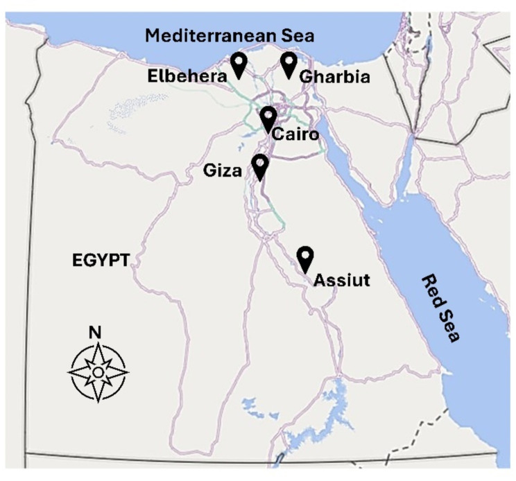
Background: Sarcocystosis is a parasitic disease found worldwide, resulting from various Sarcocystis species. The current research was carried out in three significant economic areas in Egypt: Greater Cairo, the Nile Delta, and Upper Egypt. It aimed to investigate the occurrence of Sarcocystis spp. in locally bred water buffaloes Bubalus bubalis.
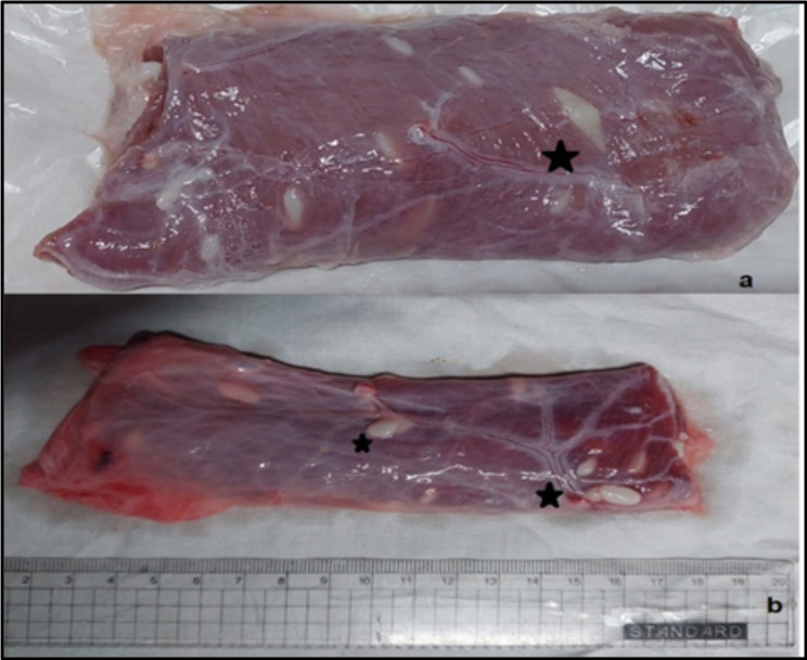
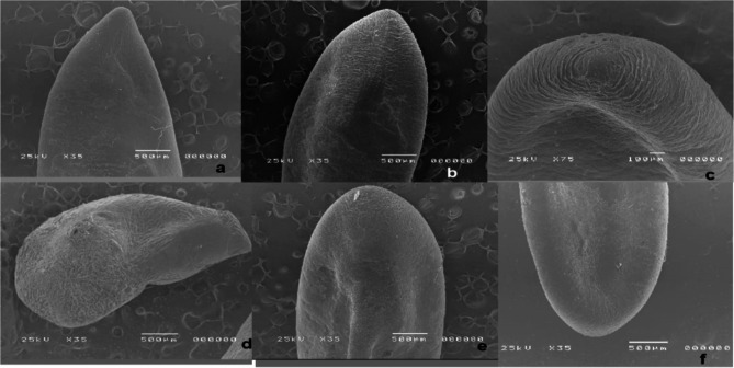
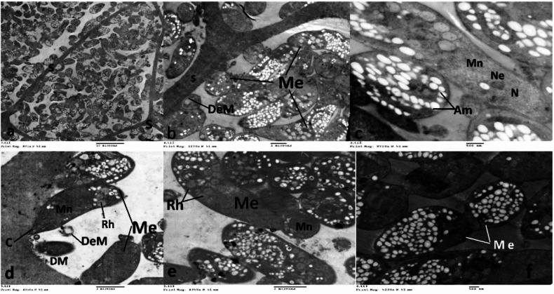
Methods: To achieve this objective, 317 buffalos were slaughtered in different slaughterhouses in various regions of Egypt. Samples of heart, skeletal muscle, esophagus, and tongue were assessed using macroscopic and microscopic tests. Examination methods included direct optical observation of tissues as well as digestion and examination of the sediment obtained from the tissues. Additionally, ultrastructural features were analyzed using scanning and transmission electron microscopy. Molecular characterization was conducted through PCR, followed by nucleotide sequencing and phylogenetic analysis.
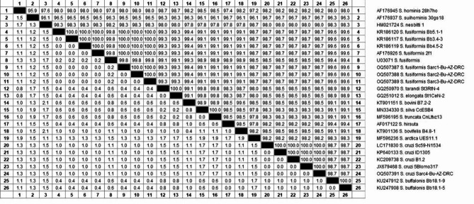
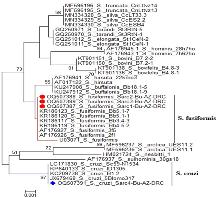
Results: A total of 317 slaughtered buffaloes were examined for Sarcocystis during the period from September 2021 to October 2023. The prevalence of infection was recorded with 229 out of 317 (72.2%) infected with Sarcocystis spp. The results also showed that the prevalence of Sarcocystis species in females was higher than males. Based on the age of carcasses, adults (> 2 years) had a higher infection rate compared to young ones (< 2 years). Regarding seasonal variation, the highest prevalence of infection was recorded during the summer followed by spring, and then autumn, while winter had the lowest prevalence of infection. Additionally, the skeletal muscle was the most susceptible organ to sarcocystosis (87.3%) followed by the esophageal muscle (8.3%), the tongue (4.4%), and no infection in the heart muscle. The use of scanning and transmission electron microscopy allowed the identification of S. fusiformis and S. cruzi in buffaloes in Egypt. Furthermore, the Sarcocystis 18 S rRNA genes from skeletal tissue samples were cloned and sequenced under accession numbers OQ507387, OQ507388, and OQ507389 for S. fusiforms, and one OQ507391 for S. cruzi.
Conclusion: The findings revealed a notably high prevalence of Sarcocystis infection (72.2%) in buffaloes from Egypt, with skeletal muscle identified as the organ most susceptible to the parasite. Two Sarcocystis species were detected: S. fusiformis and S. cruzi.
Keywords: Sarcocystis cruzi; Sarcocystis fusiformis; Buffaloes; Molecular; Pathogenicity; Sarcocystosis; Ultrastructural.
© 2024. The Author(s).
Conflict of interest statement
Declarations. Ethics approval and consent to participate: The research was conducted exclusively on carcasses obtained from animals that had already been slaughtered within licensed slaughterhouses. All slaughterhouses in Egypt adhere to the Animal Protection Law (Decision 517 of 86), which is aligned with international veterinary medical regulations and met the standards set by the ARRIVE criteria. Consent for publication: Not applicable. Competing interests: The authors declare no competing interests.
Figures


References
-
- Prakas P P, Strazdaitė-Žielienė Ž, Januškevičius V, Chiesa F, Baranauskaitė A, Rudaitytė-Lukošienė E, Servienė E, Petkevičius S, Butkauskas D. Molecular identification of four Sarcocystis species in cattle from Lithuania, including S. Hominis, and development of a rapid molecular detection method. Parasit Vectors. 2020;13(1):610. 10.1186/s13071-020-04473-9. - DOI - PMC - PubMed
MeSH terms
LinkOut - more resources
Full Text Sources
Miscellaneous
