. 2024 Dec 23;12(1):201.
doi: 10.1186/s40478-024-01912-x.

JMJD3 deficiency disturbs dopamine biosynthesis in midbrain and aggravates chronic inflammatory pain

Affiliations

JMJD3 deficiency disturbs dopamine biosynthesis in midbrain and aggravates chronic inflammatory pain

Xi-Biao He et al. Acta Neuropathol Commun. .

Abstract

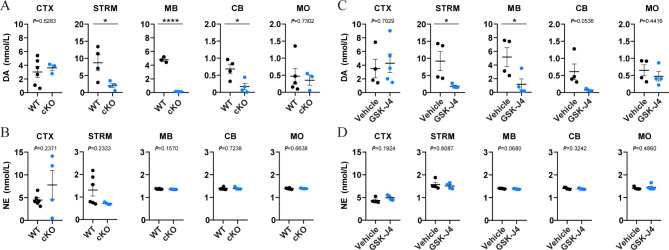
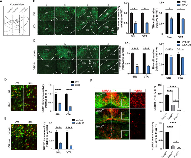
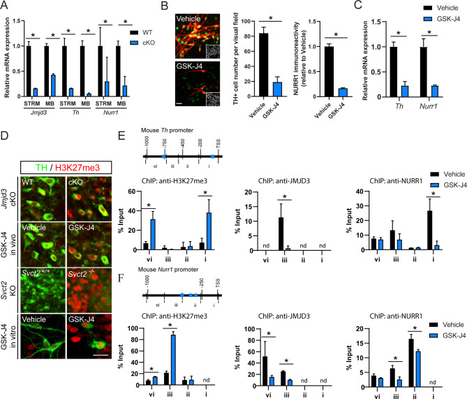
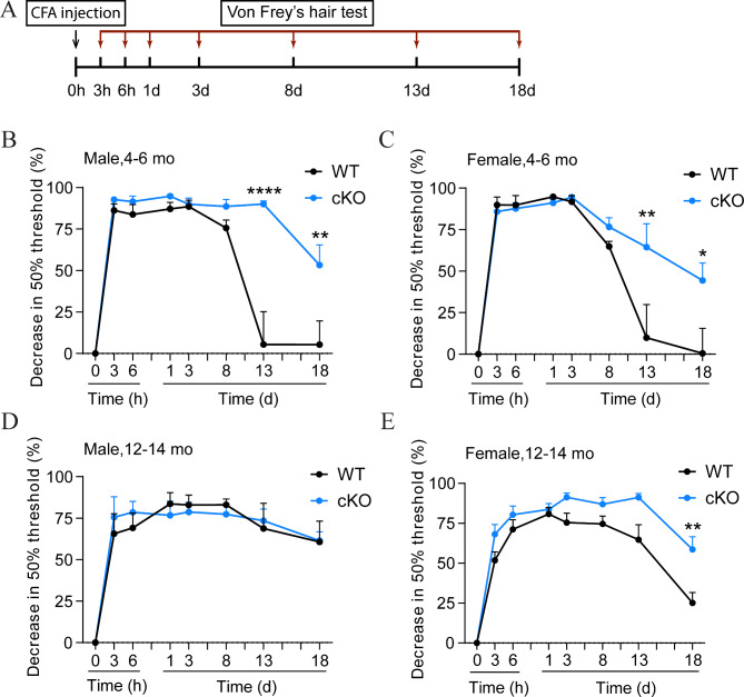
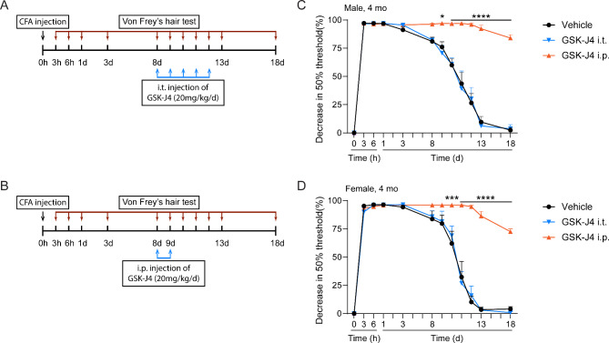
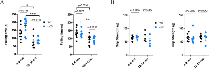
Midbrain dopamine (mDA) neurons participate in a wide range of brain functions through an intricate regulation of DA biosynthesis. The epigenetic factors and mechanisms in this process are not well understood. Here we report that histone demethylase JMJD3 is a critical regulator for DA biosynthesis in adult mouse mDA neurons. Mice carrying Jmjd3 conditional knockout or undergoing pharmaceutical inhibition of JMJD3 showed consistent reduction of DA content in midbrain and striatum. Histological examination of both mice confirmed that TH and NURR1, two key molecules in DA biosynthesis pathway, were decreased in mDA neurons. Mechanistic experiments in vivo and in vitro further demonstrated that the transcriptions of Th and Nurr1 in mDA neurons were suppressed by JMJD3 deficiency, because of increased repressive H3K27me3 and attenuated bindings of JMJD3 and NURR1 on the promoters of both genes. On behavioral level, a significant prolonged inflammation-induced mechanical hyperalgesia was found in conditional knockout mice regardless of sex and age, whereas motor function appeared to be intact. Our findings establish a novel link between DA level in mDA neurons with intrinsic JMJD3 activity, and suggest prolonged chronic inflammatory pain as a major loss-of-function consequence.

Keywords: Chronic inflammatory pain; Dopamine biosynthesis; Epigenetic control; Midbrain dopamine neuron; Transcriptional regulation.


Conflict of interest statement

Declarations. Competing interests: The authors declare no competing interests. Ethics approval: Approval was obtained from the ethics committee of Shanghai University of Medicine & Health Sciences. The procedures used in this study adhere to the tenets of the Declaration of Helsinki. Consent to participate: Not applicable. Consent to publish: Not applicable.

Figures

Fig. 1
Fig. 1
JMJD3 expression in tyrosine hydroxylase (TH) + neurons in the central and peripheral nervous systems. Immunofluorescence co-labeling of TH (green) and JMJD3 (red) in midbrain dopamine (DA) neurons (A), non-midbrain DA neurons (B), norepinephrine (NE) neurons in locus coeruleus (C) and C-LTMR neurons in dorsal root ganglion (D) from 8-week-old C57BL/6 mice. Cell nuclei were counterstained by 4’,6-diamidino-2-phenylindole (blue). Scale bar represents 20 μm
Fig. 2
Fig. 2
Dopamine (DA) levels are decreased in the midbrain and striatum of two mouse models of JMJD3 deficiency. Brain tissues from the frontal cortex (CTX), striatum (STRM), midbrain (MB), cerebellum (CB) and medulla oblongata (MO) were micro-dissected, homogenized and the contents of DA and norepinephrine (NE) were measured by high performance liquid chromatography-mass spectrometry. (A, B) 4-month-old male conditional knockout (cKO) mice and their control littermates (WT). (C, D) 4-month-old male C57BL/6 mice underwent 5 consecutive days of intraperitoneal injection of GSK-J4 (20 mg/kg daily). In parallel, dimethyl sulfoxide was injected as Vehicle. N = 4–7 mice. Data represent mean ± SEM. *P < 0.05, ****P < 0.0001; Student’s t-test
Fig. 3
Fig. 3
Reduced expressions of tyrosine hydroxylase (TH) and NURR1 in midbrain dopamine (mDA) neurons of two JMJD3 deficiency mouse models. (A) Schematics showing the coronal view of the adult mouse midbrain compromised of ventral tegmental area (VTA), substantia nigra pars compacta (SNc) and pars reticulata (SNr). Dotted lines a-d indicate positions shown in B and C. (B) Left, representative immunofluorescence images of TH + mDA neurons in Jmjd3 conditional knockout (cKO) mice and their control littermates (WT). Scale bar represents 100 μm. Right, quantifications of TH immunoreactivity and cell number of mDA neurons in VTA and SNc in WT and cKO mice. (C) Left, representative immunofluorescence images of TH + mDA neurons in GSK-J4 injected mice and their control (Vehicle). Scale bar represents 100 μm. Right, quantifications of TH immunoreactivity and cell number of DA neurons in VTA and SNc in GSK-J4 injected mice and control. (D, E) Representative immunofluorescence images and quantifications of NURR1 immunoreactivity showing decreased NURR1 (red) expression in TH + mDA neurons (green) in SNc and VTA of cKO and GSK-J4-injected mice in comparison to WT and Vehicle, respectively. Scale bar represents 20 μm. (F) Representative immunofluorescence images and quantifications showing effect of Svct2 KO on TH and NURR1 expressions in mouse embryonic day 14.5 ventral mesencephalon. Insets show NURR1 expression in higher magnification. Scale bar represents 50 μm. For immunoreactivity analysis, at least 50 TH + cells from each mouse were measured and averaged. N = 3–5 mice. Data represent mean ± SEM. *P < 0.05, **P < 0.01, ****P < 0.0001; Student’s t-test
Fig. 4
Fig. 4
Epigenetic control of Th and Nurr1 gene transcription by JMJD3. (A) Real-time PCR analysis of mRNA levels of Jmjd3, Th and Nurr1 from striatum (STRM) and midbrain (MB) in Jmjd3 conditional knockout mice (cKO) and their control littermates (WT). N = 3 mice. Data represent mean ± SEM. (B) Representative immunofluorescence images and quantifications of NURR1 expression (green) in tyrosine hydroxylase (TH; red) + cells from primary culture of mouse midbrain dopamine (mDA) neurons with or without treatment of GSK-J4. Insets show cell nuclei. Scale bar represents 20 μm. (C) Real-time PCR analysis of Th and Nurr1 mRNA levels in primary cultured mDA neurons after GSK-J4 treatment. (D) Representative immunofluorescence images of histone 3 lysine 27 tri-methylation (H3K27me3) in TH + mDA neurons from cKO, GSK-J4 mice, Svct2 KO mouse embryos and primary cultured mDA neurons treated with GSK-J4. Scale bar represents 20 μm. (E, F) Chromatin immunoprecipitation (ChIP)-quantitative PCR analysis of H3K27me3, JMJD3 and NURR1enrichments on 1 kilo bp mouse Th (E) and Nurr1 (F) promoters from DA neuronal cell line MES23.5 with or without GSK-J4 treatment. Blue boxes represent consensus NURR1 binding sites. Nd = not detected. N = 3 independent cell cultures. Data represent mean ± SEM. *P < 0.05; Student’s t-test and one-way ANOVA with Tukey’s post-hoc test
Fig. 5
Fig. 5
Chronic inflammatory pain is exaggerated in Jmjd3 conditional knockout (cKO) mice. (A) Schematics showing the experimental procedure for the assessment of complete Freund’s adjuvant (CFA)-induced chronic inflammatory pain. Mice received an intraplantar injection of CFA and changes in 50% paw withdrawal threshold assessed by Von Frey’s hair were monitored over 18 days. The CFA test was applied to male (B, D) and female (C, E) cKO mice and their control littermates (WT) at two different ages (mature-adult, 4–6 months old (mo) and middle-aged, 12–14 mo). Data represent mean ± SEM. *P < 0.05, **P < 0.01, ****P < 0.0001; two-way ANOVA with Sidak’s multiple comparisons test
Fig. 6
Fig. 6
Peripheral inhibition of JMJD3 does not affect chronic pain response. (A, B) schematics showing the modified experimental procedure based on the complete Freund’s adjuvant (CFA)-induced chronic inflammatory pain. To induce peripheral inhibition of JMJD3, GSK-J4 was intrathecally (i.t.) injected during day 8 to day 13 into L4-L6 of 4-months old male and female C57BL/6mice (A). To induce temporary inhibition of JMJD3, GSK-J4 was intraperitoneally (i.p.) injected at day 8 and day 9 right before the pain recovery occurs (B). Dimethyl sulfoxide was injected as Vehicle. (C, D) No difference was found by peripheral inhibition of JMJD3 in male or female mice undergoing CFA-induced chronic inflammatory pain. N = 4 mice. Data represent mean ± SEM. Ns = not significant; two-way ANOVA with Sidak’s multiple comparisons test
Fig. 7
Fig. 7
Motor function is not affected in Jmjd3 conditional knockout (cKO) mice. Mature-adult (4–6months old (mo)) and middle-aged (12–14 mo) cKO and WT mice were sub-grouped into male and female, applied to the accelerating rotarod test (A) and the grip strength test (B). N = 4–10 mice. Data represent mean ± SEM. *P < 0.05, **P < 0.01, ***P < 0.001. One- or two-way ANOVA with Tukey’s post-hoc test or Sidak’s multiple comparisons test

Similar articles

References

    1. Garritsen O, Van Battum EY, Grossouw LM, Pasterkamp RJ (2023) Development, wiring and function of dopamine neuron subtypes. Nat Rev Neurosci 24:134–152 - PubMed
    1. Björklund A, Dunnett SB (2007) Dopamine neuron systems in the brain: an update. Trends Neurosci 30:194–202 - PubMed
    1. Willemsen MA, Verbeek MM, Kamsteeg E-J, de Rijk-van Andel JF, Aeby A, Blau N et al (2010) Tyrosine hydroxylase deficiency: a treatable disorder of brain catecholamine biosynthesis. Brain 133:1810–1822 - PubMed
    1. Zhou Q-Y, Quaife CJ, Palmiter RD (1995) Targeted disruption of the tyrosine hydroxylase gene reveals that catecholamines are required for mouse fetal development. Nature 374:640–643 - PubMed
    1. Korner G, Noain D, Ying M, Hole M, Flydal MI, Scherer T et al (2015) Brain catecholamine depletion and motor impairment in a th knock-in mouse with type B tyrosine hydroxylase deficiency. Brain 138:2948–2963 - PubMed

Publication types

MeSH terms

Substances

LinkOut - more resources