. 2024 Dec 23;21(1):329.
doi: 10.1186/s12974-024-03319-w.

Prostanoid signaling in retinal cells elicits inflammatory responses relevant to early-stage diabetic retinopathy

Affiliations

Prostanoid signaling in retinal cells elicits inflammatory responses relevant to early-stage diabetic retinopathy

Amy K Stark et al. J Neuroinflammation. .

Abstract

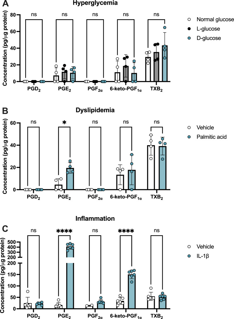
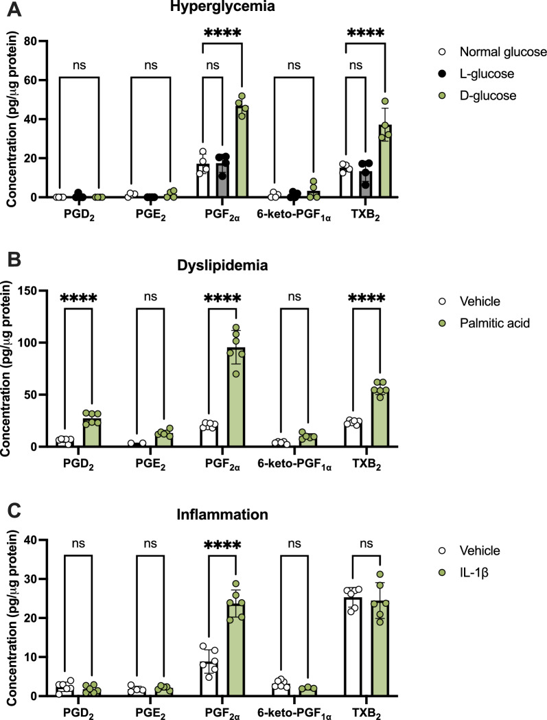
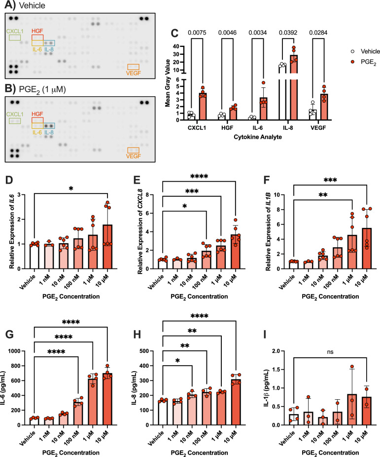
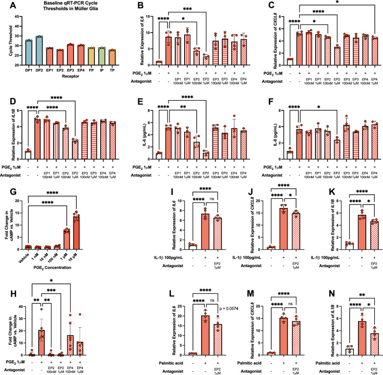
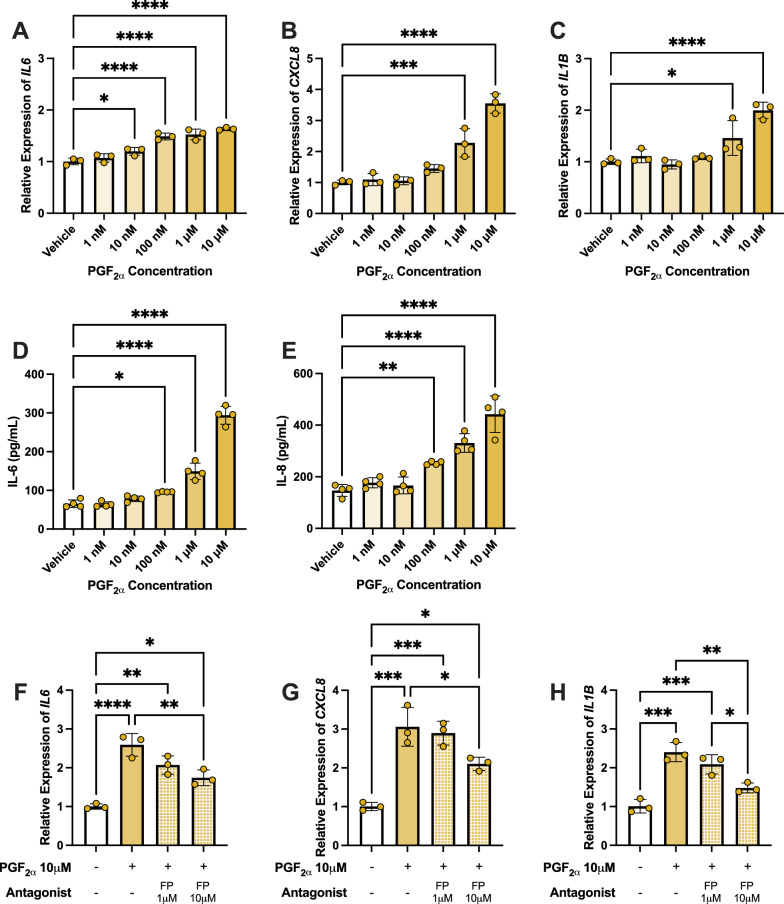
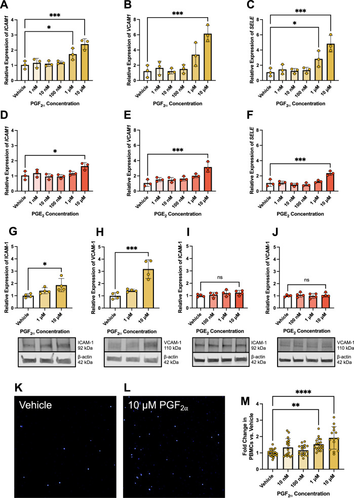
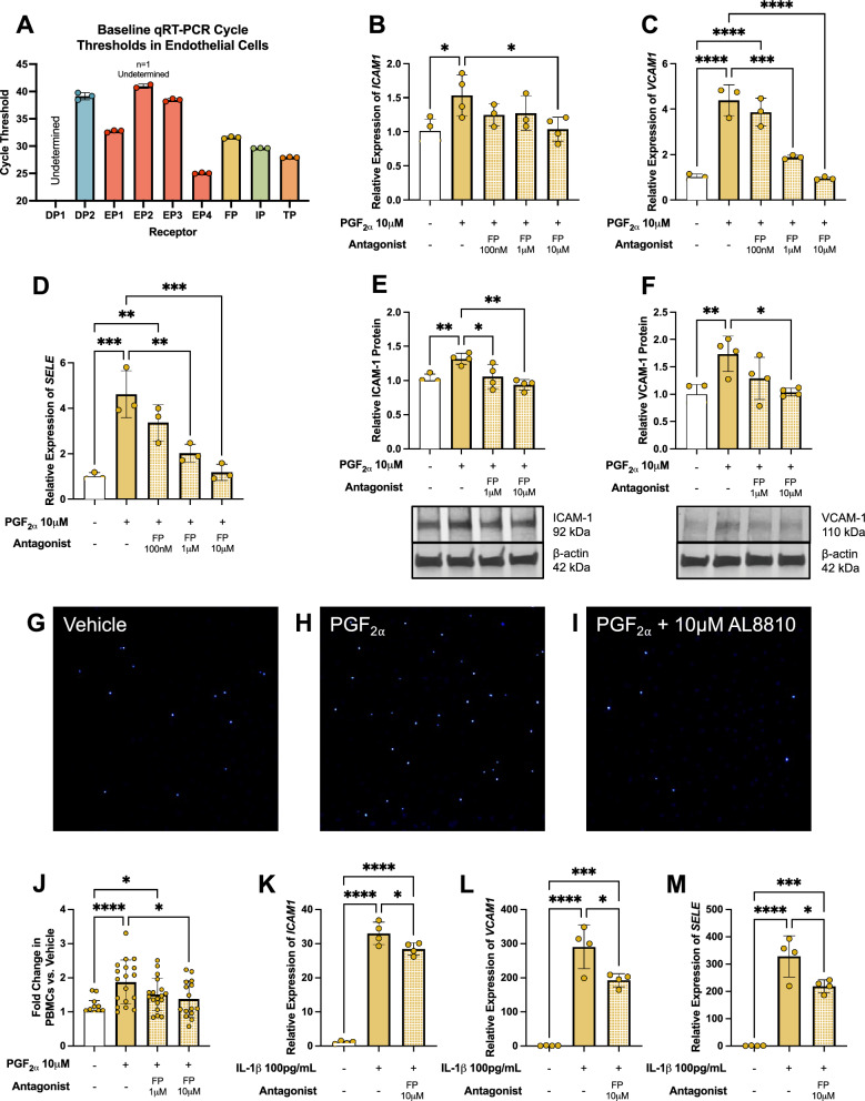
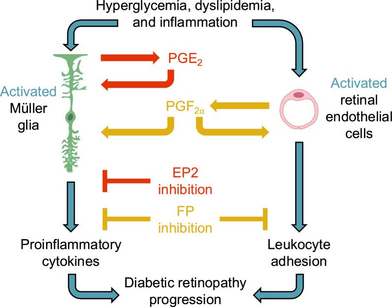
Inflammation is a critical driver of the early stages of diabetic retinopathy (DR) and offers an opportunity for therapeutic intervention before irreversible damage and vision loss associated with later stages of DR ensue. Nonsteroidal anti-inflammatory drugs (NSAIDs) have shown mixed efficacy in slowing early DR progression, notably including severe adverse side effects likely due to their nonselective inhibition of all downstream signaling intermediates. In this study, we investigated the role of prostanoids, the downstream signaling lipids whose production is inhibited by NSAIDs, in promoting inflammation relevant to early-stage DR in two human retinal cell types: Müller glia and retinal microvascular endothelial cells. When cultured in multiple conditions modeling distinct aspects of systemic diabetes, Müller glia significantly increased production of prostaglandin E2 (PGE2), whereas retinal endothelial cells significantly increased production of prostaglandin F (PGF). Müller glia stimulated with PGE2 or PGF increased proinflammatory cytokine levels dose-dependently. These effects were blocked by selective antagonists to the EP2 receptor of PGE2 or the FP receptor of PGF, respectively. In contrast, only PGF stimulated adhesion molecule expression in retinal endothelial cells and leukocyte adhesion to cultured endothelial monolayers, effects that were fully prevented by FP receptor antagonist treatment. Together these results identify PGE2-EP2 and PGF-FP signaling as novel, selective targets for future studies and therapeutic development to mitigate or prevent retinal inflammation characteristic of early-stage DR.


Conflict of interest statement

Declarations. Ethics approval and consent to participate: Not applicable. All human materials were obtained from the National Disease Research Interchange or commercial sources and de-identified. Patient identity cannot be ascertained by the investigators. Competing interests: The authors declare no competing interests.

Figures

Fig. 1
Fig. 1
Prostanoid production by hMG in conditions simulating systemic diabetes. hMG were stimulated with (A) additional 24.5 mM l-glucose or d-glucose, (B) 250 μM palmitic acid, or (C) 1 ng/mL recombinant IL-1β or relevant controls for 24 h, then media were collected for LC–MS/MS targeting PGD2, PGE2, PGF, 6-keto-PGF (PGI2 metabolite) and TXB2 (TXA2 metabolite). Data were normalized as pg prostanoid per μg of total protein from cell lysates (n = 2–6). Data represent mean ± SD. Two-way ANOVAs with Šídák post-hoc tests were used. Statistically significant differences are represented as *P < 0.05, ****P < 0.0001, ns (not significant) P > 0.05
Fig. 2
Fig. 2
Prostanoid production from hRMEC in conditions simulating systemic diabetes. hRMEC were stimulated with (A) 24.5 mM l-glucose or d-glucose, (B) 250 μM palmitic acid, or (C) 1 ng/mL recombinant IL-1β or relevant controls for 24 h, then media were collected for LC–MS/MS targeting PGD2, PGE2, PGF, 6-keto-PGF (PGI2 metabolite) and TXB2 (TXA2 metabolite). Data were normalized as pg prostanoid per μg of total protein from cell lysates (n = 2–6). Data represent mean ± SD. Two-way ANOVAs with Šídák post-hoc tests were used. Statistically significant differences are represented as ****P < 0.0001, ns (not significant) P > 0.05
Fig. 3
Fig. 3
PGE2 stimulates elevation of proinflammatory cytokine levels. Representative cytokine arrays treated with hMG-conditioned media after 6 h of stimulation with (A) vehicle or (B) 1 μM PGE2. (C) Significantly altered targets averaged from all arrays (n = 4). Multiple ratio paired T tests with Holm-Šídák post-hoc tests were used for 3C and adjusted P values are shown. (D) IL6, (E) CXCL8, and (F) IL1B qRT-PCR gene expression changes in hMG stimulated with vehicle or elevating PGE2 concentrations for 6 h (n = 3–6). (G) IL-6, (H) IL-8, and (I) IL-1β ELISA protein level changes from media of hMG stimulated with vehicle or elevating PGE2 concentrations for 6 h (n = 2–4). Data represent mean ± SD. One-way ANOVAs with Dunnett post-hoc tests were used for 3D-I. Statistically significant differences are represented as *P < 0.05, **P < 0.01, ***P < 0.001, ****P < 0.0001, ns (not significant) P > 0.05
Fig. 4
Fig. 4
PGE2-EP2 signaling mediates proinflammatory cytokine production in hMG. (A) qRT-PCR cycle thresholds of prostanoid receptor genes in unstimulated hMG (n = 3). (B) IL6 gene expression in hMG stimulated with vehicle or PGE2 ± prostanoid receptor antagonist for 2 h (n = 3–4). (C) IL1B and (D) CXCL8 gene expression in hMG stimulated with vehicle or PGE2 ± prostanoid receptor antagonist for 6 h (n = 3–4). (E) IL-6 protein levels in culture media from hMG stimulated with vehicle or PGE2 ± prostanoid receptor antagonist for 6 h (n = 3–4). (F) IL-8 protein levels in culture media from hMG stimulated with vehicle or PGE2 ± prostanoid receptor antagonist for 10 h (n = 3–4). (G) cAMP production from hMG stimulated with vehicle or elevating PGE2 concentrations for 15 min (n = 6). (H) cAMP production from hMG stimulated with vehicle or 1 μM PGE2 ± EP2 or EP4 antagonists for 15 min (n = 6). (I) IL6, (J) CXCL8, and (K) IL1B gene expression in hMG stimulated with vehicle or 100 pg/mL IL-1β ± EP2 antagonist for 6 h (n = 4). (L) IL6, (M) CXCL8, and (N) IL1B gene expression in hMG stimulated with vehicle or 250 μM palmitic acid ± EP2 antagonist for 24 h (n = 4). Data represent mean ± SD. One-way ANOVAs with Dunnett post-hoc tests were used for 4B-G. One-way ANOVAs with Tukey post-hoc tests were used for 4H-N. Statistically significant differences are represented as *P < 0.05, **P < 0.01, ***P < 0.001, ****P < 0.0001, ns (not significant) P > 0.05
Fig. 5
Fig. 5
PGF-FP signaling promotes proinflammatory cytokine production in hMG. (A) IL6, (B) CXCL8, and (C) IL1B qRT-PCR gene expression changes in hMG stimulated with vehicle or elevating PGF concentrations for 6 h (n = 3). (D) IL-6 and (E) IL-8 ELISA protein level changes from media of hMG stimulated with vehicle or elevating PGF concentrations for 6 h (n = 4). (F) IL6 gene expression in hMG stimulated with vehicle or PGF ± FP receptor antagonist for 2 h (n = 3). (G) CXCL8 and (H) IL1B gene expression in hMG stimulated with vehicle or PGF ± FP receptor antagonist for 6 h (n = 3). Data represent mean ± SD. One-way ANOVAs with Dunnett post-hoc tests were used for 5A-E. One-way ANOVAs with Tukey post-hoc tests were used for 5F-H. Statistically significant differences are represented as *P < 0.05, **P < 0.01, ***P < 0.001, ****P < 0.0001
Fig. 6
Fig. 6
PGF, but not PGE2, promotes leukostasis-relevant activity at gene, protein, and cell behavior levels in hRMEC. (A) ICAM1, (B) VCAM1, and (C) SELE qRT-PCR gene expression changes in hRMEC stimulated with vehicle or elevating PGF concentrations for 6 h (n = 3). (D) ICAM1, (E) VCAM1, and (F) SELE qRT-PCR gene expression changes in hRMEC stimulated with vehicle or elevating PGE2 concentrations for 6 h (n = 3). (G) ICAM-1 and (H) VCAM-1 western blot protein levels and representative blots from hRMEC stimulated with vehicle or elevating PGF concentrations for 6 h (n = 4). (I) ICAM-1 and (J) VCAM-1 protein levels and representative western blots from hRMEC stimulated with vehicle or elevating PGE2 concentrations for 6 h (n = 4). Representative images of static PBMC adhesion after (K) vehicle or (L) 10 μM PGF stimulation for 6 h. (M) Static adhesion results with vehicle or elevating PGF concentrations for 6 h (n = 14–20). Data represent mean ± SD. One-way ANOVAs with Dunnett post-hoc tests were used. Statistically significant differences are represented as *P < 0.05, **P < 0.01, ***P < 0.001, ****P < 0.0001, ns (not significant) P > 0.05
Fig. 7
Fig. 7
PGF-FP signaling mediates leukocyte adhesion in hRMEC. (A) qRT-PCR cycle thresholds of prostanoid receptor genes in unstimulated hRMEC (n = 3). (B) ICAM1, (C) VCAM1, and (D) SELE gene expression in hRMEC stimulated with vehicle or PGF ± FP receptor antagonist for 6 h (n = 3). (E) ICAM-1 and (F) VCAM-1 western blot protein levels and representative blots from hRMEC stimulated with vehicle or PGF ± FP receptor antagonist for 10 h (n = 4). Representative images of static PBMC adhesion after (G) vehicle, (H) 10 μM PGF, or (I) 10 μM PGF + 10 μM FP receptor antagonist treatment. (J) Static adhesion results with vehicle or PGF ± FP receptor antagonist for 10 h (n = 15–18). (K) ICAM1, (L) VCAM1, and (M) SELE gene expression in hRMEC stimulated with vehicle or 100 pg/mL IL-1β ± FP antagonist for 6 h (n = 4). Data represent mean ± SD. One-way ANOVAs with Tukey post-hoc tests were used. Statistically significant differences are represented as *P < 0.05, **P < 0.01, ***P < 0.001, ****P < 0.0001
Fig. 8
Fig. 8
Proposed mechanisms of DR-relevant prostanoid signaling in Müller glia and retinal endothelial cells

Similar articles

Cited by

References

    1. Kempen JH, et al. The prevalence of diabetic retinopathy among adults in the United States. Arch Ophthalmol. 2004;122:552–63. - PubMed
    1. Lundeen EA, et al. Prevalence of diabetic retinopathy in the US in 2021. JAMA Ophthalmol. 2023;141:747–54. - PMC - PubMed
    1. Yau JW, et al. Global prevalence and major risk factors of diabetic retinopathy. Diabetes Care. 2012;35:556–64. - PMC - PubMed
    1. Antonetti DA, Klein R, Gardner TW. Diabetic retinopathy. N Engl J Med. 2012. 10.1056/NEJMra1005073. - PubMed
    1. Wong TY, Cheung CMG, Larsen M, Sharma S, Simó R. Diabetic retinopathy. Nat Rev Dis Prim. 2016;2:1–17. - PubMed

MeSH terms

LinkOut - more resources