. 2024 Dec 3;34(12):bhae480.
doi: 10.1093/cercor/bhae480.

Identification of Pappa and Sall3 as Gli3 direct target genes acting downstream of cilia signaling in corticogenesis

Affiliations

Identification of Pappa and Sall3 as Gli3 direct target genes acting downstream of cilia signaling in corticogenesis

Shinjini Basu et al. Cereb Cortex. .

Abstract

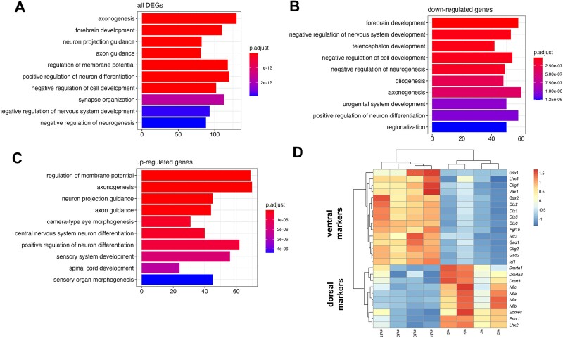
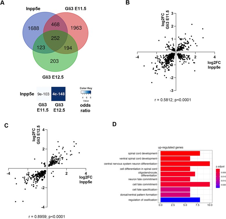
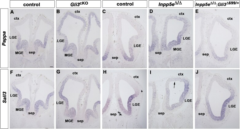
The cerebral cortex is critical for advanced cognitive functions and relies on a vast network of neurons to carry out its highly intricate neural tasks. Generating cortical neurons in accurate numbers hinges on cell signaling orchestrated by primary cilia to coordinate the proliferation and differentiation of cortical stem cells. While recent research has shed light on multiple ciliary roles in corticogenesis, specific mechanisms downstream of cilia signaling remain largely unexplored. We previously showed that an excess of early-born cortical neurons in mice mutant for the ciliary gene Inpp5e was rescued by re-introducing Gli3 repressor. By comparing expression profiles between Inpp5e and Gli3 mutants, we here identified novel Gli3 target genes. This approach highlighted the transcription factor gene Sall3 and Pappalysin1 (Pappa), a metalloproteinase involved in IGF signaling, as upregulated genes in both mutants. Further examination revealed that Gli3 directly binds to Sall3 and Pappa enhancers and suppresses their activity in the dorsal telencephalon. Collectively, our analyses provide important mechanistic insights into how primary cilia govern the behavior of neural stem cells, ultimately ensuring the production of adequate numbers of neurons during corticogenesis.

Keywords: Gli3; Inpp5e; Pappa; Sall3; corticogenesis; primary cilia.


Figures

Fig. 1
Fig. 1
Differential gene expression in the E12.5 Inpp5eΔ/Δ dorsal telencephalon. (A–C) GO analysis of all DEGs (A), and of only downregulated (B) or upregulated genes (C). (D) Heatmap comparing the expression of dorsal and ventral telencephalic markers.
Fig. 2
Fig. 2
Comparison of differential gene expression between Inpp5e and Gli3 mutants. (A) Venn diagram intersection of DEGS in E12.5 Inpp5eΔ/Δ, E11.5 and E12.5 Gli3cKO embryos. Significance and odds ratio are indicated. (B, C) Comparison of gene expression changes between Inpp5eΔ/Δ and E11.5 (B) and E12.5 (C) Gli3cKO mutants. (D) GO analysis of genes upregulated in both mutants. Statistical tests: Fisher’s exact test (A) and Spearman correlation (B, C).
Fig. 3
Fig. 3
Pappa and Sall3 expression in the Gli3cKO and Inpp5eΔ/Δ mutant telencephalon. Coronal sections of the E12.5 forebrain of the indicated genotypes were in situ hybridized with the indicated probes. (A, C) Pappa expression is confined to progenitors in the septum (sep), medial ganglionic eminence (MGE) and the ventral lateral ganglionic eminence (LGE) of control embryos. (B, D) Ectopic Pappa expression throughout the cortex (ctx) of Gli3cKO embryos and in the dorsolateral telencephalon of Inpp5e mutant embryos. (F, H) Sall3 expression is confined to the ventral telencephalon in control embryos. (G, I) Gli3cKO embryos display ectopic Sall3 transcripts in the developing cortex while Inpp5e mutants present with a more restricted up-regulation in the dorsal telencephalon (arrow). (E, J) Pappa and Sall3 expression remain confined to the ventral telencephalon in Inpp5eΔ/Δ; Gli3Δ699/+ embryos. Scale bar: 200 mm.
Fig. 4
Fig. 4
Gli3 binds to Pappa and Sall3 enhancers in vivo and in vitro. (A, B) Genome browser snapshots showing Gli3 ChIP-peaks at exon 13 of Pappa overlapping with an open chromatin region (ATACseq peak) (A) and in the first intron of Sall3 (B). The latter coincides with an H3K27ac positive region and a Gli3 ChIP-peak identified in motor neuron progenitors. (C, D) Evolutionary conservation of Gli3 binding sites in the Pappa (B) and Sall3 (D) enhancers. (E-H) Ectromobility shift assays demonstrating specific binding of a GST-Gli3 fusion protein to binding sites 1 (E) and 2 (F) of the Pappa enhancer and to sites 1 (G) and 2 (H) of the Sall3 enhancer.
Fig. 5
Fig. 5
Gli3 represses activity of Pappa and Sall3 enhancers in the dorsolateral telencephalon. Coronal forebrain sections of E14.5 embryos in utero electroporated with the indicated constructs were stained either with GFP antibodies or with X-Gal. GFP staining indicates the electroporated regions (arrows in A, C, F and H). (A-E) The Pappa and Sall3 enhancers showed weak activity in a limited number of cells in the dorsolateral telencephalon. (F, G) Mutations in the Gli3 binding sites led to strong reporter gene expression (arrow in G). Note the different staining times. (H-J) Activity of a Gli3 binding site mutant Sall3 enhancer was stronger and more widespread (arrows in J). Abbreviations: Ctx, cortex; MGE, medial ganglionic eminence; LGE, lateral ganglionic eminence; sep, septum. Scale bar: 250 mm.

Similar articles

References

    1. Akiyama R, Kawakami H, Wong J, Oishi I, Nishinakamura R, Kawakami Y. Sall4-Gli3 system in early limb progenitors is essential for the development of limb skeletal elements. Proc Natl Acad Sci USA. 2015:112:5075–5080. 10.1073/pnas.1421949112. - DOI - PMC - PubMed
    1. Aslanyan MG, Doornbos C, Diwan GD, Anvarian Z, Beyer T, Junger K, Beersum SEC, Russell RB, Ueffing M, Ludwig A, et al. . A targeted multi-proteomics approach generates a blueprint of the ciliary ubiquitinome. Front Cell Dev Biol. 2023:11:1113656. 10.3389/fcell.2023.1113656. - DOI - PMC - PubMed
    1. Ayer-le Lievre C, Stahlbom PA, Sara VR. Expression of IGF-I and -II mRNA in the brain and craniofacial region of the rat fetus. Development. 1991:111:105–115. 10.1242/dev.111.1.105. - DOI - PubMed
    1. Bachmann-Gagescu R, Ishak GE, Dempsey JC, Adkins J, O'Day D, Phelps IG, Gunay-Aygun M, Kline AD, Szczaluba K, Martorell L, et al. . Genotype-phenotype correlation in CC2D2A-related Joubert syndrome reveals an association with ventriculomegaly and seizures. J Med Genet. 2012:49:126–137. 10.1136/jmedgenet-2011-100552. - DOI - PubMed
    1. Beck KD, Powell-Braxton L, Widmer HR, Valverde J, Hefti F. Igf1 gene disruption results in reduced brain size, CNS hypomyelination, and loss of hippocampal granule and striatal parvalbumin-containing neurons. Neuron. 14:717–730. 10.1016/0896-6273(95)90216-3. - DOI - PubMed

Publication types

MeSH terms

LinkOut - more resources