Review
. 2025 May 13;148(5):1479-1506.
doi: 10.1093/brain/awae413.

From bugs to brain: unravelling the GABA signalling networks in the brain-gut-microbiome axis

Affiliations
Review

From bugs to brain: unravelling the GABA signalling networks in the brain-gut-microbiome axis

Delia Belelli et al. Brain. .

Abstract

Convergent data across species paint a compelling picture of the critical role of the gut and its resident microbiota in several brain functions and disorders. The chemicals mediating communication along these sophisticated highways of the brain-gut-microbiome (BGM) axis include both microbiota metabolites and classical neurotransmitters. Amongst the latter, GABA is fundamental to brain function, mediating most neuronal inhibition. Until recently, GABA's role and specific molecular targets in the periphery within the BGM axis had received limited attention. Yet, GABA is produced by neuronal and non-neuronal elements of the BGM, and recently, GABA-modulating bacteria have been identified as key players in GABAergic gut systems, indicating that GABA-mediated signalling is likely to transcend physiological boundaries and species. We review the available evidence to better understand how GABA facilitates the integration of molecularly and functionally disparate systems to bring about overall homeostasis and how GABA perturbations within the BGM axis can give rise to multi-system medical disorders, thereby magnifying the disease burden and the challenges for patient care. Analysis of transcriptomic databases revealed significant overlaps between GABAAR subunits expressed in the human brain and gut. However, in the gut, there are notable expression profiles for a select number of subunits that have received limited attention to date but could be functionally relevant for BGM axis homeostasis. GABAergic signalling, via different receptor subtypes, directly regulates BGM homeostasis by modulating the excitability of neurons within brain centres responsible for gastrointestinal (GI) function in a sex-dependent manner, potentially revealing mechanisms underlying the greater prevalence of GI disturbances in females. Apart from such top-down regulation of the BGM axis, a diverse group of cell types, including enteric neurons, glia, enteroendocrine cells, immune cells and bacteria, integrate peripheral GABA signals to influence brain functions and potentially contribute to brain disorders. We propose several priorities for this field, including the exploitation of available technologies to functionally dissect components of these GABA pathways within the BGM, with a focus on GI and brain-behaviour-disease. Furthermore, in silico ligand-receptor docking analyses using relevant bacterial metabolomic datasets, coupled with advances in knowledge of GABAAR 3D structures, could uncover new ligands with novel therapeutic potential. Finally, targeted design of dietary interventions is imperative to advancing their therapeutic potential to support GABA homeostasis across the BGM axis.

Keywords: GABA receptors; enteric nervous system; inflammation; neurosteroids; pre/probiotics; psychopathology.


Conflict of interest statement

The authors declare that this manuscript was composed in the absence of any commercial or financial relationships that could be construed as a potential conflict of interest. J.D.S. is in receipt of research funding from GABA Labs. D.B. works as a senior scientific consultant for GABA Labs and as a Visiting Researcher at the University of Portsmouth. All other authors report no competing interests.

Figures
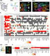
Figure 1
Figure 1
Molecular, pharmacological and expression diversity of mammalian GABA receptors throughout the body. [A(i)] The basic structure of an individual GABA type A receptor (GABAAR) subunit (viewed from the side and flattened), when anchored in the plasma membrane. These proteins consist of four transmembrane domains (TM 1–4), two intracellular loops and one extracellular loop, an extracellular amino terminus (N) and carboxyl (C) termini. TM 2 is distinguished by putatively forming the lining of the ion channel. EX = extracellular; IN = intracellular. [A(ii)] The molecular diversity of individual GABAAR subunits, as well as their general stoichiometry, assembling the pentameric GABAAR complex, which contains two α- and two β-subunits and a single auxiliary (AUX) subunit. [A(iii)] Model of the 3D crystal structure of an assembled GABAAR, viewed from the side. The image was generated by the Protein Data Bank in Europe, 8g5h, and is based on data originally published by Sun et al. [A(iv)] provides a view from the top of the GABAAR mentioned in iii, illustrating (arrows) the diversity of receptor sites at which ligands have been shown to bind. These include separate sites for the endogenous ligand and neurotransmitter GABA, the exogenous, positive allosteric modulator zolpidem (ZOL) and the endogenous neurosteroid allopregnanolone (ALLO). The data indicate that the GABA binding site is located at the interface of the α- and β-subunits, zolpidem binds to the extracellular domain interface of the α1- and 2-subunits, and the binding pockets for ALLO are located at the interface between TM 1 and 4 of the α-subunit and TM 3 of the adjacent β-subunit.B illustrates the relative mRNA expression levels of 19 different human GABAAR subunits across various tissues in the human body, based on consensus datasets from the Protein Atlas database (https://www.proteinatlas.org/). The heat map was generated using Heatmapper (http://www.heatmapper.ca/). Expression levels are represented by colour intensity: grey indicates no expression, red indicates high expression levels and white indicates low expression levels. The values are presented in normalized transcripts per million (nTPM). [C(i)] The basic structure of an individual GABA type B receptor (GABABR). The receptor is composed of two subunits, GABAB1 and GABAB2, and belongs to the super-family of metabotropic G-protein coupled receptors. [C(ii)] Model of the 3D crystal structure of the extracellular regions of an assembled GABABR, viewed from the side. The image was generated by the Protein Data Bank in Europe, 4mr7, and is based on data originally published by Geng et al. (D) The relative mRNA expression levels of the two GABABR subunits across various tissues in the human body, based on consensus datasets from the Protein Atlas database, created in a similar manner to C. The figure was prepared using BioRender (www.biorender.com).
Figure 2
Figure 2
Roles of GABAergic signalling in brain-to-gut neuronal pathways. (A) Key brain centres located within the medulla that are involved in providing motor output to the gastrointestinal tract (GIT). [A(i)] Organization between neurochemically diverse neurons of the nucleus of the solitary tract (NTS) and one of its major projection targets, the dorsal motor nucleus of the vagus (DMV). GABAergic inputs from the NTS onto DMV neurons are mediated by a range of GABA type A receptor (GABAAR) subtypes. Whilst the majority of DMV neurons express acetylcholine (ACh), they can also be divided into two subpopulations based on their co-expression of either cholecystokinin (CCK) or prodynorphin (PDYN). DMV axons project via the 10th cranial nerve (X) to make direct synaptic connections with their enteric nervous system targets in the myenteric plexus. [A(ii)] The two distinct types of DMV cholinergic neurons, CCK (blue) and PDYN (red), contact different enteric neuronal types (Chat+ and NOS1+) to induce gastric contraction and relaxation, respectively. A single-nucleus RNA sequence (snRNA-seq) analysis performed on data retrieved from a report by Tao et al., revealed the differential expression of GABAAR subunits between the two types of DMV cholinergic neurons, CCK and PDYN, in mouse models. A(iii) shows that this NTS–DMV pathway, as well as the associated GABAAR subtypes, preferentially regulates the proximal regions of the GIT. (B) Key brain centres located within the pons that are involved in providing motor output to the distal GIT (B4). [B(i)] Barrington's nucleus (BN) interacts with the locus coeruleus (LC) in regulating voiding behaviour of the gut viscera through their modulation of the distal colon. The principal noradrenergic (NA) neurons of the LC receive excitatory input from BN neurons. They also receive inhibitory inputs from GABAergic interneurons located near BN as well as within the LC. Whilst LC NA neurons have been shown to express α2/3-β-γ-GABAAR subunits, non-NA, GABAergic LC neurons express α1-β-γ-GABAAR subunits.,, [B(ii)] Single-nucleus RNA sequencing (RNA-seq) data reported by Nardone et al. were inspected in the Broad Institute Single Cell portal (https://singlecell.broadinstitute.org/single_cell), reference ‘pons_exc_neurons_snrnaseq’ to compare the gene expression of GABAAR subunits between female (F) and male (M) mice in the dorsal pontine tegmentum (DPT) region. The sizes of the dots in the dot plot indicate the percentage of cells in which gene expression was detected, and the colour scale indicates the magnitude of gene expression (scaled gene expression defines the expression maximum value as 1 and the minimum as 0). The DPT includes the pre-LC, LC, BRN and mesencephalic trigeminal nucleus (MTN) centres. [B(iii)] An analysis of mouse LC single-cell RNA-seq data reported by Luskin et al. shows the average percentage of GABAAR subunit gene expression per cell cluster. Noradrenergic (NE) neurons are shown in blue and GABA neurons in red. [B(iv)] The selective innervation of distal colon by this pathway and its involvement primarily in voiding behaviour. The figure was prepared using BioRender (www.biorender.com).
Figure 3
Figure 3
The roles of GABAergic signalling in neuronal and non-neuronal gut-to-brain pathways. (A) Schematic summary of the circuitry relaying gastrointestinal (GI) signals to the brain. [A(i)] In proximal (ORAL) regions, nodose ganglion (NG) sensory axons innervate the wall of the GI tract (GIT), with their centrally projecting axons innervating the nucleus tractus solitarii (NTS). The NTS in turn projects to neurons within the dorsal nucleus of the vagus (DMV). The NG is immunopositive for GABA type B receptors (GABABRs) but not type A receptors (GABAARs). While NG cells are devoid of synaptic inputs, GABA may act here as diffusible transmitter released non-synaptically, for example, from local satellite glial cells., [A(ii)] In distal (ANAL) regions of the GIT, sensory axons of lumbosacral dorsal root ganglion (LSDRG) neurons make a variety of contacts throughout the GIT, including the muscle, enteric nervous system (ENS) and submucosa. LSDRG centrally projecting axons innervate sacral (S) spinal neurons that project to Barrington's nucleus (BN), which in turn activates neurons in the locus coeruleus (LC). [A(iii)] An overview of different cell types located in the mucosa and the associated GABAergic molecular machinery they have been demonstrated to express, including IPANS, enteric glia, enterocytes, enteroendocrine cells (EEC) and tuft cells. MP = myenteric plexus; SP = submucosal plexus The green arrows indicate the direction of efferent information flow from the gut to the brain. B summarizes the expression and function of different GABAAR subtypes within the mouse ENS. ICC = interstitial cells of Cajal; IN = interneuron; MN = motor neuron. [B(i)] The neurochemical identity of ENS neurons that have been shown immunohistochemically to express specific GABAAR subunits.,,, [B(ii)] The effects of the pharmacological activation of different GABAAR subtypes on various parameters underlying spontaneous contractions of the mouse colon. [B(iii)] The effects of the pharmacological activation of different GABAAR subtypes on intrinsic colonic inflammation. N.D. = not determined. (C) An analysis of previously published single-cell RNA sequencing datasets of ENS GABAAR subunit mRNA expression. [C(i)] An analysis of data published by Drokhlyansky et al. The sizes of the dots in the dot plot indicate the percentage of cells in which gene expression was detected, and the colour scale indicates the magnitude of gene expression (scaled gene expression defines the expression maximum value as 1 and the minimum as 0). [C(ii)] An analysis of data published by Wright et al. depicting the proportion of cells expressing subunits. The figure was prepared using BioRender (www.biorender.com).
Figure 4
Figure 4
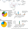
Bacterial GABA synthesis. (A) Pathways utilized by bacteria to synthesize GABA. The main synthetic route occurs via glutamic acid decarboxylase (GAD), encoded by gadA/B genes, while the putrescine pathways represent a minor route used by certain species such as Escherichia coli. These pathways are absent from Lactobacillus and Bifidobacterium strains, which instead utilize the GAD pathway. In addition to the gadA/B genes, the bacterial GAD operon also includes a gadC gene, which encodes an antiporter, allowing GABA to be exported from the cell, as reviewed by Sarasa et al., Diez-Gutierrez et al. and Yogeswara et al. Note that the measured concentrations (sub to low mM) of GABA produced by human gut bacteria fall within a physiological bioactive range. (B) An overview of human microbes expressing (i) gadA and (ii) gadB gene. Human microbes expressing either gene were identified from the Integrated Microbial Genomes & Microbiomes database (accessed on 30 June 2024). These microbes were categorized into gut microbiota, non-gut microbiota and pathogens. Pie charts illustrate the distribution, including types and relative contributions of all human microbial species from the database. Histograms depict the distribution of gut microbiota, non-gut microbiota and pathogens. The total number and percentages of human microbial species obtained from the database are described in the Supplementary material. The figure was prepared using BioRender (www.biorender.com).
Figure 5
Figure 5
Versatile sources, actions and targets of GABA in the gastrointestinal tract. GABA and putatively neurosteroids (NSs) exhibit pleiotropic actions and targets in the gastrointestinal (GI) tract. (A) GABA targets neuronal and potentially glial GABA receptors in the submucosal and myenteric plexuses of the enteric nervous system and modulates vagal transmission., GABA can act on local and systemic immune cells, (B), which express both GABA type A and B receptors (GABAARs and GABABRs). As the gut-associated lymphoid system accounts for ∼70% of the body's immune cells, the GI GABA system is ideally placed to influence immunity. Although the specific mechanisms are yet to be fully elucidated, gut GABA can also induce blood-detected exosome signalling associated with brain plasticity, e.g. in the hippocampus (C). Finally, GABA can act on bacterial communities to modulate their relative abundance [D(i)], either by acting as an essential nutrient or by affecting the expression or transfer of crucial genes, e.g. as a virulence inhibitor of invading plasmids from pathogenic species such as Agrobacterium tumefaciens., GABA specifically promotes the degradation of quorum sensing signals that control plasmid transfer. Interestingly, unique GABA binding motifs have been identified in the periplasmic bacterial protein (PBP) Atu4243 of A. tumefaciens, which functions as a selective GABA-sensor and transporter to satisfy nutritional requirements. These opposite roles of GABA suggest that GABA participates in and fine-tunes the complex relationship between hosts and biotrophic pathogens. Albeit speculative at present, GABA may also target bacterial receptors [D(ii)], akin to the identified GLIC (Gleobacter ligand-gated ion channel) and ELIC (Erwinia ligand-gated ion channel), prokaryotic GABA receptors of the Cyanobacteria phylum, closely related to some recently identified human gut bacteria. In indirect support, recent evidence has revealed that GABA modifies the bioelectrical properties of a known commensal bacterial strain Lactobacillus reuteri to impact their growth, congruent with the known bacterial use of change in membrane potential to convey and process key information. The figure was prepared using BioRender (www.biorender.com).

Similar articles

Cited by

References

    1. Cryan JF, O’Riordan KJ, Cowan CSM, et al. . The microbiota-gut-brain axis. Physiol Rev. 2019;99:1877–2013. - PubMed
    1. Morais LH, Schreiber HL 4th, Mazmanian SK. The gut microbiota-brain axis in behaviour and brain disorders. Nat Rev Microbiol. 2021;19:241–255. - PubMed
    1. Lai Y, Dhingra R, Zhang Z, Ball LM, Zylka MJ, Lu K. Toward elucidating the human gut microbiota-brain axis: Molecules, biochemistry, and implications for health and diseases. Biochemistry. 2022;61:2806–2821. - PMC - PubMed
    1. Bonaz B, Bazin T, Pellissier S. The vagus nerve at the interface of the microbiota-gut-brain axis. Front Neurosci. 2018;12:49. - PMC - PubMed
    1. Schroeder BO, Bäckhed F. Signals from the gut microbiota to distant organs in physiology and disease. Nat Med. 2016;22:1079–1089. - PubMed

Substances