

Human umbilical cord mesenchymal stem cells enhance liver regeneration and decrease collagen content in fibrosis mice after partial hepatectomy by activating Wnt/β-catenin signaling
- PMID: 39716885
- PMCID: PMC12040747
- DOI: 10.3724/abbs.2024207
Human umbilical cord mesenchymal stem cells enhance liver regeneration and decrease collagen content in fibrosis mice after partial hepatectomy by activating Wnt/β-catenin signaling
Abstract
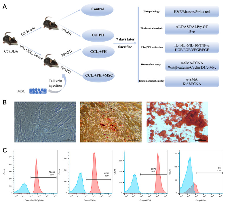
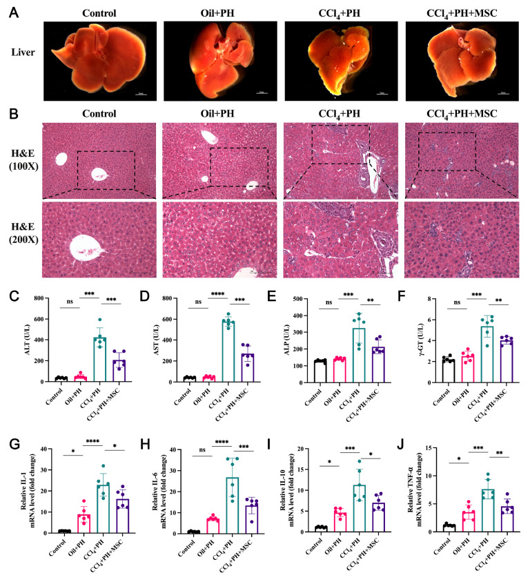
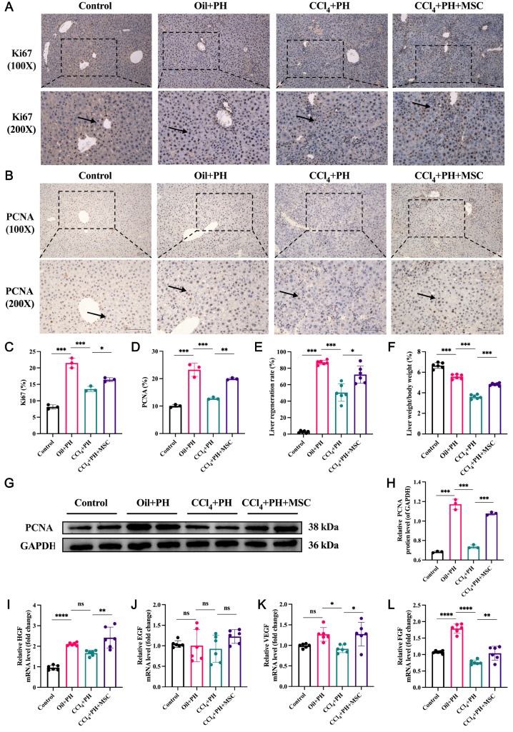
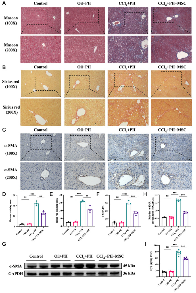
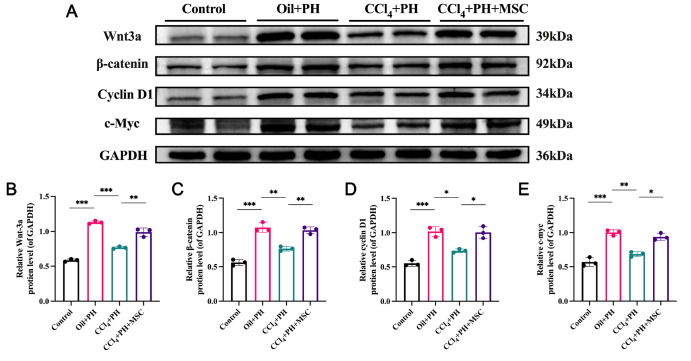
Liver fibrosis is a critical stage in the progression of various chronic liver diseases to cirrhosis and liver cancer. Early inhibition of liver fibrosis is crucial for the treatment of liver disease. Hepatectomy, a common treatment for liver-related diseases, promotes liver regeneration. However, in the context of liver fibrosis, liver regeneration is hindered. Many studies have shown that mesenchymal stem cells (MSCs) can promote liver regeneration after partial hepatectomy (PH). However, there are few reports on the impact of MSC therapy on liver regeneration post-PH in the context of hepatic fibrosis. The objective of this study is to examine the impact of MSCs on liver regeneration following PH in the fibrotic liver and uncover the related molecular mechanisms. This study reveals that MSC therapy significantly enhances liver function and mitigates liver inflammation after PH in the context of hepatic fibrosis. MSCs also significantly promote liver regeneration and alleviate liver fibrosis. In addition, this study identifies the role of MSCs in promoting liver regeneration and alleviating liver fibrosis via the activation of Wnt/β-catenin signaling. The combination of MSCs with hepatectomy may offer a novel approach for the treatment of liver fibrotic diseases.
Keywords: liver fibrosis; liver regeneration; mesenchymal stem cells; partial hepatectomy.
Conflict of interest statement
The authors declare that they have no conflict of interest.
Figures
Similar articles
-
Human bone marrow mesenchymal stem cells-derived exosomes alleviate liver fibrosis through the Wnt/β-catenin pathway.Stem Cell Res Ther. 2019 Mar 18;10(1):98. doi: 10.1186/s13287-019-1204-2. Stem Cell Res Ther. 2019. PMID: 30885249 Free PMC article.
-
Local but not systemic administration of mesenchymal stromal cells ameliorates fibrogenesis in regenerating livers.J Cell Mol Med. 2019 Sep;23(9):6238-6250. doi: 10.1111/jcmm.14508. Epub 2019 Jun 27. J Cell Mol Med. 2019. PMID: 31245923 Free PMC article.
-
Human umbilical cord-derived mesenchymal stromal cells alleviate liver cirrhosis through the Hippo/YAP/Id1 pathway and macrophage-dependent mechanism.Int Immunopharmacol. 2023 Oct;123:110456. doi: 10.1016/j.intimp.2023.110456. Epub 2023 Jul 24. Int Immunopharmacol. 2023. PMID: 37494836
-
[Umbilical cord mesenchymal stem cells and their association with liver fibrosis].Zhonghua Gan Zang Bing Za Zhi. 2017 Jan 20;25(1):65-68. doi: 10.3760/cma.j.issn.1007-3418.2017.01.019. Zhonghua Gan Zang Bing Za Zhi. 2017. PMID: 28297787 Review. Chinese.
-
Strategies to improve the efficiency of mesenchymal stem cell transplantation for reversal of liver fibrosis.J Cell Mol Med. 2019 Mar;23(3):1657-1670. doi: 10.1111/jcmm.14115. Epub 2019 Jan 11. J Cell Mol Med. 2019. PMID: 30635966 Free PMC article. Review.
Cited by
-
Exploring Cirrhosis: Insights into Advances in Therapeutic Strategies.Int J Mol Sci. 2025 Jul 25;26(15):7226. doi: 10.3390/ijms26157226. Int J Mol Sci. 2025. PMID: 40806358 Free PMC article. Review.
References
-
- Ohtani N, Kawada N. Role of the gut–liver axis in liver inflammation, fibrosis, and cancer: a special focus on the gut microbiota relationship. Hepatol Commun. . 2019;3:456–470. doi: 10.1002/hep4.1331. - DOI - PMC - PubMed
-
- Tateishi R, Uchino K, Fujiwara N, Takehara T, Okanoue T, Seike M, Yoshiji H, et al. A nationwide survey on non-B, non-C hepatocellular carcinoma in Japan: 2011–2015 update. J Gastroenterol. . 2019;54:367–376. doi: 10.1007/s00535-018-1532-5. - DOI - PMC - PubMed
-
- Kisseleva T, Brenner DA. Mechanisms of fibrogenesis. Exp Biol Med (Maywood) . 2008;233:109–122. doi: 10.3181/0707-MR-190. - DOI - PubMed
-
- Sun M, Kisseleva T. Reversibility of liver fibrosis. Clin Res Hepatol Gastroenterol. . 2015;39:S60–S63. doi: 10.1016/j.clinre.2015.06.015. - DOI - PMC - PubMed
-
- Kisseleva T, Brenner D. Molecular and cellular mechanisms of liver fibrosis and its regression. Nat Rev Gastroenterol Hepatol. . 2021;18:151–166. doi: 10.1038/s41575-020-00372-7. - DOI - PubMed
MeSH terms
Substances
LinkOut - more resources
Full Text Sources
Medical
