. 2025 Feb;12(7):e2413891.
doi: 10.1002/advs.202413891. Epub 2024 Dec 24.

Triple-Combination Therapy with a Multifunctional Yolk-Shell Nanozyme Au@CeO2 Loaded with Dimethyl Fumarate for Periodontitis

Affiliations

Triple-Combination Therapy with a Multifunctional Yolk-Shell Nanozyme Au@CeO2 Loaded with Dimethyl Fumarate for Periodontitis

Tiancheng Li et al. Adv Sci (Weinh). 2025 Feb.

Abstract

Periodontitis, a chronic inflammatory disease, is the leading cause of tooth loss in adults and is one of the most prevalent and complex oral conditions. Oxidative stress induced by the excessive generation of reactive oxygen species (ROS) leads to periodontitis, which is closely associated with pathological processes, including mitochondrial dysfunction of periodontal cells and local immune dysregulation. However, current treatment modalities that target single pathological processes have limited long-term therapeutic effects. Herein, a multifunctional Yolk-Shell nanozyme, Au@CeO2-dimethyl fumarate (DMF), which comprehensively addresses the oxidative stress-induced pathophysiological processes of periodontitis through antioxidant activity, mitochondrial maintenance, and immune modulation mechanisms, is described. For material design logic, functionally complementary Au and CeO2 formed an excellent photothermally regulated high-efficiency nanozyme, which also provided an ideal drug carrier for DMF. As for the therapeutic logic, Au@CeO2-DMF restores mitochondrial dysfunction and immune dysregulation, which also contributes to endogenous ROS elimination, thereby achieving long-term stable therapeutic effects. In a rat model, local Au@CeO2-DMF photothermal therapy effectively alleviated ROS-induced tissue damage and restored periodontal homeostasis. Altogether, this study presents a novel antioxidant nanozyme for managing alveolar bone loss under prolonged oxidative stress and demonstrates the importance of comprehensive intervention in key pathological processes in periodontitis treatment design.

Keywords: dimethyl fumarate; nanozymes; periodontitis; photothermal therapy; reactive oxygen species.


Conflict of interest statement

The authors declare no conflict of interest.

Figures

Scheme 1
Scheme 1
Material design logic of Au@CeO2‐DMF and its triple‐combination therapy for periodontitis through antioxidant effect, mitochondrial maintenance, and immunomodulatory mechanisms.
Figure 1
Figure 1
Preparation and characterization of Au@CeO2 YSNs. A) SEM and B) TEM Images of YSNs. C) HRTEM images of CeO2 (200) facets, as well as the corresponding E) FFT image, F) IFFT image, and G) lattice distance plot. D) HRTEM images of CeO2 (111) facets, as well as the corresponding H) FFT image, I) IFFT image, and J) lattice distance plot. K) Sum spectrum of elemental analysis. L) XRD patterns of Au@CeO2. M) DLS hydrodynamic diameter. N) Zeta potential of Au@CeO2‐DMF measured by DLS. O) N2 adsorption/desorption isotherms.
Figure 2
Figure 2
Photothermal properties and enzyme‐mimicking activities of Au@CeO2‐DMF YSNs. A) UV—vis–NIR Spectroscopy of Au@CeO2. B) Photothermal curves of Au@CeO2 (50 µg mL−1) under laser intensities ranging from 0.4 to 1.2 W cm−2 for 5 min. C) Photothermal curves of Au@CeO2 at concentrations from 6.25 to 200 µg mL−1 upon 0.8 W cm−2 laser irradiation for 5 min. D) Photothermal curves of PBS, AuNP, Au@CeO2, and Au@CeO2‐DMF (50 µg mL−1) under 0.8 W cm−2 laser for 5 min. E) Cyclic heating profiles of Au@CeO2 and Au@CeO2‐DMF under 0.8 W cm−2 laser for three cycles. F) Heating and cooling curves of Au@CeO2‐DMF aqueous solution (50 µg mL−1, 1 mL) under 635 nm (0.8 W cm−2) laser irradiation, linear time data obtained from the cooling period. G) IR thermal images of different solutions with laser irradiation. G) Cumulative release curves of Au@CeO2‐DMF with/without laser irradiation. H) Cumulative release curves of Au@CeO2‐DMF with/without laser irradiation. Enzyme‐mimicking activities of Au@CeO2‐DMF YSNs, including I) SOD‐like, J) GPx‐like, and K) total antioxidant capacity (n = 3). L) Photographs of O2 bubbles generated after incubating with 100 µm H2O2 for 1 h. M) H2O2 degradation curves in the presence of PBS, AuNP, and Au@CeO2‐DMF, with/without 635 nm laser irradiation (0.8 W cm−2). N) Time‐dependent O2 generation curves in 100 µm H2O2 solution under various conditions.
Figure 3
Figure 3
Osteogenic effect of Au@CeO2‐DMF in PDLCs under laser irradiation. A) Live/Dead staining assessed PDLCs viability with varying Au@CeO2‐DMF concentrations. Scale: 200 µm. B) Quantitative analysis based on Live/Dead staining. C) CCK‐8 assay evaluated PDLCs proliferation over 1, 2, and 3 days. D) Gene expression of Runx2, Osx, Col1, and Spp1 in Control, LPS, Au@CeO2, and Au@CeO2‐DMF with or without laser groups (n = 3). E) Immunofluorescence staining showed COL1 expression (red), cytoskeleton (green), and nucleus (blue). Scale: 50 µm. F) Western blot measured Runx2 and OPN protein expression. G) ALP assay on day 5 and ARS staining on day 14, with H) quantitative analysis of ALP activity and ARS staining (n = 3). Scale: 200 µm. Results were shown as mean ± SD. * p < 0.05, ** p < 0.01, *** p < 0.001.
Figure 4
Figure 4
Antioxidant and mitochondrial evaluations of PDLCs upon Au@CeO2‐DMF photothermal treatment. A) Intracellular ROS levels of PDLCs cultured with LPS and Au@CeO2‐DMF YSNs ± laser irradiation. ROS‐DCFH‐DA reaction produces green fluorescence. Nuclei stained with Hoechst 33342 (blue). Scale bar = 100 µm. Mitochondrial ROS was visualized using B) MitoSOX and C) quantified, n = 3. Scale bar = 25 µm. MMP staining with D) TMRE and E) quantified, n = 3. Scale bar = 25 µm. F) A heatmap illustrates the expression of mitochondrial genes in response to LPS and Au@CeO2‐DMF treatments. G) Western blot analysis of p‐DRP1 and TOMM20 proteins. Quantification of H) p‐DRP1 and I) TOMM20 protein changes, n = 3. Relative expression of J) Sod1, K) Nrf2, L) Cat, and M) Gpx1 genes detected by qPCR, n = 3. N) Schematic of Au@CeO2‐DMF YSNs protection for PDLCs. Data presented as mean ± SD. * p < 0.05, ** p < 0.01, *** p < 0.001.
Figure 5
Figure 5
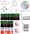
Au@CeO2‐DMF modulates bioactivities of PDLCs via the NRF2‐NLRP3 axis. A) KEGG enrichment analysis identifies key pathways involved. B) Circos plot illustrates overlapping genes from vital processes and pathways. C) Immunofluorescence staining shows NRF2 localization under various conditions. NRF2, red; cytoskeleton, green; nucleus, blue. Scare bar = 50 µm. D) Western blot analysis of NRF2 and NLRP3 protein expression. E) Immunofluorescence imaging of NLRP3 during Au@CeO2‐DMF treatment with NRF2 inhibition. NLRP3, red; cytoskeleton, green; nucleus, blue. Scare bar = 50 µm. F) ALP activity and ARS staining on day 5 and 14, respectively. G) Quantitative analysis of ALP activity and ARS Staining. H) Western blot analysis of NLRP3, p‐DRP1, TOMM20, Runx2, and OPN protein expression. Results were shown as mean ± SD. * p < 0.05, ** p < 0.01, *** p < 0.001.
Figure 6
Figure 6
The effects of Au@CeO2‐DMF treatment on macrophage polarization and inflammation‐related gene expression in vitro. A) Macrophage viability was assessed using Live/Dead staining across various concentrations of Au@CeO2‐DMF. Scale bar: 200 µm. B) Quantitative analysis was conducted based on Live/Dead staining images. C) Macrophage proliferation was evaluated using a CCK‐8 assay over 1, 2, and 3 days. D) qPCR was used to measure the expression of IL‐1β, IL‐6, IL‐10, and TGF‐β genes (n = 3). E) iNOS (red), cytoskeleton (green), and nucleus (blue) were visualized through immunofluorescence staining. Scale bar: 100 µm. F) Arg1 (red), cytoskeleton (green), and nucleus (blue) were visualized through immunofluorescence staining. G) Western blot analysis was performed to assess CCR7 and iNOS protein expression. H) Western blot analysis was conducted for Arg1 and CD206 protein expression. I) ELISA analysis was used to measure the secretion of inflammatory cytokines, IL‐6, and TNF‐α. Results were shown as mean ± SD. * p < 0.05, ** p < 0.01, *** p < 0.001.
Figure 7
Figure 7
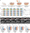
The Au@CeO2‐DMF treatment attenuated periodontitis progression in vivo. A) Schematic diagram of ligation‐induced periodontitis model and therapeutic approach in rats. B) IR thermal images of rats after periodontal injection of AuNP, Au@CeO2, and Au@CeO2‐DMF solutions. C) Photothermal curves of PBS, AuNP, Au@CeO2, and Au@CeO2‐DMF groups under 5‐min laser irradiation (635nm, 0.8 W cm−2). D) Representative 3D reconstructed sections (upper) and micro‐CT bucco‐palatal images (lower) along the maxilla's longitudinal axis. E) Quantitative assessment of CEJ‐ABC distance (mm) and micro‐CT analysis of F) BV/TV, G) Tb. Th, H) Tb. N, and I) Tb. Sp (n = 4). Data presented as mean ± SD. * p < 0.05, ** p < 0.01, *** p < 0.001.
Figure 8
Figure 8
The Au@CeO2‐DMF treatment inhibited inflammation and promoted alveolar bone regeneration during periodontitis. A) H&E, B) Masson's trichrome, and C) TRAP staining was conducted between the upper first and second molars. TRAP+ cells are indicated by red arrows. Row 1: overall view of the maxillary alveolar bone is presented (black line delineates the shape of the alveolar bone and roots, scale bar = 200 µm); Row 2: magnified interdental area view (scale bar = 100 µm). Immunohistochemical staining of D) iNOS and E) OPN (scale bar = 50 µm). Red arrows indicating positive stained cells. F) Schematic of bone remodeling via Au@CeO2‐DMF in periodontitis. Quantification of G) TRAP‐positive cells, H) iNOS, and I) OPN protein changes (n = 4). Results are mean ± SD. * p < 0.05, ** p < 0.01, *** p < 0.001. M1, first molar; M2, second molar; AB, alveolar bone; PDL, periodontal ligament.

Similar articles

Cited by

References

    1. Kinane D. F., Stathopoulou P. G., Papapanou P. N., Nat. Rev. Dis. Primers 2017, 3, 1. - PubMed
    1. Albandar J. M., Periodontol. 2000 2014, 65, 7. - PubMed
    1. Sczepanik F. S. C., Grossi M. L., Casati M., Goldberg M., Glogauer M., Fine N., Tenenbaum H. C., Periodontol. 2000 2020, 84, 45. - PubMed
    1. a) Scheibye‐Knudsen M., Fang E. F., Croteau D. L., Wilson D. M. 3rd, Bohr V. A., Trends Cell Biol. 2015, 25, 158; - PMC - PubMed
    2. b) Forrester S. J., Kikuchi D. S., Hernandes M. S., Xu Q., Griendling K. K., Circul. Res. 2018, 122, 877. - PMC - PubMed
    1. Locati M., Curtale G., Mantovani A., Annu. Rev. Pathol. 2020, 15, 123. - PMC - PubMed

LinkOut - more resources