. 2025 Feb;12(6):e2406570.
doi: 10.1002/advs.202406570. Epub 2024 Dec 24.

Light-Activatable Ubiquitin for Studying Linkage-Specific Ubiquitin Chain Formation Kinetics

Affiliations

Light-Activatable Ubiquitin for Studying Linkage-Specific Ubiquitin Chain Formation Kinetics

Sudakshina Banerjee et al. Adv Sci (Weinh). 2025 Feb.

Abstract

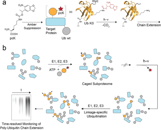
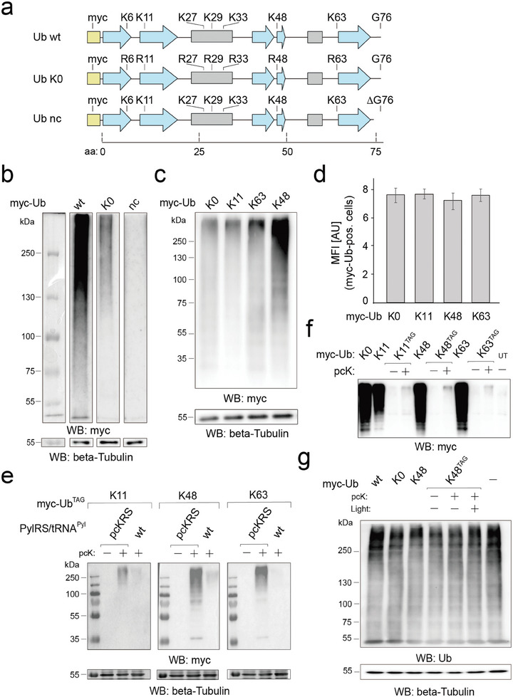
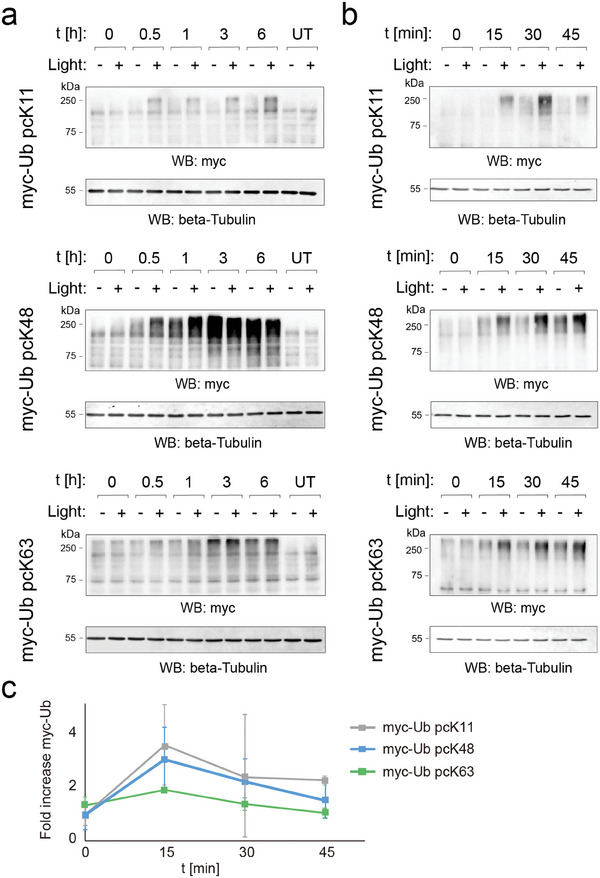
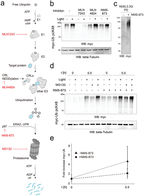
Ubiquitination is a dynamic post-translational modification governing protein abundance, function, and localization in eukaryotes. The Ubiquitin protein is conjugated to lysine residues of target proteins, but can also repeatedly be ubiquitinated itself, giving rise to a complex code of ubiquitin chains with different linkage types. To enable studying the cellular dynamics of linkage-specific ubiquitination, light-activatable polyubiquitin chain formation is reported here. By incorporating a photocaged lysine at specific sites within ubiquitin through amber codon suppression, light-dependent activation of ubiquitin chain extension is enabled for the monitoring of linkage-specific polyubiquitination. The studies reveal rapid, minute-scale ubiquitination kinetics for K11, K48, and K63 linkages. The role of individual components of the ubiquitin-proteasome system in K48-initiated chain synthesis is further studied by small molecule inhibition. The approach expands current perturbation strategies with the ability to control linkage-specific ubiquitination with high temporal resolution and should find broad application for studying ubiquitinome dynamics.

Keywords: genetic code expansion; optochemical biology; small molecule inhibitors; ubiquitin.


Conflict of interest statement

The authors declare no conflict of interest.

Figures

Figure 1
Figure 1
Light‐activated ubiquitination using a genetically encoded, photocaged lysine. a) Incorporation of photocaged lysine (pcK) into ubiquitin via amber suppression, its charging to target proteins, and its subsequent light‐decaging leads to polyubiquitination initiated by specific linkages. Ub K0: Ub with K→R mutations in all lysine sites except for the pcK site. b) Biosynthesis and light‐activation of a caged Ub subproteome that acts within the ubiquitin‐proteasome system (UPS). Inhibiting individual UPS components by small molecule inhibitors enables studying their function in early phases of de novo (poly)ubiquitination from specific linkages by rapid activation and time‐resolved monitoring.
Figure 2
Figure 2
Analysis of caged Ub proteome subpopulation for light‐activation of linkage‐specific downstream ubiquitination a) Domain structure of Ub constructs used in this study. Blue arrows: b‐strands, grey boxes: a‐helices. b) SDS PAGE/anti‐myc blots of proteomes from HEK293T cells expressing indicated Ub variants for 24 h (all lanes are from the same blot with identical exposure time). Cells were treated with 10 µm MG132 5 h before harvesting (also applies to panels (c–g)). c) Experiment as in Figure 2b with indicated Ub linkage variants. d) Anti‐myc immunostaining and FACS analysis of cells from Figure 2c reveal similar total expression levels of Ub variants. MFI: Mean fluorescence intensity; AU: arbitrary units. e) High fidelity incorporation of pcK at three different Ub lysine sites in HEK293T cells expressing the indicated amber myc‐Ub variants along with the indicated pyrrolysyl‐tRNA‐synthetase pair for 24 h (individual contrast settings for each linkage). f) SDS PAGE/anti‐myc blots of proteomes from HEK293T cells expressing myc‐Ub variants with or without amber codon and in presence or absence of pcK for 24 h reveal minimal expression levels of caged Ub variants. g) SDS PAGE/anti‐Ub blots of proteomes from HEK293T cells expressing Ub variants for 24 h with or without amber codon and in presence or absence of pcK reveal minimal expression levels of Ub variants compared to the endogenous Ub pool. Cells expressing non amber Ub variants were harvested directly after 24 h and those expressing amber Ub variants were further treated with light and harvested 24 h later.
Figure 3
Figure 3
Light activation reveals rapid, proteome‐wide ubiquitination kinetics initiated by K11, K48, and K63 linkages. a) Long‐term, linkage‐specific ubiquitination kinetics after light activation of caged myc‐Ub variants (t = time after light). HEK293T cells were cultivated in the presence of 0.32 mm pcK for 24 h after transfection. After light activation (as stated in text), cells were grown in full media containing 25 µm MG132, lacking pcK, and harvested at each corresponding time‐point after light (also applies to panels (b,c)). b) Rapid, initial ubiquitination kinetics (<45 min after light). c) Plots of rapid, initial ubiquitination kinetics (myc‐Ub proteome intensities normalized to beta‐tubulin blot and no light control of each lane; error bars from N ≥ 2).
Figure 4
Figure 4
Ubiquitin light activation in combination with small molecule UPS inhibitors enables studying the involvement of individual UPS components on rapid, K48‐linked de novo polyubiquitination. a) Scheme of UPS synthesis/degradation pathway with highlighted UPS factors targeted by used inhibitors. b) Effects of UPS inhibitors on early, K48‐specific de novo ubiquitinome synthesis (t = 0.5 h) in presence of MG132. HEK293T cells were cultivated in the presence of 0.32 mm pcK for 24 h after transfection. The inhibitors were added to the media in concentrations as mentioned. Following the pretreatment times, light activation was performed and cells were grown in full media containing 25 µm MG132 and the respective inhibitors, lacking pcK, and harvested at t = 0.5 h after light (also applies to panel (e)). c) Enrichment of branched ubiquitin with agarose‐immobilized NbSL3.3Q nanobody from HEK293T cells with light‐decaged myc‐Ub pcK48, with or without prior addition of the p97 inhibitor NMS‐873 (5 µm for 0.5 h). Eluates were analyzed by western blotting for Myc. d,e) Effect of NMS873 on early, K48‐specific de novo ubiquitinome synthesis in presence and absence of MG132.

Similar articles

Cited by

References

    1. Pohl C., Dikic I., Science 2019, 366, 818. - PubMed
    1. Clague M. J., Urbé S., Komander D., Nat. Rev. Mol. Cell Biol. 2019, 20, 338. - PubMed
    1. Ciechanover A., Stanhill A., Bba Mol. Cell Res. 2014, 1843, 86. - PubMed
    1. Lutz J., Hollmuller E., Scheffner M., Marx A., Stengel F., Angew. Chem. Int. Ed. Engl. 2020, 59, 12371. - PMC - PubMed
    1. a) Kim W., Bennett E. J., Huttlin E. L., Guo A., Li J., Possemato A., Sowa M. E., Rad R., Rush J., Comb M. J., Harper J. W., Gygi S. P., Mol. Cell. 2011, 44, 325; - PMC - PubMed
    2. b) Ross A. B., Langer J. D., Jovanovic M., Mol. Cell. Proteomics 2021, 20, 100016. - PMC - PubMed

LinkOut - more resources