. 2024 Dec 3:29:101374.
doi: 10.1016/j.mtbio.2024.101374. eCollection 2024 Dec.

Sequential delivery of IL-10 and icariin using nanoparticle/hydrogel hybrid system for prompting bone defect repair

Affiliations

Sequential delivery of IL-10 and icariin using nanoparticle/hydrogel hybrid system for prompting bone defect repair

Xiaojun Li et al. Mater Today Bio. .

Abstract

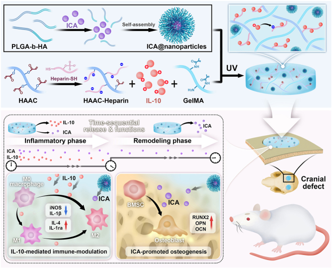
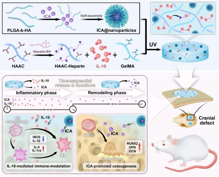
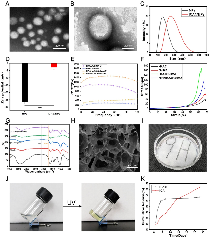
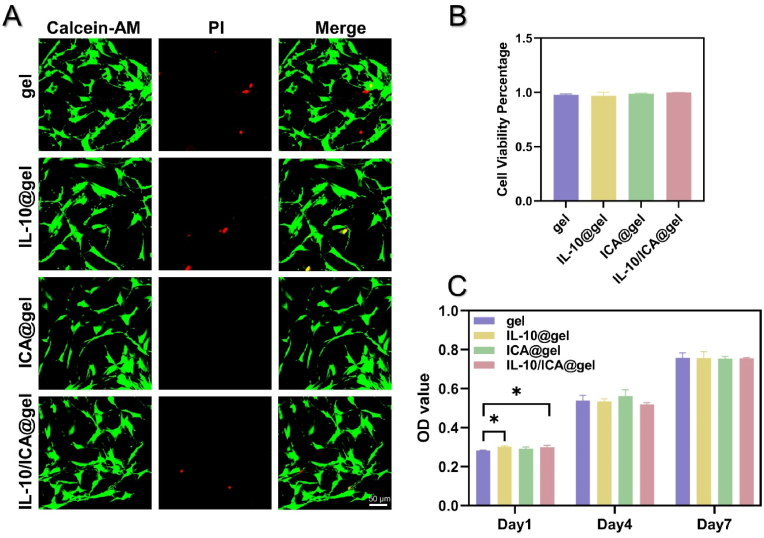
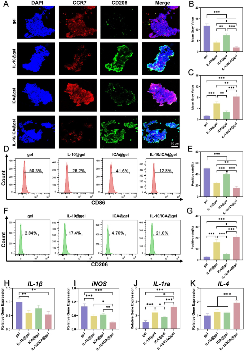
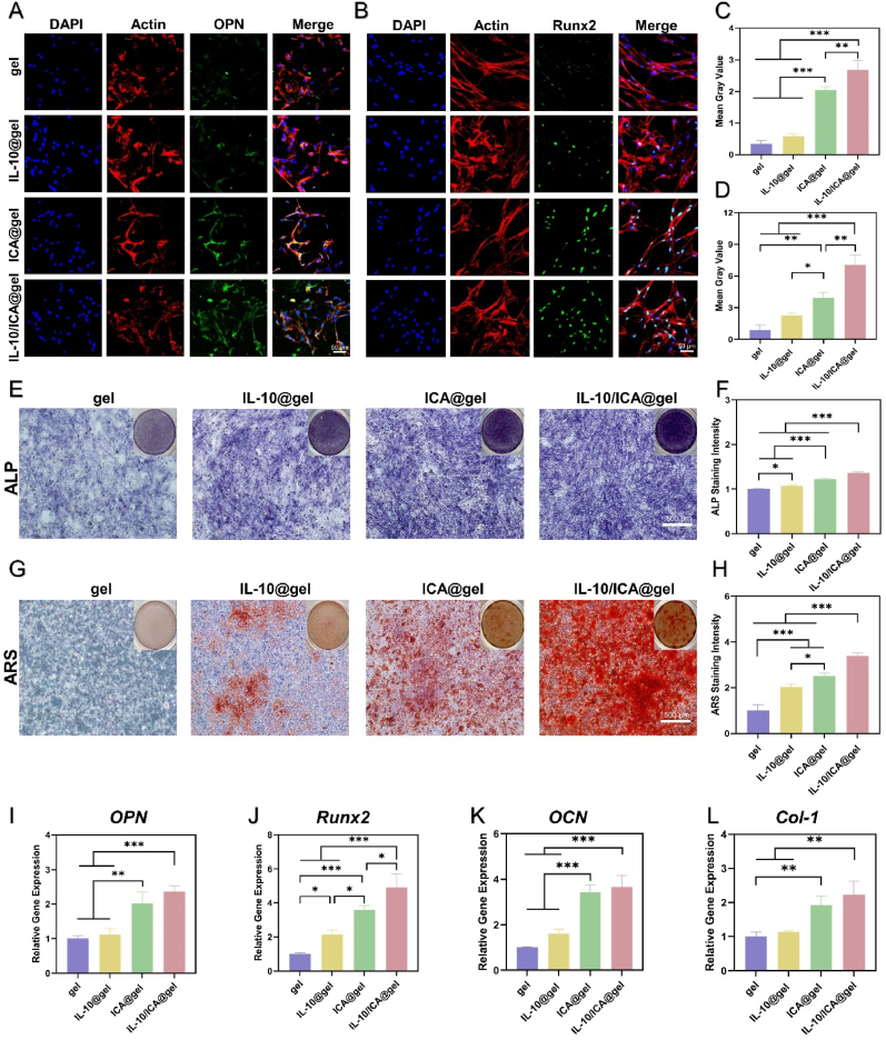
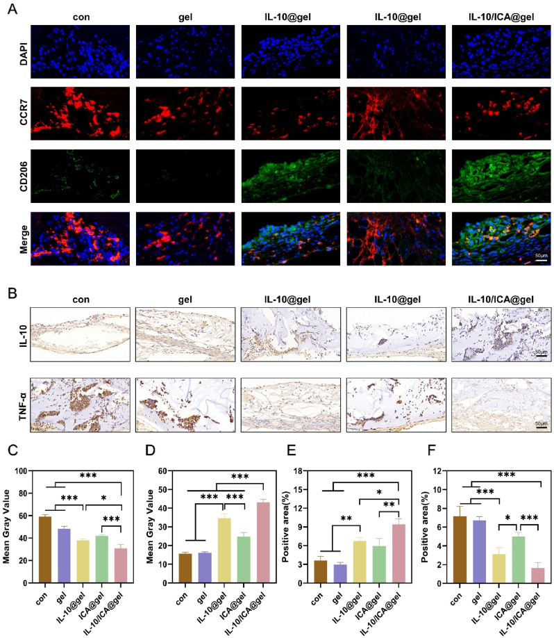
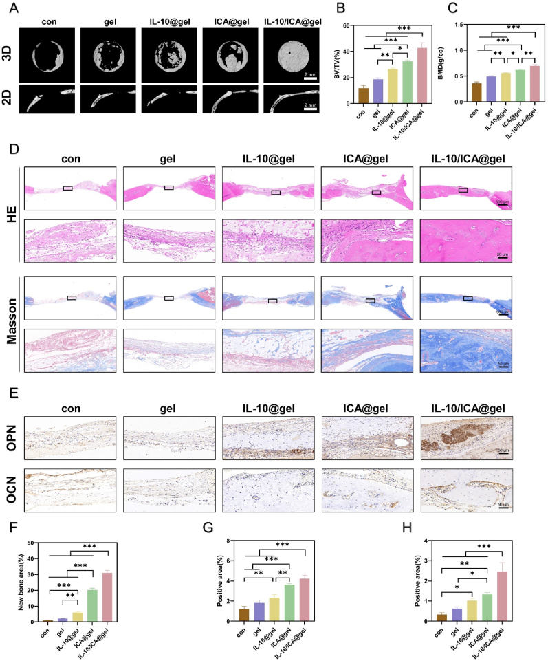
The treatment of large bone defects remains challenging due to the lack of spatiotemporal management of the immune microenvironment, inflammation response and bone remodeling. To address these issues, we designed and developed a nanoparticle/hydrogel hybrid system that can achieve the combined and sequential delivery of an anti-inflammatory factor (IL-10) and osteogenic drug (icariin, ICA). A photopolymerizable composite hydrogel was prepared by combining gelatin methacryloyl (GelMA) and heparin-based acrylated hyaluronic acid (HA) hydrogels containing IL-10, and poly(dl-lactide-co-glycolide) (PLGA)-HA nanoparticles loaded with ICA were incorporated into the composite hydrogels. The nanoparticle/hydrogel hybrid system demonstrates an array of features including mechanical strength, injectability and photo-crosslinking. The rapid release of IL-10 from the hydrogel effectively exerts immunomodulatory activity, whereas the long-term sustained release of icariin from the PLGA-HA nanoparticles significantly triggers the osteogenic differentiation of bone marrow-derived mesenchymal stem cells (BMSCs). Notably, the combined delivery of IL-10 and ICA from the hybrid system exhibit a synergistic effect for bone remodeling in a critical cranial defect rat model. Our findings indicate the importance of the immunomodulatory microenvironment and osteogenic differentiation for high-quality skull remodeling, and thus the dual-factor releasing nanoparticle/hydrogel hybrid system could be a promising candidate for repairing bone defects.

Keywords: Immunomodulation; Injectable hydrogel; Nanoparticles; Osteogenesis; Sequential release.


Conflict of interest statement

The authors declare that they have no known competing financial interests or personal relationships that could have appeared to influence the work reported in this paper.

Figures

Image 1
Graphical abstract
Scheme 1
Scheme 1
Schematic diagram of the preparation of a multi-functional composite hydrogel with time-sequential IL-10 and ICA release and its immune-modulation and bone regeneration property.
Fig. 1
Fig. 1
Preparation and characterization of the nanoparticles and the hydrogel. A) TEM image of NPs. B) TEM image of ICA@NPs. C) Size distribution of NPs. D) Zeta potential of NPs and ICA@NPs (n = 3). E) Study of frequency-responsive rheological characteristics of the hydrogel under a 1 % strain condition. F) Compressive stress-strain curves. G) FTIR analysis of hydrogel. H) The SEM image of the hydrogel. I) Images reflecting the injectability and moldability of the hydrogel. J) Images showing the gelation process after UV. K) Cumulative release of IL-10 and ICA from the hydrogel. Data are shown as mean ± SD (∗∗∗p < 0.001).
Fig. 2
Fig. 2
Biocompatibility of the hydrogels in vitro. A) Live/dead assay of BMSCs cultured for 1 day of co-cultured on hydrogels. The green fluorescence indicates live cells stained by Calcein-AM. The red fluorescence indicates dead cells stained by PI. B) The quantitative data of live/dead assay. C) Cell proliferation of BMSCs for 1, 4 and 7 days of co-cultured on hydrogels were tested using CCK-8 analysis (n = 3) (∗p < 0.05).
Fig. 3
Fig. 3
Regulation of the macrophage-polarization phenotype in vitro. A) Representative CLSM images of surface markers CCR7 (red, M1 phenotype) and CD206 (green, M2 phenotype) expressions of RAW264.7 cells cultured with hydrogels for 3d by immunofluorescence analysis. B, C) Mean gray value of CCR7 and CD206 staining (n = 3); D, E) Flow cytometry analysis of CD86 (M1 phenotype) and CD206 expression of RAW264.7 cells cultured with hydrogels. F, G) The quantitative analysis of the percentage of M1 macrophages and M2 macrophages. H-K) Real-time PCR of M1 polarization-related IL-1β (H), iNOS (I) and M2 polarization-related IL-1ra (J) and IL-4 (K) (n = 3) (∗p < 0.05, ∗∗p < 0.01, ∗∗∗p < 0.001).
Fig. 4
Fig. 4
Osteogenic differentiation of BMSCs stimulated by the hydrogel. A, B) Immunofluorescence staining images of OPN and Runx2 of BMSCs cultured with hydrogels for 7d. C, D) Quantification of the immunofluorescence density of OPN and Runx2 (n = 3). E, F) Alkaline phosphatase (ALP) of BMSCs cultured for 7d and quantitative evaluation of ALP staining intensity. G, H) Alkaline phosphatase S (ARS) staining of BMSCs cultured for 21 d and quantitative evaluation of ARS staining intensity. I-L) Relative mRNA expression of the osteogenic genes OPN, Runx2, OCN and Col-1 in BMSCs (n = 3) (∗p < 0.05, ∗∗p < 0.01, ∗∗∗p < 0.001).
Fig. 5
Fig. 5
In vivo immunomodulatory evaluations of the hydrogels. A) Representative images of surface markers CCR7 (red, M1 phenotype) and CD206 (green, M2 phenotype) expressions by immunofluorescence analysis; B) Immunohistochemistry of the IL-10 and TNF-α; C, D) The qualitative analysis of CCR7 and CD206; E, F) The qualitative analysis of IL-10 and TNF-α.(∗p < 0.05, ∗∗p < 0.01, ∗∗∗p < 0.001).
Fig. 6
Fig. 6
In vivo osteogenesis capability of the hydrogel. A) Representative micro-CT images of skull defects implanted with the hydrogels. B, C) Quantitative evaluation of the osteogenic parameters, BV represents the amount of new bone volume, TV represents the amount of the total defect volume and BMD represents the new bone density. D) Representative H&E and Masson staining images of regenerative bone tissue at 8 week after surgery. E) Representative IHC staining of osteogenic marker (OPN and OCN); F) Quantitative analysis of new bone area; G, H) Quantitative analysis of OPN and OCN positive area. (∗p < 0.05, ∗∗p < 0.01, ∗∗∗p < 0.001).

Similar articles

Cited by

References

    1. Shen X., Zhang Y., Gu Y., Xu Y., Liu Y., Li B., Chen L. Sequential and sustained release of SDF-1 and BMP-2 from silk fibroin-nanohydroxyapatite scaffold for the enhancement of bone regeneration. Biomaterials. 2016;106:205–216. - PubMed
    1. Wang J., Wu Y., Li G., Zhou F., Wu X., Wang M., Liu X., Tang H., Bai L., Geng Z., Song P., Shi Z., Ren X., Su J. Engineering large-scale self-mineralizing bone organoids with bone matrix-inspired hydroxyapatite hybrid bioinks. Adv. Mater. 2024 - PubMed
    1. Zhou J., See C.W., Sreenivasamurthy S., Zhu D. vol. 6. Research (Wash D C); 2023. p. 239. (Customized Additive Manufacturing in Bone Scaffolds-The Gateway to Precise Bone Defect Treatment). - PMC - PubMed
    1. Dimitriou R., Jones E., McGonagle D., Giannoudis P.V. Bone regeneration: current concepts and future directions. BMC Med. 2011;9:66. - PMC - PubMed
    1. Bose S., Sarkar N. Natural medicinal compounds in bone tissue engineering. Trends Biotechnol. 2020;38(4):404–417. - PMC - PubMed

LinkOut - more resources