. 2024 Dec 4;27(12):111437.
doi: 10.1016/j.isci.2024.111437. eCollection 2024 Dec 20.

Screening and molecular mechanism research on bile microRNAs associated with chemotherapy efficacy in perihilar cholangiocarcinoma

Affiliations

Screening and molecular mechanism research on bile microRNAs associated with chemotherapy efficacy in perihilar cholangiocarcinoma

Shijie Fu et al. iScience. .

Abstract

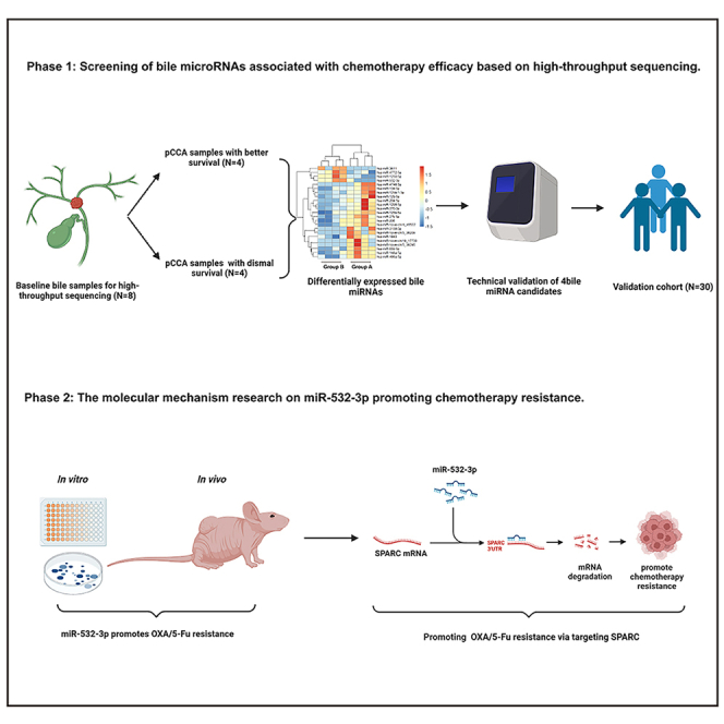
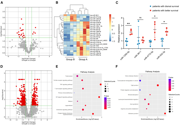
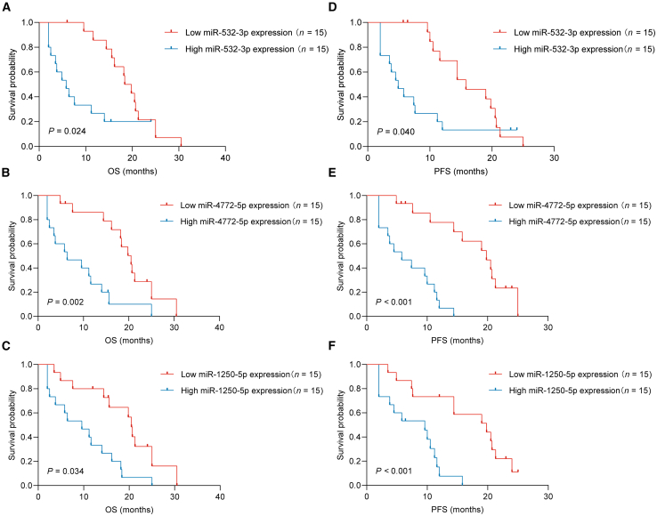
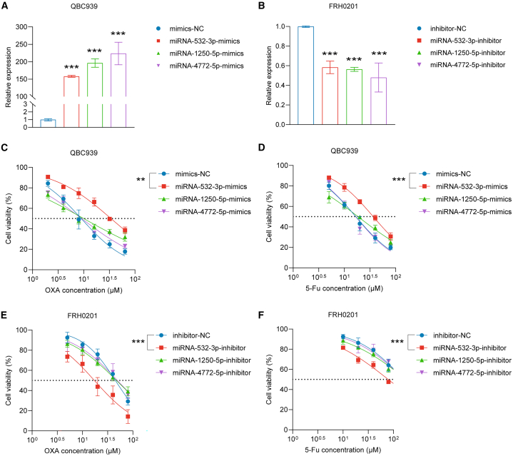
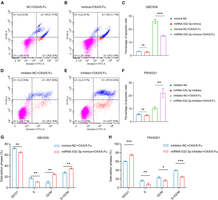
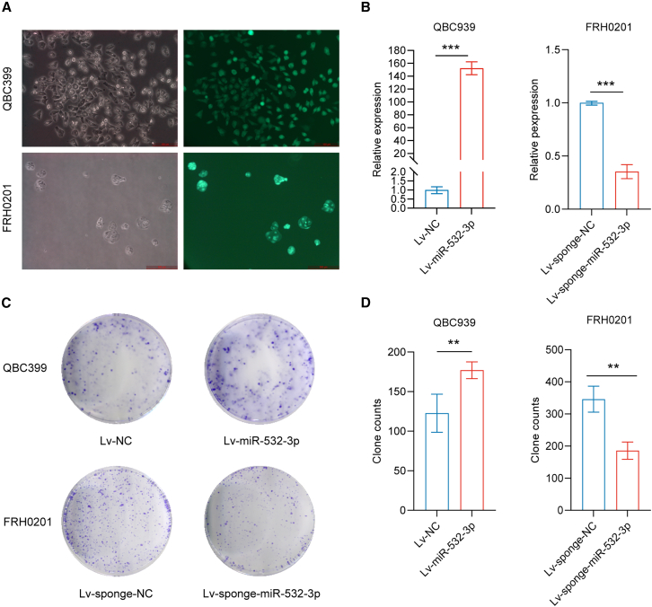
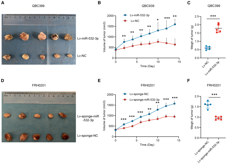
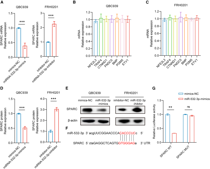
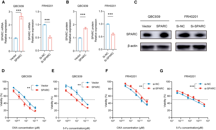
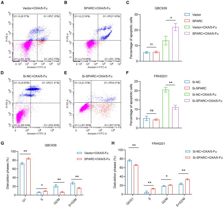
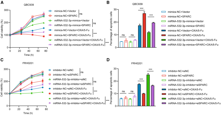
The efficacy of hepatic arterial infusion chemotherapy (HAIC) with oxaliplatin (OXA) and 5-fluorouracil (5-Fu) for treating advanced perihilar cholangiocarcinoma (pCCA) has been demonstrated, yet the survival benefits of HAIC for pCCA patients vary. Here, we aimed to screen out HAIC resistance-related bile microRNAs (miRNAs) and explore the functions of specific bile miRNAs in pCCA based on high-throughput sequencing. Levels of bile miR-532-3p, miR-1250-5p, and miR-4772-5p were related to the survival of advanced pCCA patients after HAIC. However, only overexpression of miR-532-3p promoted OXA/5-Fu resistance, and downregulation of its expression improved sensitivity to OXA/5-Fu. Mechanistic investigations revealed secreted protein acidic and rich in cysteine (SPARC) as the direct target of miR-532-3p. Our study reveals that bile miR-532-3p, miR-1250-5p, and miR-4772-5p may serve as survival biomarkers in advanced pCCA patients after HAIC and that bile miR-532-3p promotes resistance to HAIC with OXA and 5-Fu via negatively regulating SPARC expression.

Keywords: Cancer; Cell biology; Molecular biology.


Conflict of interest statement

The authors declare no competing interests.

Figures

None
Graphical abstract
Figure 1
Figure 1
High-throughput sequencing results (A–C) Sequencing and qPCR validation of bile miRNAs. (D) Sequencing of bile mRNAs. (E and F) KEGG pathway analysis of the upregulated mRNAs in the bile of patients with dismal prognosis (E) and better prognosis (F). Data are represented as mean ± SD. n = 4, ∗p < 0.05, ∗∗p < 0.01, ∗∗∗p < 0.001, ns: no significance, miRNAs microRNAs, qPCR quantitative polymerase chain reaction, KEGG Kyoto Encyclopedia of Genes and Genomes.
Figure 2
Figure 2
miR-532-3p, miR-1250-5p, and miR-4772-5p serve as survival biomarkers for advanced pCCA patients after HAIC (A–C) The relationship of miR-532-3p (A)/miR-1250-5p (B)/miR-4772-5p (C) expression with OS. (D–F) The relationship of miR-532-3p (D)/miR-1250-5p (E)/miR-4772-5p (F) expression with PFS. pCCA perihilar cholangiocarcinoma, HAIC hepatic arterial infusion chemotherapy, OS overall survival, PFS progression-free survival.
Figure 3
Figure 3
miR-532-3p influences the IC50 values of OXA and 5-Fu in vitro (A) miR-1250-5p, miR-4772-5p, and miR-532-3p levels were upregulated in QBC939 cells following transient transfection. (B) MiR-1250-5p, miR-4772-5p, and miR-532-3p levels were downregulated in FRH0201 cells following transient transfection. (C and D) The IC50 values of OXA (C) and 5-Fu (D) in QBC939 cells. (E andF) The IC50 values of OXA (E) and 5-Fu (F) in FRH0201 cells. Data are represented as mean ± SD. n = 3, ∗∗p < 0.01, ∗∗∗p < 0.001, IC50 half inhibitory concentration, OXA oxaliplatin, 5-Fu 5-fluorouracil, NC negative control.
Figure 4
Figure 4
miR-532-3p influences cell apoptosis and cell cycle distribution (A–C) The percentage of total apoptotic QBC939 cells after incubation with OXA (5 μmol/L) and 5-Fu (10 μmol/L) for 48 h. (D–F) The percentage of total apoptotic FRH0201 cells after incubation with OXA (20 μmol/L) and 5-Fu (80 μmol/L) for 48 h. (G) Cell cycle distribution of QBC939 cells after incubation with OXA (5 μmol/L) and 5-Fu (10 μmol/L) for 48 h. (H) Cell cycle distribution of FRH0201 cells after incubation with OXA (20 μmol/L) and 5-Fu (80 μmol/L) for 48 h. Data are represented as mean ± SD. n = 3, ∗p < 0.05, ∗∗p < 0.01, ∗∗∗p < 0.001, ns: no significance, OXA oxaliplatin, 5-Fu 5-fluorouracil, NC negative control.
Figure 5
Figure 5
miR-532-3p promotes colony formation (A and B) The successful lentivirus infection of QBC939 (stable miR-532-3p overexpression) and FRH cells (stable miR-532-3p underexpression). (C and D) Colony-forming assays on QBC cells (after incubation with OXA at a concentration of 5 μmol/L and 5-Fu at a concentration of 10 μmol/L for 14 days) and FRH0201 cells (after incubation with OXA at a concentration of 20 μmol/L and 5-Fu at a concentration of 80 μmol/L for 14 days). Data are represented as mean ± SD. n = 3, ∗∗p < 0.01, ∗∗∗p < 0.001,OXA oxaliplatin, 5-Fu 5-fluorouracil, NC negative control.
Figure 6
Figure 6
miR-532-3p promotes OXA/5-Fu resistance in vivo (A–C) Tumor volumes and weights of mice bearing QBC939 cells (stably overexpressing miR-532-3p or control) 2 weeks following the first drug injection. (D–F) Tumor volumes and weights of mice bearing FRH0201 cells (stably underexpressing miR-532-3p or control) after 2 weeks following the first drug injection. Data are represented as mean ± SD. n = 5, ∗p < 0.05, ∗∗p < 0.01, ∗∗∗p < 0.001, OXA oxaliplatin, 5-Fu 5-fluorouracil, NC negative control.
Figure 7
Figure 7
miR-532-3p pairs with 3′-UTR of SPARC (A–C) Overexpression or decreased expression of miR-532-3p affected SPARC mRNA levels but did not affect the mRNA expression of other genes. (D and E) SPARC protein levels were affected by miR-532-3p. (F) Binding sites between the SPARC 3′UTR and miR-532-3p. (G) Results of luciferase reporter assays. Data are represented as mean ± SD. n = 3, ∗∗p < 0.01, ∗∗∗p < 0.001, ns: no significance, 3′-UTRs 3′-untranslated regions, SPARC secreted protein acidic and rich in cysteine, NC negative control.
Figure 8
Figure 8
SPARC influences the IC50 values of OXA/5-Fu in vitro (A–C) Successful transfection of SPARC plasmids on QBC939 cells and siRNA against SPARC on FRH cells. (D and E) IC50 values of OXA (D) and 5-Fu (E) on QBC939 cells. (F and G) IC50 values of OXA (F) and 5-Fu (G) on FRH0201 cells. Data are represented as mean ± SD. n = 3, ∗∗p < 0.01, ∗∗∗p < 0.001, SPARC secreted protein acidic and rich in cysteine, IC50 half inhibitory concentration, OXA oxaliplatin, 5-Fu 5-fluorouracil, siRNA small interfering RNA, NC negative control.
Figure 9
Figure 9
miR-532-3p influences cell apoptosis and cell cycle distribution (A–C) Percentage of total apoptotic QBC939 cells (following the transfection of SPARC plasmids or control) after incubation with OXA (5 μmol/L) and 5-Fu (10 μmol/L) for 48 h. (D–F) Percentage of total apoptotic FRH0201 cells (following the transfection of siRNA against SPARC or control, 2 μg) after incubation with OXA (20 μmol/L) and 5-Fu (80 μmol/L) for 48 h. (G) Cell cycle distribution of QBC939 cells (following the transfection of SPARC plasmids or control) after incubation with OXA (5 μmol/L) and 5-Fu (10 μmol/L) for 48 h. (H) Cell cycle distribution of FRH0201 (following the transfection of siRNA of SPARC or control) cells after incubation with OXA (20 μmol/L) and 5-Fu (80 μmol/L) for 48 h. Data are represented as mean ± SD. n = 3, ∗p < 0.05, ∗∗p < 0.01, ns: no significance. SPARC secreted protein acidic and rich in cysteine, OXA oxaliplatin, 5-Fu 5-fluorouracil, siRNA small interfering RNA.
Figure 10
Figure 10
SPARC partially reverses the function of miR-532-3p in vitro (A) Cell proliferation was reversed with co-transfection of miR-532-3p mimics (50 nM) and SPARC (2 μg) on QBC939 cells after incubation with OXA (5 μmol/L) and 5-Fu (10 μmol/L). (B) Similarly, cell apoptosis was reversed on QBC939 cells after incubation with OXA (5 μmol/L) and 5-Fu (10 μmol/L) for 48 h. (C) Cell proliferation was reversed with co-transfection of miR-532-3p inhibitor (100 nM) and siRNA (100 nM) on FRH0201 cells after incubation with OXA (20 μmol/L) and 5-Fu (80 μmol/L). (D) Cell apoptosis was reversed on FRH0201 cells after incubation with OXA (20 μmol/L) and 5-Fu (80 μmol/L) for 48 h. Data are represented as mean ± SD. n = 3, ∗∗∗p < 0.001, ns: no significance, SPARC secreted protein acidic and rich in cysteine, OXA oxaliplatin, 5-Fu 5-fluorouracil, siRNA small interfering RNA, NC negative control.

Similar articles

References

    1. Greten T.F., Schwabe R., Bardeesy N., Ma L., Goyal L., Kelley R.K., Wang X.W. Immunology and immunotherapy of cholangiocarcinoma. Nat. Rev. Gastroenterol. Hepatol. 2023;20:349–365. doi: 10.1038/s41575-022-00741-4. - DOI - PubMed
    1. Mauro E., Ferrer-Fàbrega J., Sauri T., Soler A., Cobo A., Burrel M., Iserte G., Forner A. New challenges in the management of cholangiocarcinoma: the role of liver transplantation, locoregional therapies, and systemic therapy. Cancers. 2023;15 doi: 10.3390/cancers15041244. - DOI - PMC - PubMed
    1. Rizvi S., Khan S.A., Hallemeier C.L., Kelley R.K., Gores G.J. Cholangiocarcinoma - evolving concepts and therapeutic strategies. Nat. Rev. Clin. Oncol. 2018;15:95–111. doi: 10.1038/nrclinonc.2017.157. - DOI - PMC - PubMed
    1. Soares K.C., Jarnagin W.R. The landmark series: hilar cholangiocarcinoma. Ann. Surg Oncol. 2021;28:4158–4170. doi: 10.1245/s10434-021-09871-6. - DOI - PMC - PubMed
    1. Chan E., Berlin J. Biliary tract cancers: understudied and poorly understood. J. Clin. Oncol. 2015;33:1845–1848. doi: 10.1200/jco.2014.59.7591. - DOI - PubMed