Review
. 2024 Nov 15:8:100317.
doi: 10.1016/j.crmicr.2024.100317. eCollection 2025.

Multifaceted virus-like particles: Navigating towards broadly effective influenza A virus vaccines

Affiliations
Review

Multifaceted virus-like particles: Navigating towards broadly effective influenza A virus vaccines

Jaffar Ali Muhamad Norizwan et al. Curr Res Microb Sci. .

Abstract

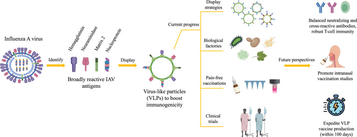
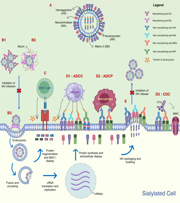
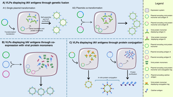
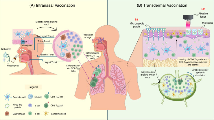
The threat of influenza A virus (IAV) remains an annual health concern, as almost 500,000 people die each year due to the seasonal flu. Current flu vaccines are highly dependent on embryonated chicken eggs for production, which is time consuming and costly. These vaccines only confer moderate protections in elderly people, and they lack cross-protectivity; thereby requiring annual reformulation to ensure effectiveness against contemporary circulating strains. To address current limitations, new strategies are being sought, with great emphasis given on exploiting IAV's conserved antigens for vaccine development, and by using different vaccine technologies to enhance immunogenicity and expedite vaccine production. Among these technologies, there are growing pre-clinical and clinical studies involving virus-like particles (VLPs), as they are capable to display multiple conserved IAV antigens and augment their immune responses. In this review, we outline recent findings involving broadly effective IAV antigens and strategies to display these antigens on VLPs. Current production systems for IAV VLP vaccines are comprehensively reviewed. Pain-free methods for administration of IAV VLP vaccines through intranasal and transdermal routes, as well as the mechanisms in stimulating immune responses are discussed in detail. The future perspectives of VLPs in IAV vaccine development are discussed, particularly concerning their potentials in overcoming current immunological limitations of IAV vaccines, and their inherent advantages in exploring intranasal vaccination studies. We also propose avenues to expedite VLP vaccine production, as we envision that there will be more clinical trials involving IAV VLP vaccines, leading to commercialization of these vaccines in the near future.

Keywords: Broadly reactive; Clinical trial; Conserved antigens; Influenza A virus; Pain-free; Production system; Vaccine; Virus-like particle.


Conflict of interest statement

The authors declare that they have no known competing financial interests or personal relationships that could have appeared to influence the work reported in this paper.

Figures

Image, graphical abstract
Graphical abstract
Fig 1:
Fig. 1
The immune responses induced by the hemagglutinin (HA), neuraminidase (NA), matrix 2 (M2) and nucleoprotein (NP) of influenza A virus (IAV). A schematic diagram depicting HA, NA, M2 and NP in IAV (A). The immune responses stimulated by the antigens include virus neutralization (B1, B2, B3), epitope-specific CD8 T cell cytotoxicity (C), Fc-mediated antibody effector functions (D1, D2, D3), and prevention of virus budding (E). Neutralizing anti-HA antibodies bind to HA, causing viral aggregations that prevent interaction with host's sialic acid (SA) receptor for viral infection (B1). Neutralizing anti-NA antibodies interact with NA's catalytic sites, inhibiting the virus release from sialylated mucins (B2) and sialylated “decoy” receptors on the cell surface (B3), thereby preventing efficient IAV infection. IAV epitopes originated from HA, NA, M2e and NP are displayed on major histocompatibility complex (MHC I), and its interaction with the epitope-specific CD8 T cells (C) causes cell lysis through cytolytic molecules (perforins and granzymes). Non-neutralizing anti-HA, anti-NA, anti-M2e and anti-NP interact with respective antigens displayed on the surface of the infected cell. The fragment crystallizable (Fc) region of the antibodies interacts with Fc receptors on natural killer cells (NK cells), resulting in antibody-dependent cellular cytotoxicity (ADCC) (D1) through secretion of perforins and granzymes. Fc region of non-neutralizing anti-HA and anti-M2e antibodies can also interact with macrophages to eliminate the infected cells through antibody-dependent cellular phagocytosis (ADCP) (D2), or engage with C1q protein that leads to complement-dependent cytotoxicity (CDC) (D3) through membrane attack complex (MAC). The binding of non-neutralizing anti-HA, anti-NA or anti-M2e antibodies on their respective antigens on the cell surface inhibits virus release (E) by preventing NA cleavage of sialic acid from HA (anti-NA), prevention of M2 mediated membrane scission (anti-M2e) and HA cross-linking (anti-HA).
Fig 2:
Fig. 2
A schematic diagram summarizing the dynamics of displaying IAV antigens on VLPs. Through genetic fusion (A), single (A1) or different IAV antigens (A2) can be displayed on the viral protein monomers (which form the VLPs) by transforming the expression system with a single or different plasmids, respectively. Alternatively, IAV antigens can be displayed on VLPs by co-expression with viral protein monomers (B). This is succeeded by co-transforming the expression system with different plasmids encoding the viral protein monomer and those encoding IAV antigens. IAV antigens can also be displayed on VLPs through protein conjugation (C). Viral protein monomers displaying the conjugation tag are produced in one expression system, while the IAV antigens with complementary catcher are produced in a separate expression system. The VLPs and catcher-antigens are purified and conjugated in vitro.
Fig 3:
Fig. 3
Pain-free IAV VLP vaccine administrations through intranasal and transdermal routes. In intranasal vaccination (A), IAV VLPs are inoculated into the nostrils using either a nasal spray or nebulizer. Upon inoculation, the VLPs are transported across the follicle-associated epithelium (FAE) via transcytosis by microfold (M) cells, and engulfed by nearby dendritic cells (DCs). The activated DCs then migrate into the draining nasal-associated lymphoid tissues (NALT), comprising of pharyngeal tonsil, tubal tonsil, palatine tonsil and lingual tonsil. The DCs then stimulated epitope-specific T cells and B cells, leading to lymphocyte proliferation and differentiation. A subset of CD4 and CD8 T cells differentiates into tissue-resident memory T cells (TRM), which reside at the nasal tissues and lungs. Upon next encounter with the same antigen, these TRM will rapidly proliferate and eliminate the IAV-infected cells. The activation of B cells results in production of secretory IgA (sIgA), which is secreted into the mucous lining to bind with IAV and prevent viral infection right at the viral entry site. Meanwhile, in transdermal vaccination (B), IAV VLP vaccines can be inoculated either through a microneedle patch (MP) (B1) or an ablative laser (B2). For MP, IAV VLPs are loaded onto the MP and applied onto the skin, which releases them slowly into the dermis and epidermis. Meanwhile, an ablative laser is used to create micropores on the epidermis, which acts as depots for sustained IAV VLPs release into the skin. Upon VLPs entry into the epidermis and dermis, the particles are engulfed by Langerhan cells (LCs) and dermal dendritic cell (dDC), which are abundantly present in these regions. The activated LCs and dDCs migrate towards the draining lymph node (LN), and results in activation, proliferation and differentiation of epitope-specific T cells and B cells. Subsets of CD4 and CD8 T cells differentiate into TRM, which migrate and reside in the skin, while other subsets such as T-effector memory cells (TEM) migrate to the peripheral organs for immunosurveillance. Meanwhile, activated B cells differentiate into plasma cells (to produce antibodies) and memory B cells (migrate to the peripheral organs for immunosurveillance).

Similar articles

References

    1. Adorini L., Nagy Z.A. Peptide competition for antigen presentation. Trends Immunol. 1990;11(1):21–24. doi: 10.1016/0167-5699(90)90006-U. - DOI - PubMed
    1. Air G.M. Influenza neuraminidase. Influenza Other Respir. Viruses. 2012;6(4):245–256. doi: 10.1111/J.1750-2659.2011.00304.X. - DOI - PMC - PubMed
    1. AstraZeneca . 2022. FluenzⓇ Tetra product insert.https://www.ema.europa.eu/en/documents/product-information/fluenz-tetra-... (accessed 7 May 2024)
    1. Avcil M., Çelik A. Microneedles in drug delivery: progress and challenges. Micromachines. 2021;12(11):1321. doi: 10.3390/MI12111321. - DOI - PMC - PubMed
    1. Ball P. The lightning-fast quest for COVID vaccines - and what it means for other diseases. Nature. 2021;589:16–18. doi: 10.1038/D41586-020-03626-1. - DOI - PubMed

LinkOut - more resources