

A Rare Case of Polymicrogyria in an Elderly Individual With Unique Polygenic Underlining
- PMID: 39717325
- PMCID: PMC11665267
- DOI: 10.7759/cureus.74300
A Rare Case of Polymicrogyria in an Elderly Individual With Unique Polygenic Underlining
Abstract
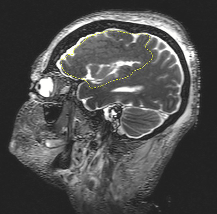
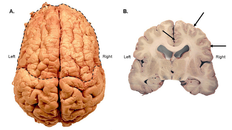
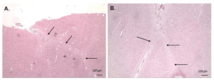
Polymicrogyria (PMG) is the most common malformation of cortical development (MCD) and presents as an irregularly patterned cortical surface with numerous small gyri and shallow sulci leading to various neurological deficits including developmental delays, intellectual disability, epilepsy, and language and motor issues. The presentation of PMG varies and is often found in conjunction with other congenital anomalies. Histologically, PMG features an abnormal cortical structure and dyslamination, resulting in its classification as a defect of neuronal migration and organization. Due in part to a variety of etiologies, little is known about the molecular mechanism(s) underlining PMG. To address this gap in knowledge, a case study is presented where an elderly individual with a medical history of unspecified PMG was examined postmortem by using a combination of anatomical, magnetic resonance imaging (MRI), histopathological, and genetic techniques. The results of the study allowed the classification of this case as bifrontal PMG. The genetic screening by whole exome sequencing (WES) on the Illumina Next Generation Sequencing (NGS) platform yielded 83 rare (minor allele frequency, MAF ≤ 0.01) pathological/deleterious variants where none of the respective genes has been previously linked to PMG. However, a subsequent analysis of those variants revealed that a significant number of affected genes were associated with most of the biological processes known to be impaired in PMG thereby pointing toward a polygenic nature in the present case. One of the notable features of the WES dataset was the presence of rare pathological/deleterious variants of genes (ADGRA2, PCDHA1, PCDHA12, PTK7, TPGS1, and USP4) involved in the regulation of Wnt signaling potentially highlighting the latter as an important PMG contributor in the present case. Notably, ADGRA2 warrants a closer look as a candidate gene for PMG because it not only regulates cortical patterning but has also been recently linked to two cases of bifrontal PMG with multiple congenital anomalies through its compound heterozygous mutations.
Keywords: bifrontal; malformation of cortical development; next-generation sequencing; polymicrogyria; whole exome sequencing.
Copyright © 2024, Frolov et al.
Conflict of interest statement
Human subjects: Consent for treatment and open access publication was obtained or waived by all participants in this study. Conflicts of interest: In compliance with the ICMJE uniform disclosure form, all authors declare the following: Payment/services info: All authors have declared that no financial support was received from any organization for the submitted work. Financial relationships: All authors have declared that they have no financial relationships at present or within the previous three years with any organizations that might have an interest in the submitted work. Other relationships: All authors have declared that there are no other relationships or activities that could appear to have influenced the submitted work.
Figures
Similar articles
-
The Genetic Landscape of Polymicrogyria.Ann Indian Acad Neurol. 2022 Jul-Aug;25(4):616-626. doi: 10.4103/aian.aian_97_22. Epub 2022 May 5. Ann Indian Acad Neurol. 2022. PMID: 36211152 Free PMC article.
-
Bilateral polymicrogyria associated with dystonia: A new neurogenetic syndrome?Am J Med Genet A. 2020 Oct;182(10):2207-2213. doi: 10.1002/ajmg.a.61795. Epub 2020 Aug 17. Am J Med Genet A. 2020. PMID: 33001581
-
Polymicrogyria: a common and heterogeneous malformation of cortical development.Am J Med Genet C Semin Med Genet. 2014 Jun;166C(2):227-39. doi: 10.1002/ajmg.c.31399. Epub 2014 May 28. Am J Med Genet C Semin Med Genet. 2014. PMID: 24888723 Review.
-
Surgical management of medically refractory epilepsy in patients with polymicrogyria.Epilepsia. 2016 Jan;57(1):151-61. doi: 10.1111/epi.13264. Epub 2015 Dec 9. Epilepsia. 2016. PMID: 26647903 Free PMC article.
-
Epilepsy surgery for polymicrogyria: a challenge to be undertaken.Epileptic Disord. 2018 Oct 1;20(5):319-338. doi: 10.1684/epd.2018.1004. Epileptic Disord. 2018. PMID: 30378553 Review.
References
-
- Polymicrogyria: a common and heterogeneous malformation of cortical development. Stutterd CA, Leventer RJ. Am J Med Genet C Semin Med Genet. 2014;166C:227–239. - PubMed
-
- Clinical and imaging features of cortical malformations in childhood. Leventer RJ, Phelan EM, Coleman LT, Kean MJ, Jackson GD, Harvey AS. Neurology. 1999;53:715–722. - PubMed
Publication types
LinkOut - more resources
Full Text Sources
Miscellaneous