

Thoracic endovascular aortic repair (TEVAR) in a combined penetrating thoracic aortic and spinal cord injury
- PMID: 39717414
- PMCID: PMC11663410
- DOI: 10.1093/jscr/rjae771
Thoracic endovascular aortic repair (TEVAR) in a combined penetrating thoracic aortic and spinal cord injury
Abstract
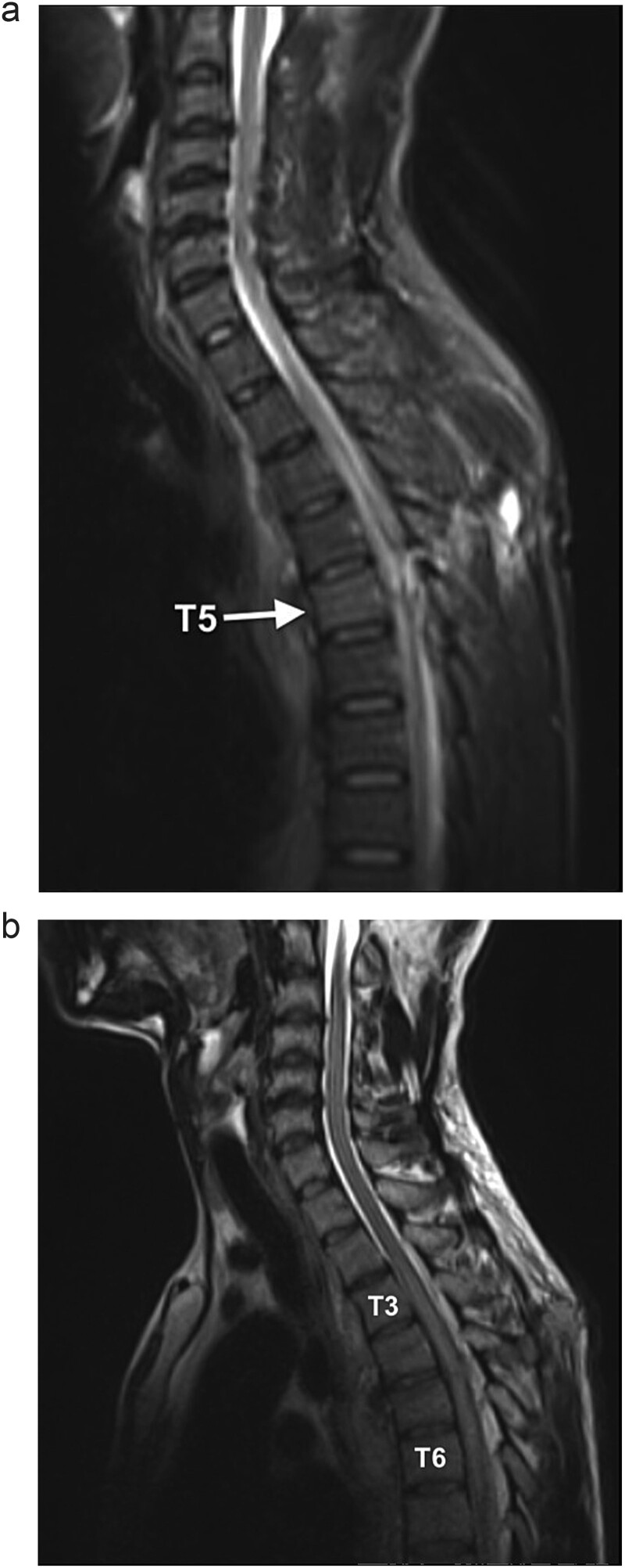
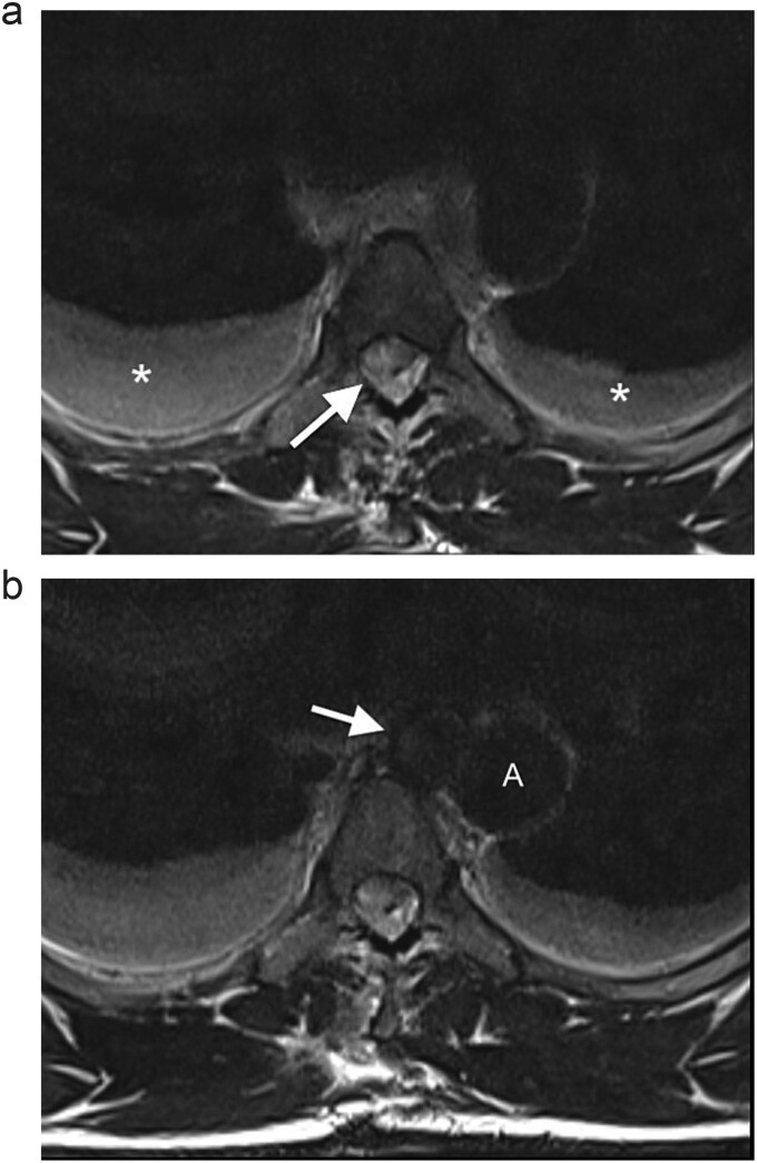
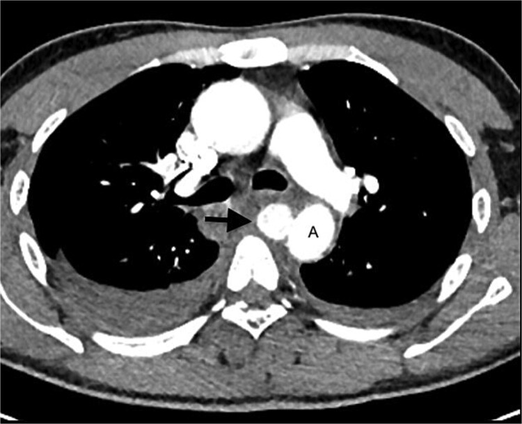
Endovascular repair of aortic injuries secondary to blunt trauma has been widely described. However, literature on endovascular management in penetrating aortic injuries is scarce. The patient in this case report, a victim of penetrating thoracic aortic trauma, presented 5 days after injury with Brown-Sequard syndrome and a contained aortic injury (pseudoaneurysm) and was haemodynamically stable. Therefore, thoracic endovascular aortic repair was an option in this case. Endovascular repair carries a lower peri-operative morbidity and mortality rate than open repair. However, because most cases of penetrating thoracic vascular injury have haemodynamic instability, open surgery is considered the standard of care. This case demonstrates successful management of an aortic injury with a minimally invasive procedure.
Keywords: Brown–Sequard syndrome; aortic pseudoaneurysm; case report; penetrating injury; thoracic aortic injury; thoracic endovascular aortic repair.
Published by Oxford University Press and JSCR Publishing Ltd. © The Author(s) 2024.
Conflict of interest statement
There are no conflicts of interest.
Figures
Similar articles
-
Outcomes following thoracic endovascular aortic repair for blunt thoracic aortic injury stratified by Society for Vascular Surgery grade.J Vasc Surg. 2023 Jul;78(1):38-47.e2. doi: 10.1016/j.jvs.2023.03.021. Epub 2023 Mar 15. J Vasc Surg. 2023. PMID: 36931613 Free PMC article.
-
Patterns, management options and outcome of blunt thoracic aortic injuries: a 20-year experience from a Tertiary Care Hospital.Eur J Trauma Emerg Surg. 2022 Oct;48(5):4079-4091. doi: 10.1007/s00068-022-01930-1. Epub 2022 Mar 14. Eur J Trauma Emerg Surg. 2022. PMID: 35286404 Free PMC article.
-
Solving Intraoperative Complications During Endovascular Repair of Late Contained Ruptured Aortic Pseudoaneurysm After Surgical De-coarctation: Case Report and Systematic Review of Literature.J Endovasc Ther. 2025 Apr;32(2):290-302. doi: 10.1177/15266028231177047. Epub 2023 Jun 4. J Endovasc Ther. 2025. PMID: 37271989
-
Nonoperative management of grade III blunt thoracic aortic injuries.J Vasc Surg. 2016 Dec;64(6):1580-1586. doi: 10.1016/j.jvs.2016.05.070. Epub 2016 Jul 25. J Vasc Surg. 2016. PMID: 27461999
-
A Systematic Review on Thoracic Endovascular Repair Outcomes in Blunt Thoracic Aortic Injuries.J Endovasc Ther. 2024 Feb 18:15266028241233163. doi: 10.1177/15266028241233163. Online ahead of print. J Endovasc Ther. 2024. PMID: 38369733 Review.
References
-
- Veller M (ed). Thoracic aortic interventions. In: Vascular Society of Southern Africa. Johannesburg: Vascular Society of Southern Africa; 2023. Available from: https://www.vascularsociety.co.za/wp-content/uploads/2024/03/Thoracic-ao...
-
- Bhana M, Fru P, Plani F. A long walk to freedom: the epidemiology of penetrating trauma in South Africa - analysis of 4 697 patients over a six-year period at Chris Hani Baragwanath academic hospital. S Afr J Surg 2022;60:77–83. - PubMed
-
- D'Souza D, Chieng R, Sharma R. et al. Thoracic Aortic Injury. Australia: Radiopaedia.org; 2023. 10.53347/rID-2171 (18 November 2023, date last accessed). - DOI
Publication types
LinkOut - more resources
Full Text Sources
