. 2024 Dec 4:13:100617.
doi: 10.1016/j.ejro.2024.100617. eCollection 2024 Dec.

Multidisciplinary quantitative and qualitative assessment of IDH-mutant gliomas with full diagnostic deep learning image reconstruction

Affiliations

Multidisciplinary quantitative and qualitative assessment of IDH-mutant gliomas with full diagnostic deep learning image reconstruction

Christer Ruff et al. Eur J Radiol Open. .

Abstract

Rationale and Objectives: Diagnostic accuracy and therapeutic decision-making for IDH-mutant gliomas in tumor board reviews are based on MRI and multidisciplinary interactions.

Materials and methods: This study explores the feasibility of deep learning-based reconstruction (DLR) in MRI for IDH-mutant gliomas. The research utilizes a multidisciplinary approach, engaging neuroradiologists, neurosurgeons, neuro-oncologists, and radiotherapists to evaluate qualitative aspects of DLR and conventional reconstructed (CR) sequences. Furthermore, quantitative image quality and tumor volumes according to Response Assessment in Neuro-Oncology (RANO) 2.0 standards were assessed.

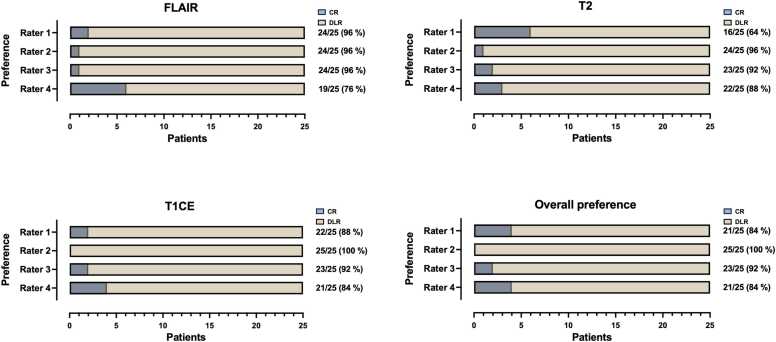
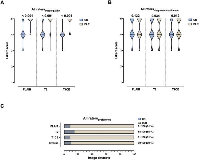
Results: All DLR sequences consistently outperformed CR sequences (median of 4 for all) in qualitative image quality across all raters (p < 0.001 for all) and revealed higher SNR and CNR values (p < 0.001 for all). Preference for all DLR over CR was overwhelming, with ratings of 84 % from the neuroradiologist, 100 % from the neurosurgeon, 92 % from the neuro-oncologist, and 84 % from the radiation oncologist. The RANO 2.0 compliant measurements showed no significant difference between the CR and DRL sequences (p = 0.142).

Conclusion: This study demonstrates the clinical feasibility of DLR in MR imaging of IDH-mutant gliomas, with significant time savings of 29.6 % on average and non-inferior image quality to CR. DLR sequences received strong multidisciplinary preference, underscoring their potential for enhancing neuro-oncological decision-making and suitability for clinical implementation.

Keywords: Deep learning; Diagnostic accuracy; IDH-mutant gliomas; Image reconstruction; Magnetic resonance imaging; Multidisciplinary; Visual perception preference.


Conflict of interest statement

The authors declare that they have no known competing financial interests or personal relationships that could have appeared to influence the work reported in this paper.

Figures

Fig. 1
Fig. 1
Study flowchart and patient enrollment.
Fig. 2
Fig. 2
Thirty-six-year-old man with histologically confirmed oligodendroglioma in the left frontal lobe with O6-Methylguanine-DNA-methyltransferase (MGMT) promoter methylation. Deep learning reconstructed (DLR) fluid-attenuated inversion recovery images (FLAIR), T2-weighted images, and T1-weighted contrast-enhanced (CE) images showed non-inferior image quality, image sharpness, and tumor conspicuity as well as less image noise compared to conventional reconstruction (CR). Furthermore, DLR images demonstrate less image noise.
Fig. 3
Fig. 3
Follow-up MRI of a fifty-nine-year-old man after radiation therapy and current therapy with Bevacizumab of a left frontal histologically confirmed astrocytoma grade 2. Deep learning reconstructed (DLR) fluid-attenuated inversion recovery images (FLAIR), T2-weighted images, and T1-weighted contrast-enhanced (CE) images revealed sharper and better delineation of the intra-ventricular tumor structures in the DLR T2-weighted images compared to the standard image technique. Note the DLR algorithm's tendency to produce images of the tumor's internal structure that appear more uniform and softer in contrast to conventionally reconstructed (CR) images. Note minimal (banding) artifacts in T2- and T1-weighted DLR images.
Fig. 4
Fig. 4
Follow-up MRI of an astrocytoma grade 4 without O6-Methylguanine-DNA-methyltransferase (MGMT) promoter methylation in the left frontal lobe in a fifty-eight-year-old man after partial resection, radiation therapy, and temozolomide. The accelerated deep learning reconstructed (DLR) images presented non-inferior overall image quality and preferred visual perception compared to conventional reconstructed images (CR). CR: conventional reconstruction; DLR: deep learning reconstructed technique; FLAIR = fluid-attenuated inversion recovery images; CE = contrast-enhanced.
Fig. 5
Fig. 5
Visual preferences in the review of 25 paired magnetic resonance image sets containing astrocytoma grade 2 – 4 and oligodendroglioma by four experienced raters. Raters consist of a neuroradiologist (Rater 1), a neurosurgeon (Rater 2), a neuro-oncologist (Rater 3), and a radiation oncologist (Rater 4). CR = conventional reconstruction; DLR = deep learning reconstructed technique; FLAIR = fluid-attenuated inversion recovery images; T2 = T2-weighted images; T1CE = T1-weighted contrast-enhanced images.
Fig. 6
Fig. 6
RANO-compliant measurements of non-enhancing non-target lesions (NENT) through the utilization of conventional reconstructed (CR) and deep learning reconstructed (DLR) FLAIR sequences of 25 paired image sets. FLAIR = fluid-attenuated inversion recovery images.
Fig. 7
Fig. 7
Multidisciplinary rating of image quality (A) and diagnostic confidence (B) of deep learning reconstructed images and conventional recorded images by four experienced raters in a pooled data analysis. Likert scale ranging from 1 to 5 with 5 being the best rating. Significant differences are indicated. The dotted lines represent the median. (C) displays the visual preferences of the four 25 paired magnetic resonance image sets containing astrocytoma grade 2 – 4 and oligodendroglioma of all raters in a pooled data analysis. CR = conventional reconstruction; DLR = deep learning reconstructed technique; FLAIR = fluid-attenuated inversion recovery images; T2 = T2-weighted images; T1CE = T1-weighted contrast-enhanced images.

References

    1. Reuss D.E. Updates on the WHO diagnosis of IDH-mutant glioma. J. Neurooncol. 2023;162(3):461–469. - PMC - PubMed
    1. Pellerino A., et al. Epidemiology, risk factors, and prognostic factors of gliomas. Clin. Transl. Imaging. 2022;10(5):467–475.
    1. Niyazi M., et al. ESTRO-ACROP guideline "target delineation of glioblastomas. Radio. Oncol. 2016;118(1):35–42. - PubMed
    1. Srinivasan S., et al. The promise of magnetic resonance imaging in radiation oncology practice in the management of brain, prostate, and GI malignancies. JCO Glob. Oncol. 2022;8 - PMC - PubMed
    1. Alkanhal H., et al. Differentiating nonenhancing grade II gliomas from grade III gliomas using diffusion tensor imaging and dynamic susceptibility contrast MRI. World Neurosurg. 2021;146:e555–e564. - PubMed

LinkOut - more resources