. 2024 Dec 23:28:0118.
doi: 10.34133/bmr.0118. eCollection 2024.

Custom-Made Ce-Mn Bimetallic Nanozyme for the Treatment of Intervertebral Disc Degeneration by Inhibiting Oxidative Stress and Modulating Macrophage M1/M2 Polarization

Affiliations

Custom-Made Ce-Mn Bimetallic Nanozyme for the Treatment of Intervertebral Disc Degeneration by Inhibiting Oxidative Stress and Modulating Macrophage M1/M2 Polarization

Jianwei Wu et al. Biomater Res. .

Abstract

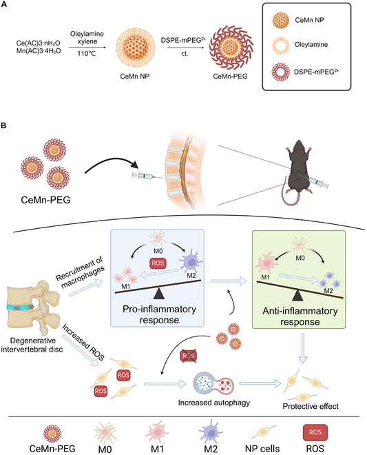
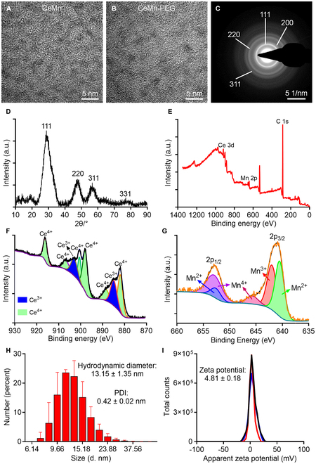
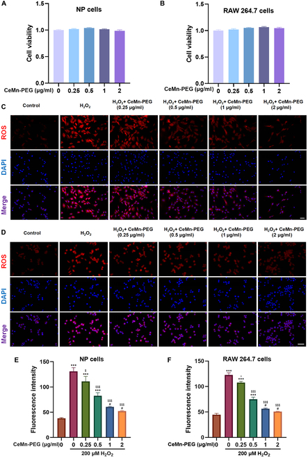
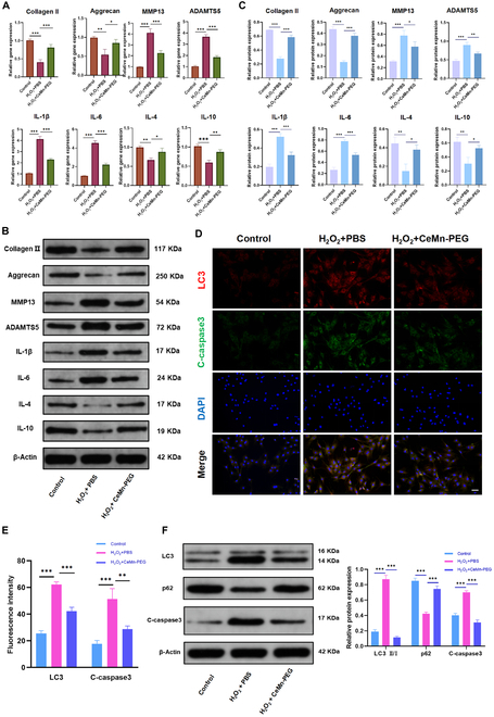
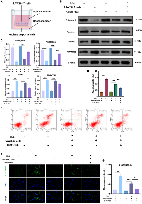
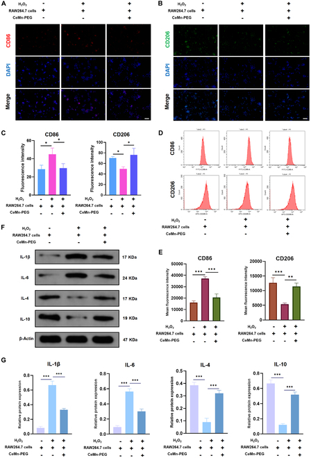
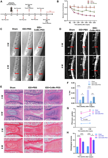
Intervertebral disc degeneration (IDD)-induced lower back pain (LBP) brings heavy burden worldwide. In the degenerated intervertebral disc, there is an increase in the accumulation of reactive oxygen species (ROS) and the infiltration of M1 macrophages, which leads to abnormal local inflammatory microenvironment and exacerbates IDD. In this study, we developed a novel injectable polyethylene glycol (PEG)-capped cerium ion-manganese ion (Ce-Mn) bimetallic nanozyme (CeMn-PEG) with strong ROS scavenging and M2-type macrophage polarizing abilities to efficiently alleviate IDD. In vitro experiments demonstrated that CeMn-PEG effectively scavenged excess ROS in both nucleus pulposus (NP) and RAW264.7 cells. In addition, we found that CeMn-PEG markedly protected NP cells from H2O2-induced overproduction of inflammatory cytokines, excessive cell apoptosis and autophagy, and imbalance between extracellular matrix (ECM) degradation. Moreover, CeMn-PEG induced macrophages to transition from the M1 phenotype to the M2 phenotype and the increased M2-type macrophages could alleviate H2O2-induced ECM degradation and cell apoptosis in NP cells. In a puncture-induced mouse IDD model, CeMn-PEG treatment could effectively ameliorate the progression of disc degeneration and mitigate puncture-induced mechanical hyperalgesia. Thus, our study demonstrated the effectiveness of CeMn-PEG as a novel treatment strategy for the treatment of IDD and a range of other inflammatory diseases.


Conflict of interest statement

Competing interests: The authors declare that they have no competing interests.

Figures

Fig. 1.
Fig. 1.
(A) Diagram of synthesis of CeMn NP and CeMn-PEG. (B) Schematic diagram of CeMn-PEG for the treatment of IDD in mouse.
Fig. 2.
Fig. 2.
Characterization of CeMn NP and CeMn-PEG. Representative TEM image of CeMn NP (A) and CeMn-PEG (B). SAED pattern (C) and XRD (D) of CeMn. (E) Survey scan XPS spectrum of CeMn-NP. (F) XPS spectrum of Ce3d revealed the mixed valence states of Ce3+ (884.88 and 902.89 eV) and Ce4+ (881.89, 888.38, 897,88, 900.48, 906.66, and 916.03 eV). (G) XPS spectrum of Mn2p revealed the mixed valence states of Mn2+ (640.51 and 652.6 eV), Mn3+ (641.99 eV), and Mn4+ (645.69 and 652.91 eV). Hydrodynamic diameter (H) and zeta potential (I) of CeMn-PEG.
Fig. 3.
Fig. 3.
The biocompatibility and ROS scavenging properties of CeMn-PEG in vitro. (A and B) Effects of various concentrations of CeMn-PEG (0, 0.25, 0.5, 1, and 2 μg/ml) on the viability of both NP and RAW264.7 cells after incubation for 24 h by using CCK8 assay. (C to F) Representative ROS fluorescence microscopy images and relative fluorescence intensity of NP cells (C and E) and RAW264.7 cells (D and F) pretreated with various concentrations of CeMn-PEG (0, 0.25, 0.5, 1, and 2 μg/ml) after the treatment of H2O2 (200 μM) for 6 h. Scale bar, 25 μm. Data are presented as the mean ± SEM. #P > 0.05, *P < 0.05, **P < 0.01, ***P < 0.001 relative to the control group; ^P > 0.05, $P < 0.05, $$P < 0.01, $$$P < 0.001 relative to the H2O2-treated group, n = 3.
Fig. 4.
Fig. 4.
CeMn-PEG ameliorated H2O2-induced inflammation and ECM degradation and reduced H2O2-induced apoptosis and autophagy in vitro. (A) The relative mRNA expression of IL-1β, IL-4, IL-6, IL-10, collagen II, aggrecan, MMP13, and ADAMTS5 was determined by using qPCR. (B and C) Representative images and quantification data of Western blot results of IL-1β, IL-4, IL-6, IL-10, collagen II, aggrecan, MMP13, and ADAMTS5. (D and E) Representative images and relative fluorescence intensity of double immunofluorescence of LC3 (red) and C-caspase3 (green) in NP cells. (F) Representative images and quantification data of Western blot results of LC3, p62, and C-caspase3. Scale bar, 25 μm. Data are presented as the mean ± SEM. *P < 0.05, **P < 0.01, ***P < 0.001, n = 3.
Fig. 5.
Fig. 5.
RAW264.7 cells pretreated with CeMn-PEG could alleviate H2O2-induced ECM degradation and apoptosis in NP cells. (A) Diagram of RAW264.7 cells (apical chamber) and NP cells (basal chamber) cocultured by using Transwell dish. RAW264.7 cells were pretreated with CeMn-PEG and then cocultured with NP cells. (B and C) Representative images and quantification data of Western blot results of collagen II, aggrecan, MMP13, and ADAMTS5 in NP cells in the coculture system. (D) Representative images of flow cytometry analysis of NP cells in the coculture system. (E) Apoptosis rate of NP cells in the coculture system estimated by flow cytometry analysis. (F and G) Representative images and relative fluorescence intensity of C-caspase3 (green) in NP cells in the coculture system. Scale bar, 25 μm. Data are presented as the mean ± SEM. *P < 0.05, **P < 0.01, ***P < 0.001, n = 3.
Fig. 6.
Fig. 6.
CeMn-PEG could promote the polarization of macrophages to M2 type. (A to C) Representative images and relative fluorescence intensity of CD86 and CD206 in RAW264.7 cells in the coculture system. (D and E) Representative images of cytometry analysis (D) and mean fluorescence intensity (MFI) quantification (E) of CD86 and CD206 in RAW264.7 cells in the coculture system. (F and G) Representative images and quantification data of Western blot results of IL-1β, IL-4, IL-6, and IL-10 in RAW264.7 cells in the coculture system. Scale bar, 25 μm. Data are presented as the mean ± SEM. *P < 0.05, **P < 0.01, ***P < 0.001, n = 3.
Fig. 7.
Fig. 7.
CeMn-PEG could ameliorate the puncture-induced degeneration of mouse IVD in vivo. (A) Schematic graph exhibits in vivo experimental procedure including the establishment of the puncture-induced mouse IVD model with injection of 5 μl of PBS or CeMn-PEG (1 μg/ml) and arrangement of behavior test (von Frey test) and imaging examination (MRI and x-ray). Mice were harvested at 3 and 6 weeks after surgery. (B) Paw withdrawal threshold of mice by using von Frey test. Data are presented as the mean ± SEM. #P > 0.05, *P < 0.05, **P < 0.01, ***P < 0.001 relative to the IDD + PBS group, n = 8. (C) Representative radiographs of needle-punctured mouse lumbar disc of each group at 3 and 6 weeks after surgery (red arrows). (D) Representative T2-weighted MRI images of needle-punctured mouse lumbar disc of each group at 3 and 6 weeks after surgery (red arrows). (E) Representative H&E staining and Safranin O/Fast Green staining of disc samples in each group at 3 and 6 weeks after the operation. Scale bar, 100 μm. (F) Disc height index (%DHI) of needle-punctured mouse lumbar disc evaluated according to radiographs in 3 groups at 3 and 6 weeks after surgery. Data are presented as the mean ± SEM. *P < 0.05, **P < 0.01, ***P < 0.001, n = 8. (G) Pfirrmann grade scores of needle-punctured mouse lumbar disc evaluated according to MRI images in 3 groups at 3 and 6 weeks after surgery. Data are presented as the mean ± SEM. ***P < 0.001 relative to the IDD + PBS group, n = 8. $P < 0.05, $$P < 0.01 relative to the sham group, n = 8. (H) Histological grades of needle-punctured mouse lumbar disc evaluated according to H&E staining and Safranin O/Fast Green staining in 3 groups at 3 and 6 weeks after surgery. Data are presented as the mean ± SEM. *P < 0.05, **P < 0.01, ***P < 0.001, n = 8.

Similar articles

References

    1. Katz JN. Lumbar disc disorders and low-back pain: Socioeconomic factors and consequences. J Bone Joint Surg Am. 2006;88(Suppl 2):21–24. - PubMed
    1. GBD 2016 Disease and Injury Incidence and Prevalence Collaborators. Global, regional, and national incidence, prevalence, and years lived with disability for 328 diseases and injuries for 195 countries, 1990-2016: A systematic analysis for the Global Burden of Disease Study 2016. Lancet. 2017;390(10100):1211–1259. - PMC - PubMed
    1. Hudson KD, Alimi M, Grunert P, Härtl R, Bonassar LJ. Recent advances in biological therapies for disc degeneration: Tissue engineering of the annulus fibrosus, nucleus pulposus and whole intervertebral discs. Curr Opin Biotechnol. 2013;24(5):872–879. - PubMed
    1. Urban JP, Roberts S. Degeneration of the intervertebral disc. Arthritis Res Ther. 2003;5(3):120–130. - PMC - PubMed
    1. Jin H, Wang Q, Wu J, Han X, Qian T, Zhang Z, Wang J, Pan X, Wu A, Wang X. Baicalein inhibits the IL-1β-induced inflammatory response in nucleus pulposus cells and attenuates disc degeneration in vivo. Inflammation. 2019;42(3):1032–1044. - PubMed

LinkOut - more resources