. 2024 Sep 8;7(1):101215.
doi: 10.1016/j.jhepr.2024.101215. eCollection 2025 Jan.

Biliary atresia susceptibility gene EFEMP1 regulates extrahepatic bile duct elastic fiber formation and mechanics

Affiliations

Biliary atresia susceptibility gene EFEMP1 regulates extrahepatic bile duct elastic fiber formation and mechanics

Kapish Gupta et al. JHEP Rep. .

Abstract

Background & aims: EGF-containing fibulin extracellular matrix protein 1 (EFEMP1, also called fibulin-3) is an extracellular matrix protein linked in a genome-wide association study to biliary atresia, a fibrotic disease of the neonatal extrahepatic bile duct. Fibulin-3 is deposited in most tissues and Efemp1 null mice have decreased elastic fibers in visceral fascia; however, fibulin-3 does not have a role in the development of large elastic fibers and its overall function in the extrahepatic bile ducts remains unclear.

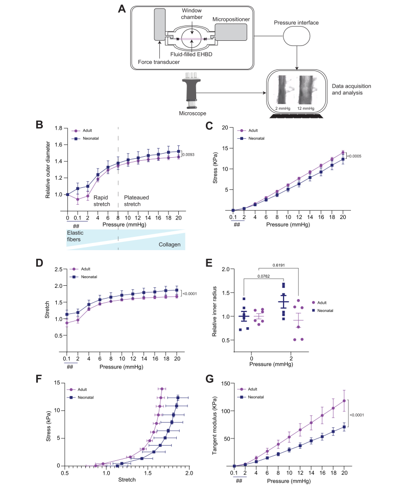
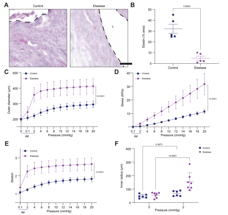
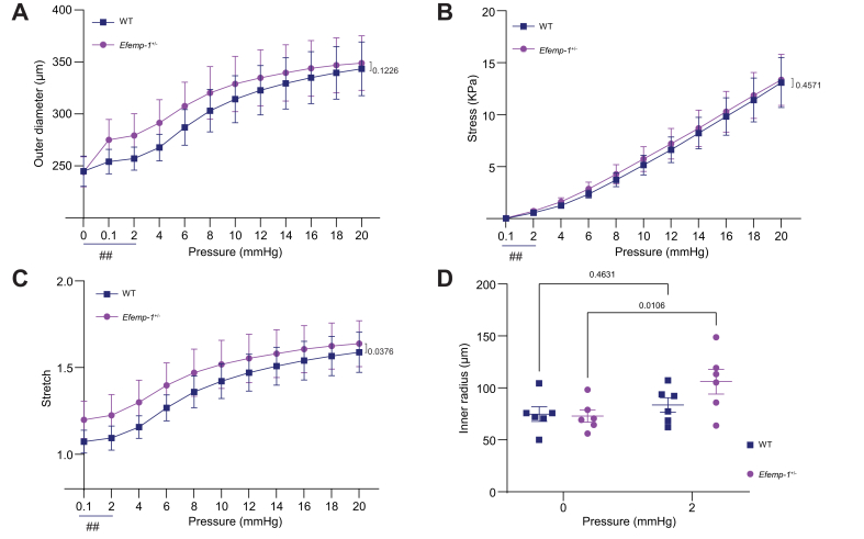
Methods: We used staining and histology to define the amount and organization of key extracellular matrix components in the extrahepatic bile ducts. We also repurposed pressure myography, a technique heretofore applied to the vasculature, to determine the contribution of elastin and fibulin-3 to extrahepatic bile duct mechanics. We examined extrahepatic bile duct structure and mechanics in three models: neonatal vs. adult rat ducts (n = 6 each), elastase-treated adult rat ducts (n = 6-7 each), and Efemp1 +/- vs. wild-type mouse ducts (n = 6 each).

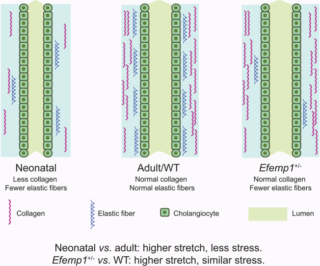
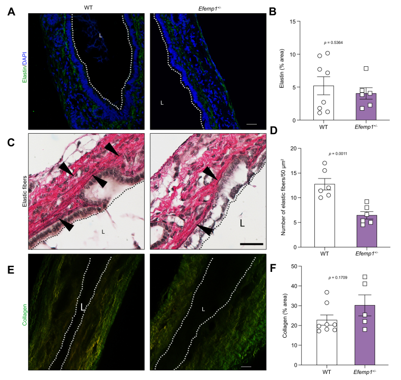
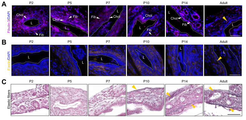
Results: We demonstrated that fibulin-3 is expressed in the submucosa of both neonatal and adult mouse, rat and human extrahepatic bile ducts and that, in adult Efemp1 +/- mouse ducts, elastin organization into fibers is decreased by approximately half. Pressure myography showed that Efemp1 +/- ducts have altered mechanics compared to control ducts, with Efemp1 +/- ducts displaying significant stretch compared to controls (p = 0.0376); these changes in stretch are similar to those observed in elastase-treated vs. normal ducts (p <0.0001) and in neonatal ducts vs. adult ducts (p <0.0001).

Conclusion: Fibulin-3 has an important role in the formation of elastic fibers and the mechanical properties of the extrahepatic bile duct. This provides functional relevance for the biliary atresia susceptibility gene EFEMP1.

Impact and implications: The gene EFEMP1 was found via a genome-wide association study to be a susceptibility gene for the neonatal disease biliary atresia. EFEMP1 encodes the protein fibulin-3, which regulates elastic fiber organization in the extrahepatic bile duct (EHBD), the major site of disease in biliary atresia. We showed that neonatal EHBDs as well as mice heterozygous for Efemp1 have decreased numbers of elastic fibers, and that this alters EHBD mechanics. This work is important for understanding the mechanism of biliary atresia, in particular susceptibility to obstruction.

Keywords: Pressure myography; bile duct mechanics; collagen; elastin; mechanobiology; stiffness.


Conflict of interest statement

The authors declare no conflict of interest. Please refer to the accompanying ICMJE disclosure forms for further details.

Figures

Image 1
Graphical abstract
Fig. 1
Fig. 1
Elastic fibers are reduced in adult Efemp1+/- EHBDs but elastin expression is unchanged. (A) Representative staining of elastin in WT and Efemp1+/- EHBDs. (B) Quantification of percent area of staining. (C) Representative elastic fiber staining of WT and Efemp1+/- EHBDs. Arrowheads show examples of elastic fibers. Note organized fibers under the basement membrane in WT. Additional examples shown in Fig. S2. (D) Quantification of the number of elastic fibers per 50 μm2. (E) Representative images of collagen by SHG imaging in WT and Efemp1+/- EHBDs. (F) Quantification of percent collagen area of SHG signal. Forward signal in red and backward signal in green. Dotted lines and L denote lumens. 6-8 ducts were analyzed per genotype for all stains, with 3-5 images taken per duct. All scale bars 50 μm. Data shown are mean ± SEM. Significance in B and D determined by unpaired t-test. Significance in F determined using the Mann-Whitney test since the data were not normally distributed. EHBD, extrahepatic bile duct; SHG, second harmonic generation; WT, wild-type.
Fig. 2
Fig. 2
Fibulin-3 expression and elastin organization during postnatal development of the mouse EHBD. (A) Representative images of fibulin-3 staining in neonatal (P2, 5, 7, 10, 14) and adult mouse EHBD. White arrows show cholangiocytes (Chol) and likely fibroblasts (Fib) expressing fibulin-3. Yellow arrow shows fibulin-3 localization under the cholangiocyte monolayer in adult. (B) Representative images of elastin staining in mouse samples. Yellow arrow shows elastin localization under the cholangiocyte monolayer in the adult EHBD. In all immunohistochemistry stains, nuclei are stained with DAPI (blue). For A and B, in all images, dotted lines and L denote lumens. (C) Representative images of elastic fiber staining. Yellow arrows show elastic fiber localization under the cholangiocyte monolayer. Images in insets show regions highlighted in boxes outlined with dotted lines. 3-4 samples were stained for each age, with 3-5 images taken per duct. EHBD, extrahepatic bile duct.
Fig. 3
Fig. 3
Neonatal and adult rat EHBDs have different mechanics. (A) Schematic of the pressure myography setup. The EHBD is mounted onto a pressure myography apparatus and progressively pressurized. A force transducer and micro-positioner control the pressure and position. The EHBD is imaged through a window chamber, and the data, including pressure values and microscopic images, is recorded and analyzed on a computer. (B) Relative outer diameter of neonatal (P7-10) and adult rat EHBDs at increasing pressures. Rapid stretch was observed up to 8 mmHg pressure followed by a plateau phase. Blue triangles under graph represent estimate of the relative contribution of elastin and collagen to the curves, consistent with data from the vascular literature. (C) Circumferential stress profile of neonatal vs. adult EHBD. (D) Circumferential stretch profile of neonatal vs. adult EHBD as pressure is increased. (E) Relative change in inner radius of neonatal and adult EHBD as pressure is increased from unloaded (0 mmHg) to physiological pressure (2 mmHg). (F) Calculated circumferential stress and circumferential stretch of neonatal and adult EHBD under increasing pressure from unloaded (0 mmHg) to physiological pressure (2 mmHg). Statistical analyses for circumferential stress and circumferential stretch individually are shown in panels B and C. (G) Calculated tangent modulus (stiffness) of neonatal and adult rat EHBDs. Six individual ducts were examined per group. Note uneven x axes represented by ##. Data shown are mean ± SEM. p values in graphs B-D and G represent two-way ANOVA between neonatal and adult EHBD. p values in E represent Fisher's least significant difference after two-way ANOVA. EHBD, extrahepatic bile duct.
Fig. 4
Fig. 4
Elastase treatment leads to altered EHBD mechanics. (A) Representative images of elastic fiber staining in control- and elastase-treated rat adult EHBDs stained using elastin staining solution according to Weigert. Dotted lines and L denote lumens. Scale bar, 50 μm. (B) Quantification showing significant removal of elastin after elastase treatment. (C) Measured outer diameter of control and elastase-treated rat EHBDs at increasing pressures. (D) Circumferential stress profile of control vs. elastase-treated EHBD. (E) Circumferential stretch profile of control vs. elastase-treated EHBD. (F) Relative change in inner radius of control vs. elastase-treated EHBD as pressure is increased from unloaded (0 mmHg) condition to physiological pressure (2 mmHg). Data shown are mean ± SEM. Six to seven rats used per condition. ## indicate uneven x axes. p value in graph B represents p value for t-test, in graph C-E represents two-way ANOVA between profile of elastase-vs. control-treated EHBD and in F represents Fisher's least significant difference after two-way ANOVA. EHBD, extrahepatic bile duct.
Fig. 5
Fig. 5
Mechanical properties measured by pressure myography are altered in Efemp1+/- EHBDs. (A) Measured outer diameter of WT and Efemp1+/- adult mice EHBDs at increasing pressures. (B) Calculated circumferential stress of WT and Efemp1+/- adult mice EHBDs at increasing pressures. (C) Calculated circumferential stretch of WT and Efemp1+/- adult mice EHBDs at increasing pressures. (D) Relative change in inner diameter of WT vs. Efemp1+/- treated EHBD as pressure is increased from unloaded (0 mm Hg) condition to physiological pressure (2 mmHg). Data shown are mean ± SEM. Six mice used per condition. ## represent uneven x axes. p values in graphs A-C represent two-way ANOVA between profile of WT vs. Efemp1+/-. p value in D represents Fisher's least significant difference after two-way ANOVA. EHBD, extrahepatic bile duct; WT, wild-type.

Similar articles

Cited by

References

    1. Gupta K., Chen D., Wells R.G. Microcystin-RR is a biliary toxin selective for neonatal cholangiocytes. JHEP Reports. 2024 doi: 10.1016/j.jhepr.2024.101218. - DOI - PMC - PubMed
    1. Lorent K., Gong W., Koo K.A., et al. Identification of a plant isoflavonoid that causes biliary atresia. Sci Transl Med. 2015;7:286ra67. doi: 10.1126/scitranslmed.aaa1652. - DOI - PMC - PubMed
    1. Zhao X., Lorent K., Wilkins B.J., et al. Glutathione antioxidant pathway activity and reserve determine toxicity and specificity of the biliary toxin biliatresone in zebrafish. Hepatology. 2016;64:894–907. doi: 10.1002/hep.28603. - DOI - PMC - PubMed
    1. Koo K.A., Lorent K., Gong W., et al. Biliatresone, a reactive natural toxin from dysphania glomulifera and D. littoralis: discovery of the toxic moiety 1,2-Diaryl-2-propenone. Chem Res Toxicol. 2015;28:1519–1521. doi: 10.1021/acs.chemrestox.5b00227. - DOI - PMC - PubMed
    1. Gupta K., Xu J.P., Diamond T., et al. Low-dose biliatresone treatment of pregnant mice causes subclinical biliary disease in their offspring: evidence for a spectrum of neonatal injury. PLoS One. 2024;19 doi: 10.1371/journal.pone.0301824. - DOI - PMC - PubMed

LinkOut - more resources