

The rat osteoarthritis bone score for histological pathology relevant to human bone marrow lesions and pain
- PMID: 39717525
- PMCID: PMC11665527
- DOI: 10.1016/j.ocarto.2024.100544
The rat osteoarthritis bone score for histological pathology relevant to human bone marrow lesions and pain
Abstract
Objectives: Histological osteochondral characteristics of inflammation, fibrosis, vascularity, cartilage islands, vessels entering cartilage, thickened trabeculae and cysts are associated with bone marrow lesions (BMLs) in human knee osteoarthritis (OA). We identified and developed a method for scoring comparable pathology in two rat OA knee pain models.
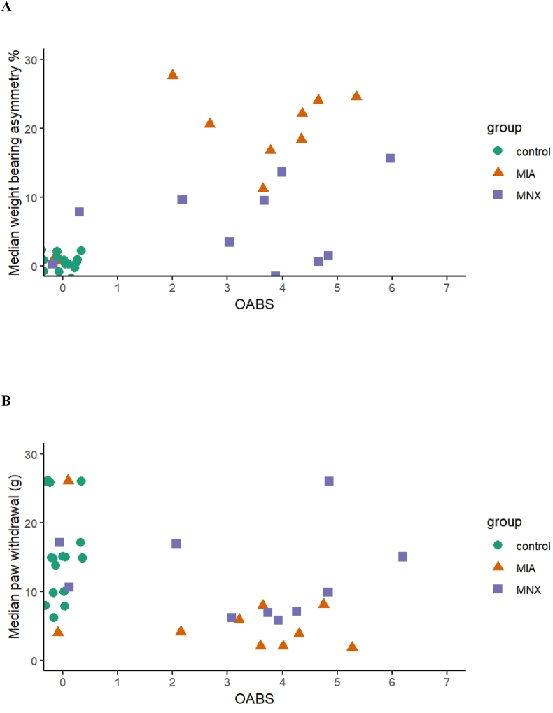
Methods: Rats (n = 8-10 per group) were injected with monoiodoacetate (MIA) or saline, or underwent meniscal transection (MNX) or sham surgery. Pain behaviour (weight bearing asymmetry and mechanical hindpaw withdrawal thresholds (PWTs)) were measured and knee samples obtained. Features associated with BMLs were evaluated using haematoxylin and eosin or Safranin-O stained knee sections. Sections were scored for chondropathy, osteophytes, synovitis and with the human OA Bone Score modified for rats (rOABS). rOABS reliability was assessed with intraclass correlation coefficient (ICC), groups were compared using Mann-Whitney U-tests, and associations examined with Spearman's rho.
Results: OABS features were more prevalent in each OA pain group than in controls. rOABS displayed good inter-rater reliability (ICC = 0.79). rOABS was higher in each model than controls; MIA 3.0 (2.3-4.0) vs vehicle 0.0 (0.0-0.0), and MNX 4.0 (2.3-4.8) vs sham 0.0 (0.0-0.0), each p < 0.003. rOABS was associated with OA cartilage involvement (rho = 0.69, p < 0.001), osteophyte (rho = 0.61, p < 0.001) and synovial inflammation (rho = 0.76, p < 0.001). Higher rOABS was associated with pain behaviour: weight bearing asymmetry (rho = 0.65, p < 0.001) and PWT (rho = -0.47, p = 0.003).
Conclusions: Subchondral pathology in rat OA models resembles human subchondral BMLs. rOABS reliably measured subchondral pathology and was associated with OA structure and pain behaviour.
Keywords: Bone marrow lesions; Histology; Meniscectomy; Monoiodoacetate; Osteoarthritis; Rat.
© 2024 The Author(s).
Conflict of interest statement
DAW has grant support from 10.13039/100004312Eli Lilly & Company, 10.13039/100009032Pfizer Ltd, GSK Ltd, 10.13039/501100024580Orion Pharma, 10.13039/100011110UCB. DAW - Consultancy fees paid to University of Nottingham; Contura International A/S, Glaxo SmithKline, AKL Research and Development Ltd, Pfizer Ltd, Abbvie Ltd, Ely Lilly & Co.Ltd, Galapagos Ltd., Reckitt Benckiser Health Ltd. DAW - Speaker fees paid to University of Nottingham; Medscape International, Pfizer Ltd. DFM has grant support from 10.13039/100004312Eli Lilly & Company, 10.13039/100009032Pfizer Ltd. NS has grant support from 10.13039/100008021Bristol Myers Squibb, 10.13039/100004312Eli Lilly & Company and 10.13039/100009032Pfizer Ltd.
Figures


References
-
- Walsh D.A., Sofat N., Guermazi A., Hunter D.J. Osteoarthritis bone marrow lesions. Osteoarthritis Cartil. 2023;31(1):11–17. - PubMed
-
- Hansen R.T., Chenu C., Sofat N., Pitsillides A.A. Bone marrow lesions: plugging the holes in our knowledge using animal models. Nat. Rev. Rheumatol. 2023;19(7):429–445. - PubMed
LinkOut - more resources
Full Text Sources
