. 2024 Dec 9:12:1514516.
doi: 10.3389/fbioe.2024.1514516. eCollection 2024.

Significant differences in knee kinematics of healthy subjects with high and low anterior tibial laxity

Affiliations

Significant differences in knee kinematics of healthy subjects with high and low anterior tibial laxity

Shiyang Chen et al. Front Bioeng Biotechnol. .

Abstract

Background: Anterior tibial laxity is considered to be a risk factor for knee injuries, including anterior cruciate ligament ruptures. The anterior cruciate ligament reconstruction also aims to restore anterior tibial laxity. While anterior tibial laxity is considered to be linked to dynamic knee stability, the mechanisms connecting anterior tibial laxity to these stability issues are not fully understood. The purpose of this study was to investigate the kinematic alterations between different anterior tibial laxity in healthy subjects. We hypothesized that anterior tibial laxity affects the anteroposterior tibial displacement during dynamic movements.

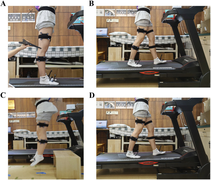
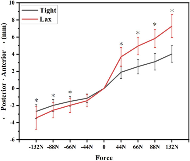
Methods: This study involved thirty-five healthy subjects. There were twenty males and fifteen females with an average age of 18.91 ± 0.78 years. Their knees were categorized into "Tight" (the smallest 50%) and "Lax" (the largest 50%) groups based on anterior tibial laxity measurements using a Kneelax3 arthrometer. Kinematic data were collected using a three-dimensional motion capture system when they performed level walking, upslope walking, and vertical jumping. The knee kinematics were recorded for statistical analysis. We used independent sample t-tests to analyze key kinematic differences between groups.

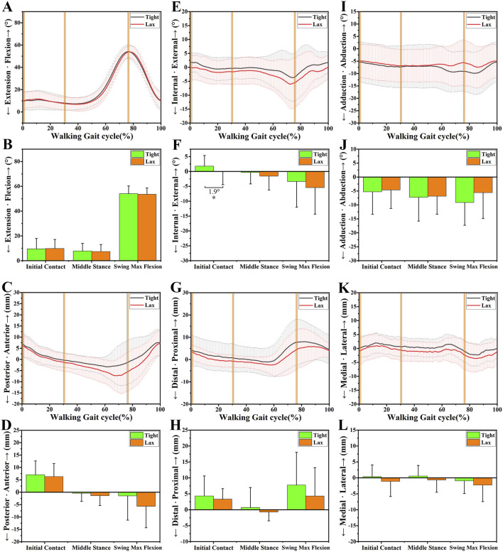
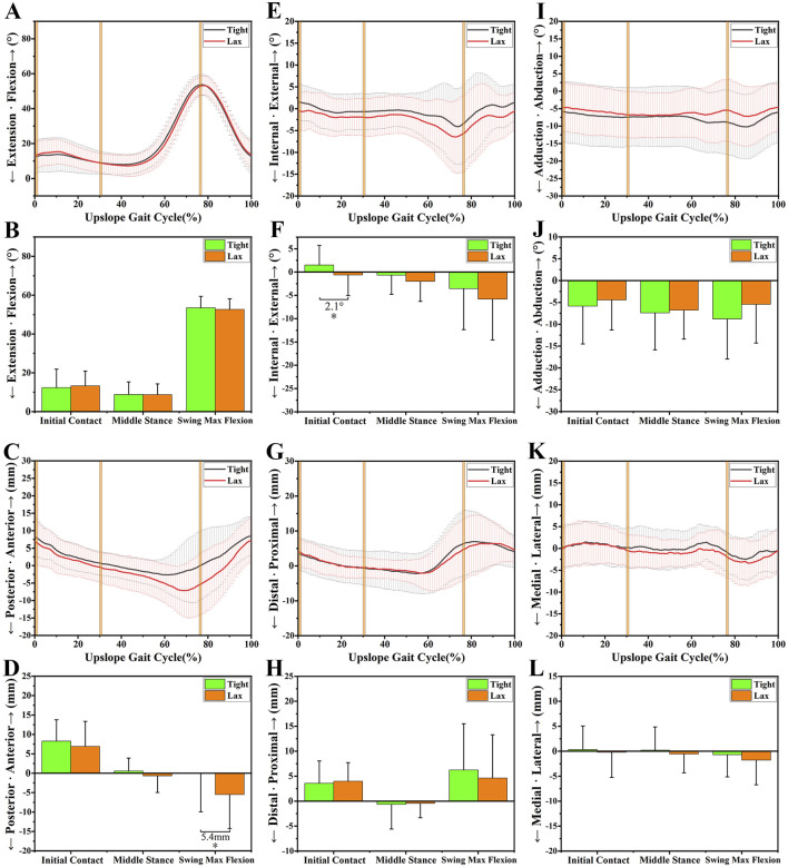
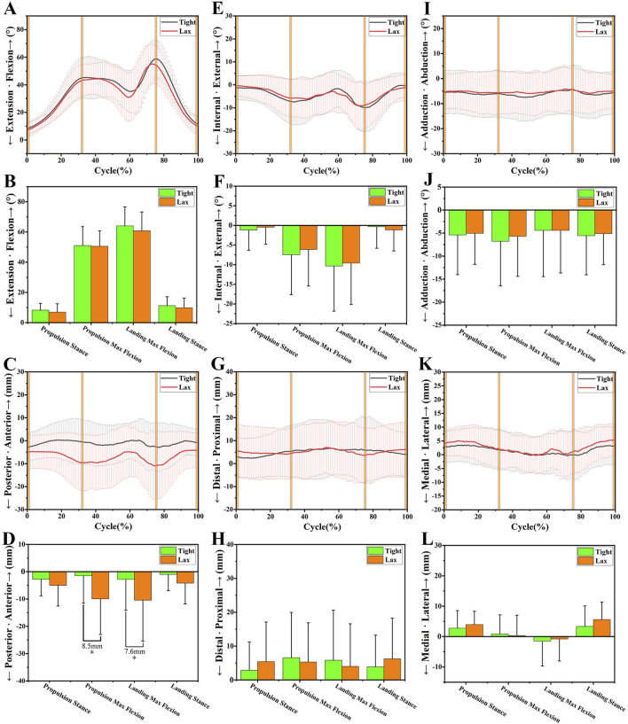
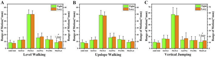
Results: The "Lax" group exhibited increased posterior tibial translation during upslope walking (5.4 ± 2.22 mm at swing max flexion, p = 0.018) and vertical jumping (8.5 ± 2.78 mm at propulsion max flexion, p = 0.003; 7.6 ± 3.17 mm at landing max flexion, p = 0.019) than the "Tight" group. Significant differences in tibial internal rotation were observed during initial contact of the gait cycle of level walking (1.9° ± 0.95°, p = 0.049) and upslope walking (2.1° ± 1.03°, p = 0.041) in the "Lax" group compared to the "Tight" group. No significant differences in adduction/abduction or medial/lateral tibial translation were found between groups.

Conclusion: The study revealed that high anterior tibial laxity resulted in increased posterior tibial translation and tibial internal rotation. High anterior tibial laxity resulted in dynamic instability of knees during motions, especially in high-demanding activities like upslope or vertical jumping. However, further research is needed to explore the clinical functional effects of knee laxity.

Keywords: anterior tibial laxity; dynamic stability; kinematic alterations; knee; knee kinematics.


Conflict of interest statement

The authors declare that the research was conducted in the absence of any commercial or financial relationships that could be construed as a potential conflict of interest.

Figures

FIGURE 1
FIGURE 1
(A) The marker sets were attached to the thigh and shin, and a handheld digital probe was used to identify patient-specific bony landmarks. (B) The level walking test. (C) The vertical jumping test. (D) The upslope walking test.
FIGURE 2
FIGURE 2
The anterior-posterior tibial laxity curves of the “Tight” group and the “Lax” group. Notes: * indicates significant differences between the “Tight” group and the “Lax” one (p < 0.05).
FIGURE 3
FIGURE 3
The 6DOF knee kinematic alterations between the “Tight” and “Lax” groups during level walking. (A-B), A shows the flexion angle alterations between two groups during a walking gait cycle and B shows their comparison at three primary time points. (C-D), C shows the anteroposterior tibial translation alterations between two groups during a walking gait cycle and D shows their comparison at three primary time points. (E-F), E shows the internal/external tibial rotation angle alterations between two groups during a walking gait cycle and F shows their comparison at three primary time points. (G-H), G shows the distal/proximal tibial translation alterations between two groups during a walking gait cycle and H shows their comparison at three primary time points. (I-J), I shows the adduction/abduction angle alterations between two groups during a walking gait cycle and J shows their comparison at three primary time points. (K-L), K shows the medial/lateral tibial translation alterations between two groups during a walking gait cycle and L shows their comparison at three primary time points. Notes: * indicates significant kinematic differences between the “Tight” group and the “Lax” one (p < 0.05).
FIGURE 4
FIGURE 4
The 6DOF knee kinematic alterations between the “Tight” and “Lax” groups during upslope walking. (A-B), A shows the flexion angle alterations between two groups during an upslope gait cycle and B shows their comparison at three primary time points. (C-D), C shows the anteroposterior tibial translation alterations between two groups during an upslope gait cycle and D shows their comparison at three primary time points. (E-F), E shows the internal/external tibial rotation angle alterations between two groups during an upslope gait cycle and F shows their comparison at three primary time points. (G-H), G shows the distal/proximal tibial translation alterations between two groups during an upslope gait cycle and H shows their comparison at three primary time points. (I-J), I shows the adduction/abduction angle alterations between two groups during an upslope gait cycle and J shows their comparison at three primary time points. (K-L), K shows the medial/lateral tibial translation alterations between two groups during an upslope gait cycle and L shows their comparison at three primary time points. Notes: * indicates significant kinematic differences between the “Tight” group and the “Lax” one (p < 0.05).
FIGURE 5
FIGURE 5
The 6DOF knee kinematic alterations between the “Tight” and “Lax” groups during vertical jumping. (A-B), A shows the flexion angle alterations between two groups during a vertical jumping cycle and B shows their comparison at four primary time points. (C-D), C shows the anteroposterior tibial translation alterations between two groups during a vertical jumping cycle and D shows their comparison at four primary time points. (E-F), E shows the internal/external tibial rotation angle alterations between two groups during a vertical jumping cycle and F shows their comparison at four primary time points. (G-H), G shows the distal/proximal tibial translation alterations between two groups during a vertical jumping cycle and H shows their comparison at four primary time points. (I-J), I shows the adduction/abduction angle alterations between two groups during a vertical jumping cycle and J shows their comparison at four primary time points. (K-L), K shows the medial/lateral tibial translation alterations between two groups during a vertical jumping cycle and L shows their comparison at four primary time points. Notes: * indicates significant kinematic differences between the “Tight” group and the “Lax” one (p < 0.05).
FIGURE 6
FIGURE 6
(A) The ROM of knee kinematics during level walking. (B) The ROM of knee kinematics during upslope walking. (C) The ROM of knee kinematics during vertical jumping. Notes: * indicates significant differences between the “Tight” group and the “Lax” one (p < 0.05).

Similar articles

References

    1. Abulhasan J. F., Grey M. J. (2017). Anatomy and physiology of knee stability. J. Funct. Morphol. Kinesiol 2, 34. 10.3390/jfmk2040034 - DOI
    1. Akbarshahi M., Schache A. G., Fernandez J. W., Baker R., Banks S., Pandy M. G. (2010). Non-invasive assessment of soft-tissue artifact and its effect on knee joint kinematics during functional activity. J. Biomech. 43, 1292–1301. 10.1016/j.jbiomech.2010.01.002 - DOI - PubMed
    1. Amano K., Pedoia V., Su F., Souza R. B., Li X., Ma C. B. (2016). Persistent biomechanical alterations after ACL reconstruction are associated with early cartilage matrix changes detected by quantitative MR. Orthop. J. Sports Med. 4, 2325967116644421. 10.1177/2325967116644421 - DOI - PMC - PubMed
    1. Boeth H., Duda G. N., Heller M. O., Ehrig R. M., Doyscher R., Jung T., et al. (2013). Anterior cruciate ligament–deficient patients with passive knee joint laxity have a decreased range of anterior-posterior motion during active movements. Am. J. Sports Med. 41, 1051–1057. 10.1177/0363546513480465 - DOI - PubMed
    1. Dejour D., Pungitore M., Valluy J., Nover L., Saffarini M., Demey G. (2019). Preoperative laxity in ACL-deficient knees increases with posterior tibial slope and medial meniscal tears. Knee Surg. sports Traumatol. Arthrosc. Off. J. ESSKA 27, 564–572. 10.1007/s00167-018-5180-3 - DOI - PubMed

LinkOut - more resources