. 2024 Dec 4;10(23):e40907.
doi: 10.1016/j.heliyon.2024.e40907. eCollection 2024 Dec 15.

vCPP2319 interacts with metastatic breast cancer extracellular vesicles (EVs) and transposes a human blood-brain barrier model

Affiliations

vCPP2319 interacts with metastatic breast cancer extracellular vesicles (EVs) and transposes a human blood-brain barrier model

Filipa D Oliveira et al. Heliyon. .

Abstract

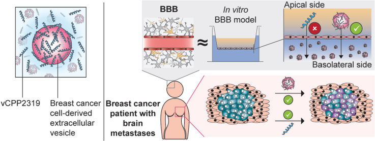
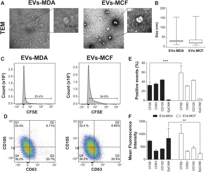
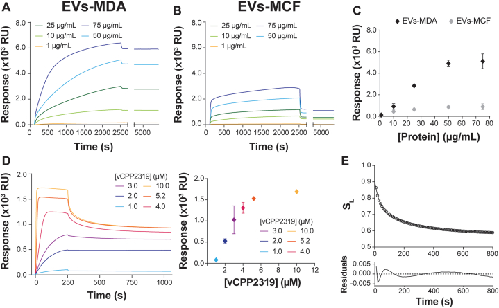
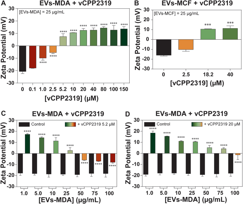
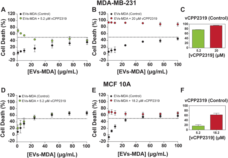
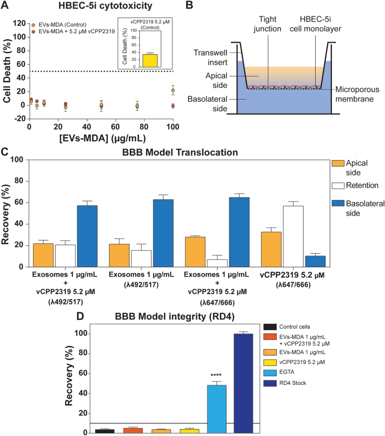
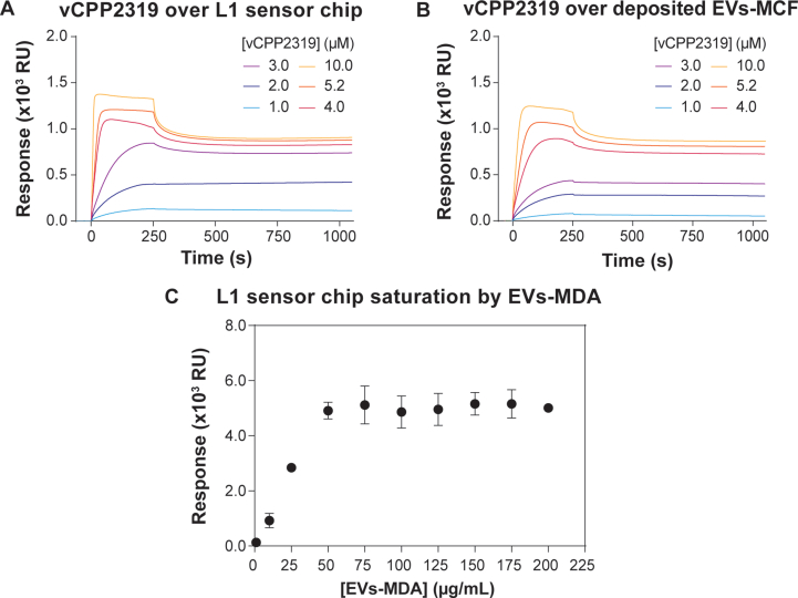
Brain metastases (BM) are frequently found in cancer patients and, though their precise incidence is difficult to estimate, there is evidence for a correlation between BM and specific primary cancers, such as lung, breast, and skin (melanoma). Among all these, breast cancer is the most frequently diagnosed among women and, in this case, BM cause a critical reduction of the overall survival (OS), especially in triple negative breast cancer (TNBC) patients. The main challenge of BM treatment is the impermeable nature of the blood-brain barrier (BBB), which shields the central nervous systems (CNS) from chemotherapeutic drugs. Extracellular vesicles (EVs) have been proposed as ideal natural drug carriers and these may exhibit some advantages over synthetic nanoparticles (NPs). In this work, we isolate breast cancer-derived EVs and study their ability to carry vCPP2319, a peptide with dual cell-penetration and anticancer activities. The selective cytotoxicity of anticancer peptide-loaded EVs towards breast cancer cells and their ability to translocate an in vitro BBB model are also addressed. Overall, it was possible to conclude that vCPP2319 naturally interacts with breast cancer-derived EVs, being retained at the surface of these vesicles. Moreover, the results revealed a cytotoxic activity for peptide-loaded EVs similar to that obtained with the peptide alone and the ability of peptide-loaded EVs to translocate an in vitro BBB model, which contrasts with the results obtained with the peptide alone. In conclusion, this work supports the use of EVs in the development of biological drug-delivery systems (DDS) capable of translocating the BBB.

Keywords: Anticancer peptide; Blood-brain barrier; Brain metastases; Drug-delivery systems; Extracellular vesicles; Metastatic breast cancer.


Conflict of interest statement

The authors declare that they have no known competing financial interests or personal relationships that could have appeared to influence the work reported in this paper.

Figures

Image 1
Graphical abstract
Fig. 1
Fig. 1
TEM imaging of extracellular vesicles (EVs) isolated from MDA-MB-231 and MCF 10A cell cultures (EVs-MDA and EVs-MCF, respectively) and molecular characterization by flow cytometry. EVs were isolated from MDA-MB-231 and MCF 10A cell cultures using the Total Exosome Isolation reagent (Invitrogen) and samples were imaged by TEM (A). The displayed images were proportionally resized from raw images with an 100 000 × magnification. The sizes of the vesicles, obtained by measuring the diameter of the vesicles in raw TEM images, are also shown (B). Five independent samples were analyzed through TEM for EVs-MDA and EVs-MCF in at least 3 different days. Significance was assessed by unpaired t-test and no significant differences were detected between the sizes of the EVs from both cell types. The EVs were also characterized by flow cytometry (C to F). CFSE was added to the cells in culture, and it was used as an intraluminal dye. The presence of CD63, CD105 and EpCAM at the surface of the vesicles was studied. Relative frequency of CFSE (C) and CD105 vs CD63 (D) are shown for EVs-MDA and EVs-MCF. Positive events (%) (E) and mean fluorescence intensity (MFI) (F) are shown for each marker. Flow cytometry characterization was repeated in three different days, with independent samples, for each type of EVs. Significance was assessed by ANOVA followed by Tukey's post-test. ∗∗ p-value ≤0.01, ∗∗∗ p-value ≤0.001.
Fig. 2
Fig. 2
Study of the interaction between vCPP2319 and extracellular vesicles derived from MDA-MB-231 through surface plasmon resonance (SPR). EVs isolated from MDA-MB-231 (EVs-MDA) and MCF 10A cells (EVs-MCF) were injected at several protein concentrations ranging from 1 to 75 μg/mL over an L1 sensor chip and the sensorgrams were recorded (A and B). A comparison of the response between the EVs origin was obtained by plotting the final response values for each protein concentration (C). Several vCPP2319 concentrations ranging from 1.0 to 10.0 μM were injected on EVs-MDA deposited over the L1 sensor chip at a concentration of 75 μg/mL, sensorgrams were recorded and the final response values were plotted as function of the peptide concentration (D). A dissociation analysis was performed, and the peptide fraction associated to the deposited EVs (SL) was plotted as a function of the dissociation time. The curve represents the two-phase decay fit to the data and the respective residual plots are represented. The sensorgrams in A, B and D and the dissociation analysis in E are representative replicates. Error bars in C and D correspond to the standard error of independent replicates. All experiments were performed in duplicate (n = 2).
Fig. 3
Fig. 3
Study of the interaction of vCPP2319 with extracellular vesicles isolated from breast cells through zeta potential. Zeta potential measurements were performed with EVs obtained from MDA-MB-231 (EVs-MDA) (A) and MCF 10A cells (EVs-MCF) (B) in the absence (n = 5 and n = 2, respectively) and presence of vCPP2319, at increasing peptide concentrations (n = 2). The zeta potential of EVs-MDA was also measured in the absence and presence of vCPP2319 at fixed concentrations of 5.2 (C, n = 3) and 20 μM (D, n = 2), in a range of protein concentration for EVs-MDA samples of 1–100 μg/mL. Error bars refer to the standard deviation. Significance was assessed by ANOVA followed by Sidak's post-test. ∗∗∗P-value ≤0.001; ∗∗∗∗P-value ≤0.0001.
Fig. 4
Fig. 4
Cytotoxic activity of breast cancer and healthy cells treated with vCPP2319 and breast cancer extracellular vesicles (EVs), added simultaneously to the well. MDA-MB-231 (A, B and C) and MCF 10A cells (D, E and F) were treated with vCPP2319 at the concentrations of 5.2 μM and 20 or 18.2 μM, in the presence (A, B, D and E) and absence (C and F) of EVs isolated from MDA-MB-231 (EVs-MDA), in concentrations ranging from 1 to 100 μg/mL. Error bars refer to the standard deviation. All experiments were performed in triplicate (n = 3).
Fig. 5
Fig. 5
Translocation of an in vitro monoculture BBB model by extracellular vesicles isolated from MDA-MB-231 cells (EVs-MDA) combined with 5.2 μM vCPP2319. The cytotoxic activity of EVs-MDA (1–100 μg/mL) towards human brain endothelial cells HBEC-5i was evaluated, with and without vCPP2319 5.2 μM (A). A scheme of the experimental setup of the BBB model is shown (B): HBEC-5i are cultured as monolayer on the apical side of a polyester (PET) transwell insert with 1.0 μm pore size. The BBB model translocation by the EVs-MDA, vCPP2319 and the combination of both was then evaluated by adding them at the apical side and measuring their presence on the apical and basolateral side after 24 h (D). EVs-MDA were labelled with CFSE and vCPP2319 was labelled with Quasar® 670 to allow for fluorescent detection. Error bars refer to standard deviation. These experiments were performed with independently grown cultures in three different days. Significance was assessed by two-way ANOVA followed by Tukey's post-test (C) and one-way ANOVA followed by Dunnett's post-test (D). ∗∗∗∗P-value ≤0.0001.
figs1
figs1
vCPP2319 interaction directly with the L1 sensor chip (A) and with previously deposited extracellular vesicles isolated MCF 10A cells (EVs-MCF) (B). L1 sensor chip saturation by EVs-MDA (1-200 μg/mL) (C). The sensorgrams in A and B are representative replicates. vCPP2319 interaction with the L1 sensor chip was repeated three times (n=3) while the interaction with deposited EVs-MCF was repeated twice (n=2). Error bars in C correspond to the standard error of independent replicates with n=4 for 75 μg/mL and n=2 for all other protein concentrations.

Similar articles

References

    1. Steindl A., Berghoff A.S. Brain metastases in metastatic cancer: a review of recent advances in systemic therapies. Expert Rev. Anticancer Ther. 2021;21:325–339. doi: 10.1080/14737140.2021.1851200. - DOI - PubMed
    1. Proescholdt M.A., Schödel P., Doenitz C., Pukrop T., Höhne J., Schmidt N.O., Schebesch K.-M. The management of brain metastases—systematic review of neurosurgical aspects. Cancers. 2021;13:1616. doi: 10.3390/cancers13071616. - DOI - PMC - PubMed
    1. Nolan C., Deangelis L.M. Overview of metastatic disease of the central nervous system. Handb. Clin. Neurol. 2018;149:3–23. doi: 10.1016/B978-0-12-811161-1.00001-3. - DOI - PubMed
    1. Achrol A.S., Rennert R.C., Anders C., Soffietti R., Ahluwalia M.S., Nayak L., Peters S., Arvold N.D., Harsh G.R., Steeg P.S., Chang S.D. Brain metastases. Nat. Rev. Dis. Prim. 2019;5:5. doi: 10.1038/s41572-018-0055-y. - DOI - PubMed
    1. Suh J.H., Kotecha R., Chao S.T., Ahluwalia M.S., Sahgal A., Chang E.L. Current approaches to the management of brain metastases. Nat. Rev. Clin. Oncol. 2020;17:279–299. doi: 10.1038/s41571-019-0320-3. - DOI - PubMed

LinkOut - more resources