. 2024 Dec:109:323-401.
doi: 10.3114/sim.2024.109.05. Epub 2024 Sep 18.

Cytospora: an important genus of canker pathogens

Affiliations

Cytospora: an important genus of canker pathogens

L Lin et al. Stud Mycol. 2024 Dec.

Abstract

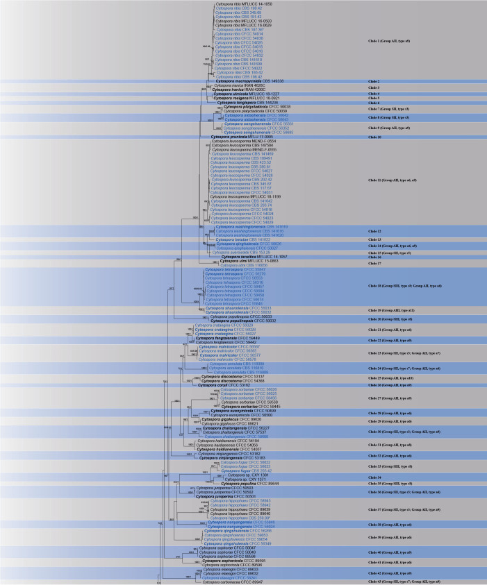
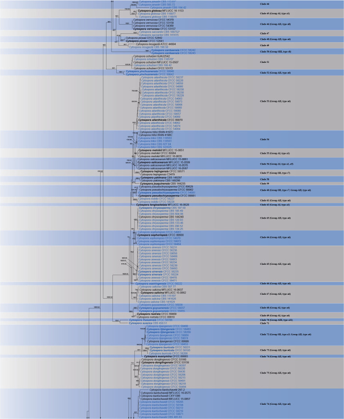
Cytospora species have commonly been reported as important plant pathogenic fungi with wide host ranges and geographic distributions. With the increase in the number of cryptic species being described, a comprehensive global taxonomic revision of the genus Cytospora is required. The present study includes 399 isolates from 32 countries. These isolates were subjected to DNA sequence analysis for five genomic loci (ITS, act1, rpb2, tef1-α and tub2). Based on these data, it could be confirmed that Cytospora, Leucostoma, Valsa, Valsella and Valseutypella are congeneric. Furthermore, 111 species of Cytospora could also be reassessed, 44 species and four combinations newly introduced, and new typifications proposed for a further three species. Three asexual morphological groups (including 13 asexual morphological types) and three sexual morphological groups (including eight sexual morphological types) were designated. The present study explored the species diversity of Cytospora and re-evaluated the identity of all cultures in the Westerdijk Fungal Biodiversity Institute (Utrecht, The Netherlands) that were deposited as either Cytospora or as one of its related genera. This is the most comprehensive phylogenetic analysis thus far conducted on Cytospora and the results contribute to an increased understanding of the taxonomy of these important fungi. It is also hoped that the findings will lead to improved management strategies for diseases associated Cytospora species. Taxonomic novelties: New species: Cytospora acericola X.L. Fan & C.M. Tian, C. adamsii Jami, Crous & M.J. Wingf., C. beijingensis L. Lin & X.L. Fan, C. betulae Jami, Crous & M.J. Wingf., C. brabeji Jami, Crous & M.J. Wingf., C. castaneicola L. Lin & X.L. Fan, C. cerebriformis L. Lin & X.L. Fan, C. conceptaculata L. Lin & X.L. Fan, C. crataegina X.L. Fan & C.M. Tian, C. deqinensis L. Lin & X.L. Fan, C. diqingensis L. Lin & X.L. Fan, C. eastringensis L. Lin & X.L. Fan, C. elaeagnina L. Lin & X.L. Fan, C. fraxinea X.L. Fan & C.M. Tian, C. guyuanensis L. Lin & X.L. Fan, C. jiufengensis L. Lin & X.L. Fan, C. lauricola L. Lin & X.L. Fan, C. lhasaensis L. Lin & X.L. Fan, C. lijiangensis L. Lin & X.L. Fan, C. lvxinensis L. Lin & X.L. Fan, C. malvicolor X.L. Fan & C.M. Tian, C. multiseriata L. Lin & X.L. Fan, C. nanyangensis X.L. Fan & C.M. Tian, C. polyspora X.L. Fan & C.M. Tian, C. pseudochrysosperma L. Lin & X.L. Fan, C. qinghaiensis L. Lin & X.L. Fan, C. qingshuiensis L. Lin & X.L. Fan, C. sanbaensis L. Lin & X.L. Fan, C. shaanxiensis L. Lin & X.L. Fan, C. shangrilaensis L. Lin & X.L. Fan, C. sidaohensis L. Lin & X.L. Fan, C. sinensis L. Lin & X.L. Fan, C. songshanensis L. Lin & X.L. Fan, C. suecica Jami, Crous & M.J. Wingf., C. syringina L. Lin & X.L. Fan, C. tenebrica L. Lin & X.L. Fan, C. tetraspora L. Lin & X.L. Fan, C. tongzhouensis X.L. Fan & C.M. Tian, C. uniloculata L. Lin & X.L. Fan, C. washingtonensis Jami, Crous & M.J. Wingf., C. xiaolongmenensis L. Lin & X.L. Fan, C. yinchuanensis L. Lin & X.L. Fan, C. yuduensis L. Lin & X.L. Fan, C. yulinensis L. Lin & X.L. Fan. New combinations: Cytospora auerswaldii (Nitschke) L. Lin & X.L. Fan, C. multicollis (Checa et al.) L. Lin, X.L. Fan & Crous, C. tristicha (De Not.) L. Lin, X.L. Fan & Crous, C. weiriana (Petr.) X.L. Fan & Crous. New replacement names: Cytospora desmazieri L. Lin, X.L. Fan & Crous, C. fuckeliana L. Lin, X.L. Fan & Crous, C. hoffmannii L. Lin, X.L. Fan & Crous, C. massarii L. Lin, X.L. Fan & Crous, C. nitschkeana L. Lin, X.L. Fan & Crous, C. saccardoi L. Lin, X.L. Fan & Crous. New synonyms: Cytospora ampulliformis Norph., Bulgakov, T.C. Wen & K.D. Hyde, C. brevispora (G.C. Adams & Jol. Roux) G.C. Adams & Rossman, C. cenisia Sacc., C. ceratospermopsis C.M. Tian & X.L. Fan, C. cotini Norph., Bulgakov & K.D. Hyde, C. ershadii Zafari & Hanifeh, C. erumpens Norph., Bulgakov, T.C. Wen & K.D. Hyde, C. fraxinigena Senan., Camporesi & K.D. Hyde, C. galegicola Q.J. Shang, E. Camporesi & K.D. Hyde, C. granati D.P. Lawr., L.A. Holland & Trouillas, C. hippophaicola Spetik, Eichmeier, Gramaje, Stuskova & Berraf-Tebbal, C. massariana Sacc., C. nivea (Hoffm.) Sacc., C. parakantschavelii Norph., Bulgakov, T.C. Wen & K.D. Hyde, C. parasitica Norph., Bulgakov & K.D. Hyde, C. paratranslucens Norph., Bulgakov, T.C. Wen & K.D. Hyde, C. pini Desm., C. populicola D.P. Lawr., L.A. Holland & Trouillas, C. predappioensis Q.J. Shang, Norph., Camporesi & K.D. Hyde, C. quercicola Senan., Camporesi & K.D. Hyde, C. rosae Senan., Camporesi & K.D. Hyde, C. salicella Sacc., C. vinacea D.P. Lawr., Travadon & Pouzoulet, Valsa germanica Nitschke, V. massariana De Not., V. nivea (Hoffm.) Fr., Valsella salicis Fuckel, Sphaeria nivea Hoffm. Typification: Lecto- and epitypifications (basionyms): Sphaeria chrysosperma Pers., Valsa eucalypti Cooke & Harkn., Valsella salicis Fuckel. Citation: Lin L, Fan XL, Groenewald JZ, Jami F, Wingfield MJ, Voglmayr H, Jaklitsch W, Castlebury LA, Tian CM, Crous PW (2024). Cytospora: an important genus of canker pathogens. Studies in Mycology 109: 323-401. doi: 10.3114/sim.2024.109.05.

Keywords: Cytosporaceae; Diaporthales; multi-gene phylogeny; taxonomy.


Conflict of interest statement

The authors declare that there is no conflict of interest.

Figures

Fig. 1
Fig. 1
Phylogram of Cytospora resulting from a maximum likelihood analysis based on combined ITS, act, rpb2, tef1-α, and tub2 genes. Numbers above the branches indicate ML bootstrap values (ML-BS ≥ 60%) and Bayesian Posterior Probabilities (BPP ≥ 0.8). Ex-type isolates are in bold. Reference strains are marked with*. Isolates from the present study are marked in blue.
Fig. 1
Fig. 1
Phylogram of Cytospora resulting from a maximum likelihood analysis based on combined ITS, act, rpb2, tef1-α, and tub2 genes. Numbers above the branches indicate ML bootstrap values (ML-BS ≥ 60%) and Bayesian Posterior Probabilities (BPP ≥ 0.8). Ex-type isolates are in bold. Reference strains are marked with*. Isolates from the present study are marked in blue.
Fig. 2
Fig. 2
Asexual morphological groups and types of Cytospora.
Fig. 3
Fig. 3
Sexual morphological groups and types of Cytospora.
Fig. 4
Fig. 4
Cytospora acericola (BJFC-S2106). A, B. Habit of conidiomata on twig. C. Transverse section through conidioma. D. Longitudinal section through conidioma. E. Conidiophores and conidiogenous cells. F. Conidia. Scale bars: A = 1 mm; B–D = 500 μm; E, F = 10 μm.
Fig. 5
Fig. 5
Cytospora adamsii (CBS 179.70). A. Conidiomata on MEA. B, C. Conidiophores and conidiogenous cells. D. Conidia. Scale bars: A = 100 μm; B, C = 10 μm; D = 5 μm.
Fig. 6
Fig. 6
Cytospora beijingensis (BJFC-S2182). A, B. Habit of pseudostromata on twig. C. Transverse section through pseudostroma with ascomata. D. Longitudinal sections through pseudostromata with ascomata. E. Asci. F. Ascospores. Scale bars: A = 1 mm; B = 250 μm; C–D = 500 μm; E–F = 10 μm.
Fig. 7
Fig. 7
Cytospora betulae (CBS 141622). A. Exuding conidial mass from conidioma on MEA. B. Conidiomata on MEA. C–F. Conidiophores and conidiogenous cells. G. Conidia. Scale bars: B = 100 μm; C–F = 10 μm; G = 5 μm.
Fig. 8
Fig. 8
Cytospora brabeji (CBS 119207). A. Conidiomata on MEA. B. Tangential section of conidiomata. C, D. Conidiophores and conidiogenous cells. E. Conidia. Scale bars: A, B = 100 μm; C, D = 10 μm; E = 5 μm.
Fig. 9
Fig. 9
Cytospora californica (CBS 141612). A. Conidiomata on MEA. B–D. Conidiophores and conidiogenous cells. E. Conidia. Scale bars: A = 100 μm; B–D = 10 μm; E = 5 μm.
Fig. 10
Fig. 10
Cytospora castaneicola (BJFC-S1704). A. Conidioma on host issue. B. Tangential section through conidiomata. C. Longitudinal section through conidioma. D, E. Conidiophores and conidiogenous cells. F. Conidia. Scale bars: A–C = 200 μm; D–F = 10 μm.
Fig. 11
Fig. 11
Cytospora ceratosperma (BJFC-S2126). A, B. Habit of conidiomata on twig. C. Transverse section through conidioma. D. Longitudinal section through conidioma. E. Conidiophores and conidiogenous cells. F. Conidia. Scale bars: A = 1 mm; B–D = 500 μm; E, F = 10 μm.
Fig. 12
Fig. 12
Cytospora ceratosperma (BJFC-S2165). A, B. Habit of conidiomata on twig. C, D. Transverse sections through conidiomata. E. Longitudinal section through conidioma. F. Conidiophores and conidiogenous cells. G. Conidia. Scale bars: A = 1 mm; B–E = 500 μm; F, G = 10 μm.
Fig. 13
Fig. 13
Cytospora cerebriformis (BJFC-S918). A, B. Habit of conidiomata on twig. C. Transverse section through conidioma. D. Longitudinal section through conidioma. E. Conidiophores and conidiogenous cells. F. Conidia. Scale bars: A = 2 mm; B–D = 200 μm; E, F = 10 μm.
Fig. 14
Fig. 14
The lectotype designated here of Cytospora chrysosperma (Herb. Univ. Upsaliensis (F-117599) 289268).
Fig. 15
Fig. 15
Cytospora conceptaculata (BJFC-S2102). A, B. Habit of conidiomata on twig. C. Transverse section through conidioma. D. Longitudinal section through conidioma. E, F. Conidiophores and conidiogenous cells. G. Conidia. Scale bars: A = 1 mm; B–D = 200 μm; E–G = 10 μm.
Fig. 16
Fig. 16
Cytospora crataegina (BJFC-S2128). A, B. Habit of conidiomata on twig. C. Transverse section through conidioma. D. Longitudinal section through conidioma. E. Conidiophores and conidiogenous cells. F. Conidia. Scale bars: A = 1 mm; B–D = 500 μm; E, F = 10 μm.
Fig. 17
Fig. 17
Cytospora deqinensis (BJFC-S2000). A, B. Habit of conidiomata on twig. C. Transverse section through conidioma. D. Longitudinal section through conidioma. E. Conidiophores and conidiogenous cells. F. Conidia. Scale bars: A = 1 mm; B = 200 μm; C, D = 500 μm; E, F = 10 μm.
Fig. 18
Fig. 18
Cytospora diqingensis (BJFC-S2083). A, B. Habit of pseudostromata on twig. C. Transverse section through pseudostroma with ascomata. D. Longitudinal section through pseudostroma with ascomata. E–G. Asci. H. Ascospores. Scale bars: A = 2 mm; B–D = 200 μm; E–H = 10 μm.
Fig. 19
Fig. 19
Cytospora eastringensis (BJFC-S2038). A, B. Habit of conidiomata on twig. C. Transverse section through conidioma. D. Longitudinal section through conidioma. E. Conidiophores and conidiogenous cells. F. Conidia. Scale bars: A = 1 mm; B = 200 μm; C, D = 500 μm; E, F = 10 μm.
Fig. 20
Fig. 20
Cytospora elaeagnina (BJFC-S2137). A–C. Habit of pseudostromata on twig. D. Transverse section through pseudostroma with ascomata. E. Longitudinal section through pseudostroma with ascomata. F. Asci. G. Ascospores. Scale bars: A = 2 mm; B–E = 500 μm; F, G = 10 μm.
Fig. 21
Fig. 21
Cytospora fraxinea (BJFC-S2141). A, B. Habit of conidiomata on twig. C. Transverse section through conidioma. D. Longitudinal section through conidioma. E. Conidiophores and conidiogenous cells. F. Conidia. Scale bars: A = 2 mm; B–D = 500 μm; E, F = 10 μm.
Fig. 22
Fig. 22
Cytospora fugax (BJFC-S2192). A, B. Habit of pseudostromata on twig. C. Transverse section through pseudostroma with ascomata. D. Longitudinal section through pseudostroma with ascomata. E. Degraded asci. F. Ascospores. Scale bars: A = 1 mm; B = 250 μm; C, D = 500 μm; E, F = 10 μm.
Fig. 23
Fig. 23
Cytospora guyuanensis (BJFC-S2177). A, C. Habit of conidiomata on twig. B. Transverse section through conidioma. D. Longitudinal section through conidioma. E. Conidiophores and conidiogenous cells. F. Conidia. Scale bars: A = 2 mm; B–D = 500 μm; E, F = 10 μm.
Fig. 24
Fig. 24
Cytospora hoffmannii (BJFC-S2049). A. Habit of conidiomata on twig. B. Transverse section through conidioma. C. Longitudinal section through conidioma. D. Conidiophores and conidiogenous cells. E. Conidia. Scale bars: A–C = 200 μm; D, E = 10 μm.
Fig. 25
Fig. 25
Cytospora jiufengensis (BJFC-S2180). A, B. Habit of conidiomata on twig. C. Transverse section through conidioma. D. Longitudinal section through conidioma. E, F. Conidiophores and conidiogenous cells. G. Conidia. Scale bars: B–D = 500 μm; E–G = 10 μm.
Fig. 26
Fig. 26
Cytospora lauricola (BJFC-S2020). A, B. Habit of conidiomata on twig. C. Transverse section through conidioma. D. Longitudinal section through conidioma. E, F. Conidiophores and conidiogenous cells. G. Conidia. Scale bars: B = 100 μm; C, D = 200 μm; E–G = 10 μm.
Fig. 27
Fig. 27
Cytospora leucosperma (BJFC-S2022). A, B. Habit of conidiomata on twig. C. Transverse section through conidioma. D. Longitudinal section through conidioma. E, F. Conidiophores and conidiogenous cells. G. Conidia. Scale bars: A = 1 mm; B–D = 200 μm; E–G = 10 μm.
Fig. 28
Fig. 28
Cytospora lhasaensis (BJFC-S2100). A, B. Habit of conidiomata on twig. C. Transverse section through conidioma. D. Longitudinal section through conidioma. E, F. Conidiophores and conidiogenous cells. G. Conidia. Scale bars: A = 1 mm; B = 100 μm; C, D = 500 μm; E–G = 10 μm.
Fig. 29
Fig. 29
Cytospora lijiangensis (BJFC-S2030). A, B. Habit of conidiomata on twig. C. Transverse section through conidioma. D. Longitudinal section through conidioma. E. Habit of pseudostroma on twig. F. Transverse section through pseudostroma with ascomata. G. Longitudinal section through pseudostroma with ascomata. H. Conidiophores and conidiogenous cells. I. Conidia. J. Ascus. K. Ascospores. Scale bars: A = 1 mm; B = 100 μm; C, E–G = 200 μm; D = 500 μm; H–K = 10 μm.
Fig. 30
Fig. 30
Cytospora lvxinensis (BJFC-S2173). A, B. Habit of conidiomata on twig. C. Transverse section through conidioma. D. Longitudinal section through conidioma. E. Conidiophores and conidiogenous cells. F. Conidia. Scale bars: A = 1 mm; B–D = 500 μm; E–F = 10 μm.
Fig. 31
Fig. 31
Cytospora malvicolor (BJFC-S2120). A, B. Habit of pseudostromata on twig. C. Transverse section through pseudostroma with ascomata. D. Longitudinal section through pseudostroma with ascomata. E. Asci. F. Ascospores. G, H. Habit of conidiomata on twig. I. Transverse section through conidioma. J. Longitudinal section through conidioma. K. Conidiophores and conidiogenous cells. L. Conidia. Scale bars: A, G = 1 mm; B–D, H–J = 500 μm; E, F, K, L = 10 μm.
Fig. 32
Fig. 32
Cytospora massarii (CBS 141473). A. Conidiomata on MEA. B–D. Conidiophores and conidiogenous cells. E. Conidia. F, H. Ascomata on dead branches of Sorbus aucuparia. G. Longitudinal section of pseudostromata with ascomata. I. Tangential section of pseudostromata with ascomata. J. Ascospores. Scale bars: A, F–I = 100 μm; B–D = 10 μm; E, J = 5 μm.
Fig. 33
Fig. 33
Cytospora multiseriata (BJFC-S2036). A, B. Habit of pseudostromata on twig. C. Transverse section through pseudostroma with ascomata. D. Longitudinal section through pseudostroma with ascomata. E, F. Asci. G. Ascospores. Scale bars: A = 1 mm; B–D = 200 μm; E–G = 10 μm.
Fig. 34
Fig. 34
Cytospora nanyangensis (BJFC-S2167). A, B. Habit of conidiomata on twig. C. Transverse section through conidioma. D. Longitudinal section through conidioma. E, F. Conidiophores and conidiogenous cells. G. Conidia. Scale bars: A = 1 mm; B–D = 500 μm; E–G = 10 μm.
Fig. 35
Fig. 35
Cytospora nitschkeana (CBS 118.22). A. Conidiomata on MEA. B, C. Conidiophores and conidiogenous cells. D. Conidia. Scale bars: A = 100 μm; B, C = 10 μm; D = 5 μm.
Fig. 36
Fig. 36
Cytospora nobilis (BJFC-S2058). A, B. Habit of conidiomata on twig. C. Transverse section through conidioma. D. Longitudinal section through conidioma. E, F. Conidiophores and conidiogenous cells. G. Conidia. Scale bars: A = 1 mm; B = 100 μm; C, D = 200 μm; E–G = 10 μm.
Fig. 37
Fig. 37
Cytospora polyspora (BJFC-S2194). A, B. Habit of pseudostromata on twig. C. Transverse section through pseudostroma with ascomata. D. Longitudinal section through pseudostroma with ascomata. E. Asci. F. Ascospores. Scale bars: A = 1 mm; B–D = 250 μm; E, F = 10 μm.
Fig. 38
Fig. 38
Cytospora populi (BJFC-S2193). A. Habit of pseudostromata and conidiomata on branch. B. Disc and ostiole of conidioma. C. Transverse section through conidiomata. D. Longitudinal section through conidioma. E. Ectostromatic disc and ostioles of pseudostroma. F. Transverse sections through pseudostroma with ascomata. G, H. Longitudinal section through pseudostroma with ascomata. I–J. Conidiophore and conidiogenous cell. K. Conidia. L, M. Ascus. N. Ascospores. Scale bars: A = 250 μm; B–H = 200 μm; I–N = 10 μm.
Fig. 39
Fig. 39
Cytospora pruinosa (BJFC-S2139). A–B. Habit of conidiomata on twig. C. Transverse section through conidioma. D. Longitudinal section through conidioma. E, F. Conidiophores and conidiogenous cells. G. Conidia. Scale bars: A = 1 mm; B–D = 250 μm; E–G = 10 μm.
Fig. 40
Fig. 40
Cytospora pseudochrysosperma (BJFC-S750). A, B. Habit of conidiomata on twig. C. Transverse section through conidioma. D. Longitudinal section through conidioma. E. Conidiophores and conidiogenous cells. F. Conidia. G, H. Habit of pseudostromata on twig. I. Transverse section of pseudostroma with ascomata. J. Longitudinal section through pseudostroma with ascomata. K, L. Asci. M. Ascospores. Scale bars: A, G = 5 mm; B–D, G, H = 1 mm; E, F, K–M = 10 μm.
Fig. 41
Fig. 41
Cytospora qinghaiensis (BJFC-S671). A. Habit of conidioma on twig. B. Transverse section through conidioma. C. Longitudinal section through conidioma. D. Conidiophores and conidiogenous cells. E. Conidia. Scale bars: A–C = 200 μm; D, E = 10 μm.
Fig. 42
Fig. 42
Cytospora qingshuiensis (BJFC-S2111). A, B. Habit of conidiomata on twig. C. Transverse section through conidioma. D. Longitudinal section through conidioma. E. Conidiophores and conidiogenous cells. F. Conidia. Scale bars: A = 1 mm; B–D = 500 μm; E, F = 10 μm.
Fig. 43
Fig. 43
Cytospora ribis (BJFC-S2059). A, B. Habit of conidiomata on twig. C. Transverse section through conidioma. D. Longitudinal section through conidioma. E. Conidiophores and conidiogenous cells. F. Conidia. Scale bars: A = 2 mm; B–D = 200 μm; E, F = 10 μm.
Fig. 44
Fig. 44
Cytospora saccardoi (CBS 141615). A. Conidioma on twig in MEA. B–E. Conidiophores and conidiogenous cells. F. Conidia. Scale bars: A = 100 μm; B–E = 10 μm; F = 5 μm.
Fig. 45
Fig. 45
Cytospora sanbaensis (BJFC-S2066). A, B. Habit of pseudostroma on twig. C. Transverse section through pseudostroma with ascomata. D. Longitudinal section through pseudostroma with ascomata. E, F. Asci. G. Ascospores. Scale bars: A = 1 mm; B = 200 μm; C, D = 500 μm; E–G = 10 μm.
Fig. 46
Fig. 46
Cytospora shaanxiensis (BJFC-S2169). A, B. Habit of conidiomata on twig. C. Transverse section through conidioma. D. Longitudinal section through conidioma. E. Conidiophores and conidiogenous cells. F. Conidia.Scale bars: A = 1 mm; B–D = 500 μm; E, F = 10 μm.
Fig. 47
Fig. 47
Cytospora shangrilaensis (BJFC-S1965). A, B. Habit of conidiomata on twig. C. Transverse section through conidioma. D. Longitudinal section through conidioma. E. Conidiophores and conidiogenous cells. F. Conidia. Scale bars: B–D = 200 μm; E, F = 10 μm.
Fig. 48
Fig. 48
Cytospora sidaohensis (BJFC-S2115). A, B. Habit of pseudostromata on twig. C. Transverse section through pseudostroma with ascomata. D. Longitudinal section through pseudostroma with ascomata. E. Asci. F. Ascospores. Scale bars: A = 2 mm; B–D = 500 μm; E, F = 10 μm.
Fig. 49
Fig. 49
Cytospora sinensis (BJFC-S2071). A. Habit of conidiomata on twig. B. Transverse section through conidioma. C. Longitudinal section through conidioma. D, E. Conidiophores and conidiogenous cells. F. Conidia. Scale bars: A = 1 mm; B, C = 200 μm; D–F = 10 μm.
Fig. 50
Fig. 50
Cytospora songshanensis (BJFC-S2220). A, B. Habit of conidiomata on twig. C. Transverse section through conidioma. D. Longitudinal section through conidioma. E. Conidiophores and conidiogenous cells. F. Conidia. Scale bars: A = 2 mm; B–D = 1 mm; E, F = 10 μm.
Fig. 51
Fig. 51
Cytospora sorbariae (BJFC-S2124). A–C. Habit of conidiomata on twig. D. Transverse section through conidioma. E. Longitudinal section through conidioma. F. Conidia. Scale bars: A = 1 mm; B–E = 500 μm; F = 5 μm.
Fig. 52
Fig. 52
Cytospora suecica (CBS 450.51). A. Conidiomata on MEA. B. Conidiophores and conidiogenous cells. C. Conidia. Scale bars: A = 100 μm; B, C = 5 μm.
Fig. 53
Fig. 53
Cytospora tenebrica (BJFC-S2197). A, B. Habit of conidiomata on twig. C. Transverse section through conidioma. D. Longitudinal section through conidioma. E. Conidiophores and conidiogenous cells. F. Conidia. Scale bars: A = 2 mm; B–D = 500 μm; E, F = 10 μm.
Fig. 54
Fig. 54
Cytospora tetraspora (BJFC-S2207). A, B. Habit of pseudostromata on twig. C. Transverse section through pseudostroma with ascomata. D. Longitudinal section through pseudostroma with ascomata. E, F. Habit of conidiomata on twig. G. Transverse section through conidioma. H. Longitudinal section through conidioma. I. Asci. J. Ascospores. K. Conidiophores and conidiogenous cells. L. Conidia. Scale bars: A, E = 2 mm; B–H = 500 μm; I–L = 10 μm.
Fig. 55
Fig. 55
Cytospora tongzhouensis (BJFC-S2213). A, B. Habit of pseudostromata on twig. C. Transverse section through pseudostroma with ascomata. D. Longitudinal section through pseudostroma with ascomata. E. Asci. F. Ascospores. Scale bars: A = 1 mm; B–D = 500 μm; E–F = 10 μm.
Fig. 56
Fig. 56
Cytospora translucens (BJFC-S2093). A, B. Habit of conidiomata on twig. C. Transverse section through conidioma. D. Longitudinal section through conidioma. E. Conidiophores and conidiogenous cells. F. Conidia. Scale bars: A = 2 mm; B–D = 200 μm; E–G = 10 μm.
Fig. 57
Fig. 57
Cytospora uniloculata (BJFC-S2035). A, B. Habit of conidiomata on twig. C. Transverse section through conidioma. D. Longitudinal section through conidioma. E, F. Conidiophores and conidiogenous cells. G. Conidia. Scale bars: A = 1 mm; B–D = 200 μm; E–G = 10 μm.
Fig. 58
Fig. 58
Cytospora washingtonensis (CBS 141619). A. Conidiomata on MEA. B–E. Conidiophores and conidiogenous cells. F. Conidia. Scale bars: A = 100 μm; B–E = 10 μm; F = 5 μm.
Fig. 59
Fig. 59
Cytospora xiaolongmenensis (BJFC-S2037). A, B. Habit of pseudostromata on twig. C. Transverse section through pseudostroma with ascomata. D. Longitudinal section through pseudostroma with ascomata. E, F. Asci. G. Ascospores. Scale bars: A = 1 mm; B–D = 200 μm; E–G = 10 μm.
Fig. 60
Fig. 60
Cytospora xylocarpi (CBS 116861). A. Conidiomata on MEA. B, C. Conidiophores and conidiogenous cells. D. Conidia. Scale bars: A = 100 μm; B–D = 10 μm.
Fig. 61
Fig. 61
Cytospora yinchuanensis (BJFC-S538). A, B. Habit of conidiomata on twig. C. Transverse section through conidioma. D. Longitudinal section through conidioma. E. Conidiophores and conidiogenous cells. F. Conidia. Scale bars: A = 2 mm; B–D = 200 μm; E, F = 10 μm.
Fig. 62
Fig. 62
Cytospora yuduensis (BJFC-S2097). A, B. Habit of conidiomata on twig. C. Transverse section through conidioma. D. Longitudinal section through conidioma. E. Conidiophores and conidiogenous cells. F. Conidia. Scale bars: A = 500 μm; B–D = 100 μm; E, F = 10 μm.
Fig. 63
Fig. 63
Cytospora yulinensis (BJFC-S979). A. Habit of conidiomata on twig. B. Transverse section through conidioma. C. Longitudinal section through conidioma. D. Conidiophores and conidiogenous cells. E. Conidia. Scale bars: A–C = 200 μm; D, E = 10 μm.
Fig. 64
Fig. 64
Cytospora zhaitangensis (BJFC-S2001). A–C. Habit of pseudostromata on twig. D. Transverse section through pseudostroma with ascomata. E. Longitudinal section through pseudostroma with ascomata. F. Ascus. G. Ascospores. Scale bars: A = 1 mm; B, C = 200 μm; D, E = 500 μm; F, G = 10 μm.

Similar articles

Cited by

  • Fungal Planet description sheets: 1697-1780.
    Crous PW, Wingfield MJ, Jurjević Ž, Balashov S, Osieck ER, Marin-Felix Y, Luangsa-Ard JJ, Mejía LC, Cappelli A, Parra LA, Lucchini G, Chen J, Moreno G, Faraoni M, Zhao RL, Weholt Ø, Borovička J, Jansen GM, Shivas RG, Tan YP, Akulov A, Alfenas AC, Alfenas RF, Altés A, Avchar R, Barreto RW, Catcheside DEA, Chi TY, Esteve-Raventós F, Fryar SC, Hanh LTM, Larsbrink J, Oberlies NH, Olsson L, Pancorbo F, Raja HA, Thanh VN, Thuy NT, Ajithkumar K, Akram W, Alvarado P, Angeletti B, Arumugam E, Khalilabad AA, Bandini D, Baroni TJ, Barreto GG, Boertmann D, Bose T, Castañeda Ruiz RF, Couceiro A, Cykowska-Marzencka B, Dai YC, Darmostuk V, da Silva SBG, Dearnaley JDW, de Azevedo Santiago ALCM, Declercq B, de Freitas LWS, De la Peña-Lastra S, Delgado G, de Lima CLF, Dhotre D, Dirks AC, Eisvand P, Erhard A, Ferro LO, García D, García-Martín A, Garrido-Benavent I, Gené J, Ghobad-Nejhad M, Gore G, Gunaseelan S, Gusmão LFP, Hammerbacher A, Hernández-Perez AT, Hernández-Restrepo M, Hofmann TA, Hubka V, Jiya N, Kaliyaperumal M, Keerthana KS, Ketabchi M, Kezo K, Knoppersen R, Kolarczyková D, Kumar TKA, Læssøe T, Langer E, Larsson E, Lodge DJ, Lynch MJ, Maciá-Vicente JG, Mahadevakumar S, Mateos A, Mehrab… See abstract for full author list ➔ Crous PW, et al. Fungal Syst Evol. 2024 Dec;14:325-577. doi: 10.3114/fuse.2024.14.19. Epub 2024 Dec 6. Fungal Syst Evol. 2024. PMID: 39830292 Free PMC article.
  • Comprehensive genome analysis of two Cytospora (Cytosporaceae, Diaporthales) species associated with canker disease of spruce: C.piceae and C.piceicola sp. nov.
    Ilyukhin E, Chen Y, Markovskaja S, Shami A, Maharachchikumbura SSN. Ilyukhin E, et al. MycoKeys. 2025 May 5;117:89-119. doi: 10.3897/mycokeys.117.145445. eCollection 2025. MycoKeys. 2025. PMID: 40364895 Free PMC article.
  • Fungal Planet description sheets: 1781-1866.
    Crous PW, Catcheside DEA, Catcheside PS, Alfenas AC, Alfenas RF, Barreto RW, Lebel T, Balashov S, Broadbridge J, Jurjević Ž, De la Peña-Lastra S, Hoffmann R, Mateos A, Riebesehl J, Shivas RG, Soliz Santander FF, Tan YP, Altés A, Bandini D, Carriconde F, Cazabonne J, Czachura P, Gryta H, Eyssartier G, Larsson E, Pereira OL, Rigueiro-Rodríguez A, Wingfield MJ, Ahmad W, Bibi S, Denman S, Esteve-Raventós F, Hussain S, Illescas T, Luangsa-Ard JJ, Möller L, Mombert A, Noisripoom W, Olariaga I, Pancorbo F, Paz A, Piątek M, Polman-Short C, Suárez E, Afshan NS, Ali H, Arzanlou M, Ayer F, Barratt J, Bellanger JM, Bidaud A, Bishop-Hurley SL, Bohm M, Bose T, Campo E, Chau NB, Çolak ÖF, Cordeiro TRL, Cruz MO, Custódio FA, Couceiro A, Darmostuk V, Dearnaley JDW, de Azevedo Santiago ALCM, de Freitas LWS, Yáñez-Morales MJ, Domnauer C, Dentinger B, Dhileepan K, De Souza JT, Dovana F, Eberhardt U, Eisvand P, Erhard A, Fachada V, García-Martín A, Groenewald M, Hammerbacher A, Harms K, Haroon S, Haqnawaz M, Henriques S, Hernández AJ, Jacobus LM, Jaen-Contreras D, Jangsantear P, Kaygusuz O, Knoppersen R, Kumar TKA, Lynch MJ, Mahiques R, Maraia GL, Marbach PAS, Mehrabi-Koushki M, Miller PR, Mongkolsamr… See abstract for full author list ➔ Crous PW, et al. Persoonia. 2025 Jun;54:327-587. doi: 10.3114/persoonia.2025.54.10. Epub 2025 Jul 8. Persoonia. 2025. PMID: 40746709 Free PMC article.
  • Roles of NADPH oxidases in regulating redox homeostasis and pathogenesis of the poplar canker fungus Cytospora chrysosperma.
    Li Q, Guo R, Li A, Wang Y. Li Q, et al. Stress Biol. 2025 May 8;5(1):33. doi: 10.1007/s44154-025-00223-y. Stress Biol. 2025. PMID: 40338399 Free PMC article.
  • Cytosporalongdensis sp. nov. and C.sinensis (Diaporthales, Valsaceae) associated with Populusalba subsp. pyramidalis canker and dieback in China.
    Li J, Li C, Wang G, Zhu L, Huang L. Li J, et al. MycoKeys. 2025 Jun 13;118:345-359. doi: 10.3897/mycokeys.118.152880. eCollection 2025. MycoKeys. 2025. PMID: 40636814 Free PMC article.

References

    1. Abebe G, Hart JH, Adams RD. (1990). The relationship of site factors to the incidence of Cytospora and Septoria cankers and poplar and willow borer in hybrid poplar plantations. General Technical Report-North Central Forest Experiment Station, USDA Forest Service: 163–171.
    1. Adams GC, Roux J, Wingfield MJ. (2006). Cytospora species (Ascomycota, Diaporthales, Valsaceae): introduced and native pathogens of trees in South Africa. Australasian Plant Pathology 35: 521–548.
    1. Adams GC, Wingfield WJ, Common R. et al. (2005). Phylogenetic relationships and morphology of Cytospora species and related teleomorphs (Ascomycota, Diaporthales, Valsaceae) from Eucalyptus. Studies in Mycology 52: 1–144.
    1. Adanson M. (1763). Familles des Plantes. Vincent, Paris, France.
    1. Ariyawansa HA, Hyde KD, Jayasiri SC. et al. (2015). Fungal diversity notes 111–252 taxonomic and phylogenetic contributions to fungal taxa. Fungal Diversity 75: 27–274.

LinkOut - more resources