. 2024 Dec:109:1-56.
doi: 10.3114/sim.2024.109.01. Epub 2024 Jun 19.

Phylogenetic diversity and morphological characterization of cordycipitaceous species in Taiwan

Affiliations

Phylogenetic diversity and morphological characterization of cordycipitaceous species in Taiwan

W Y Chuang et al. Stud Mycol. 2024 Dec.

Abstract

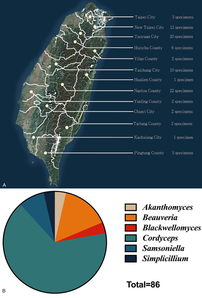
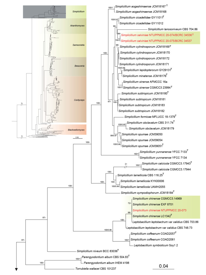
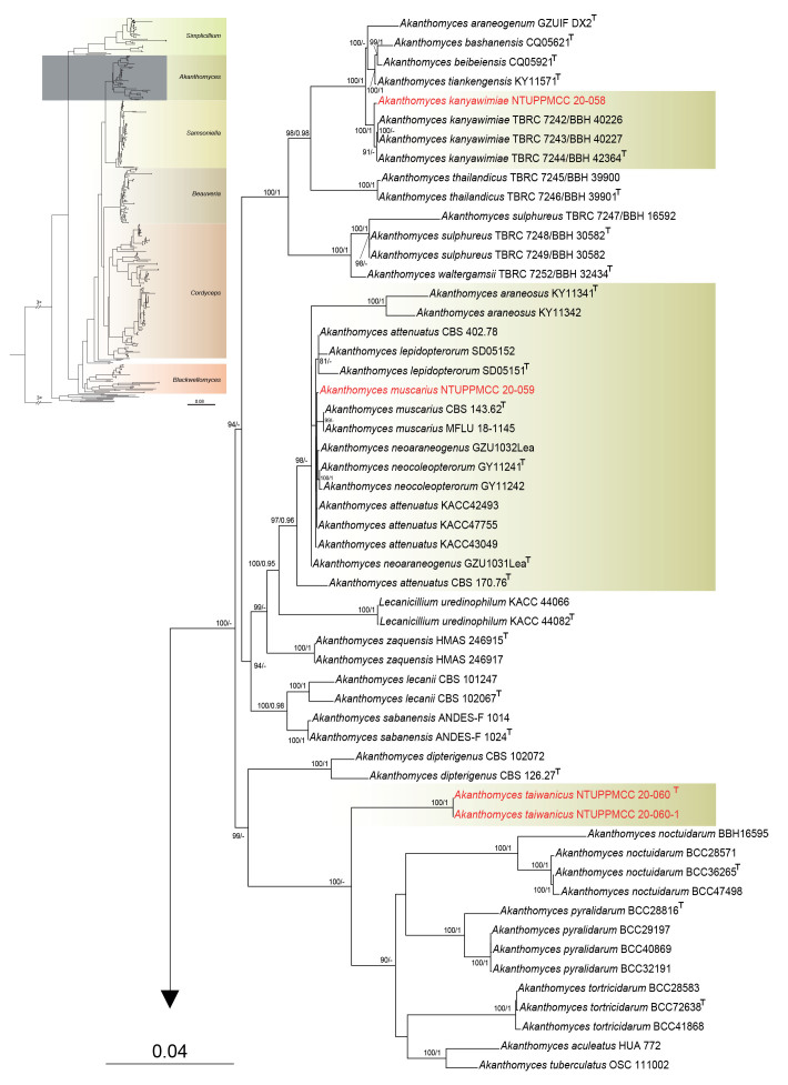
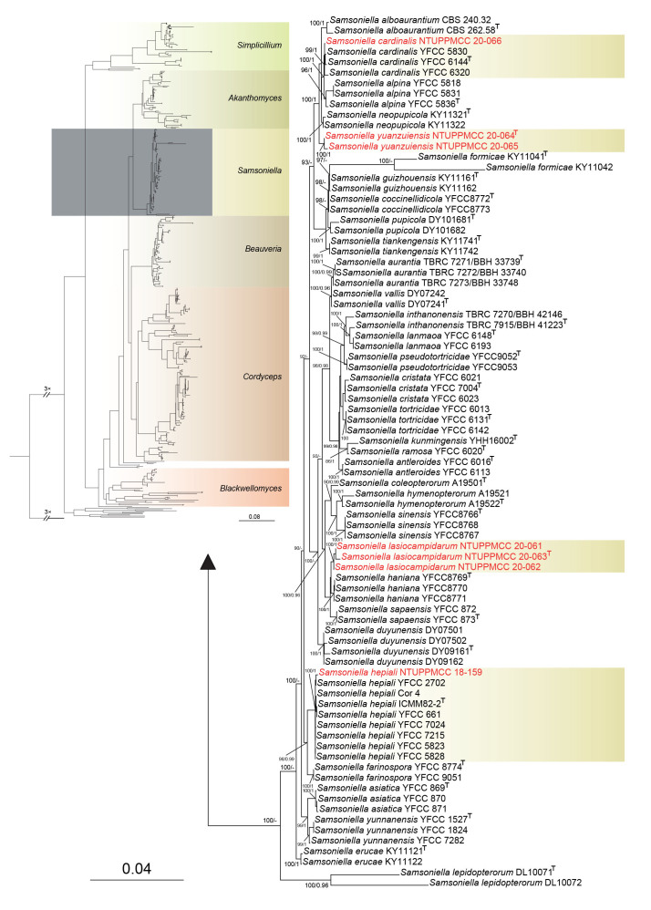
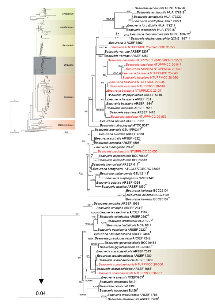
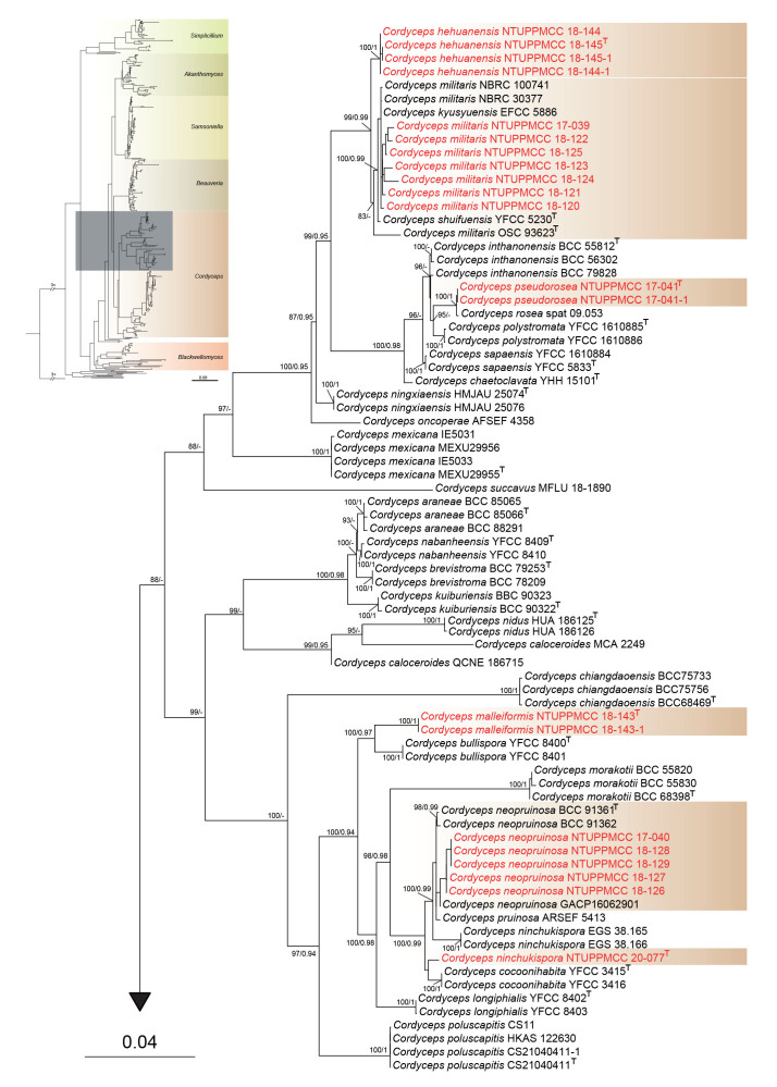
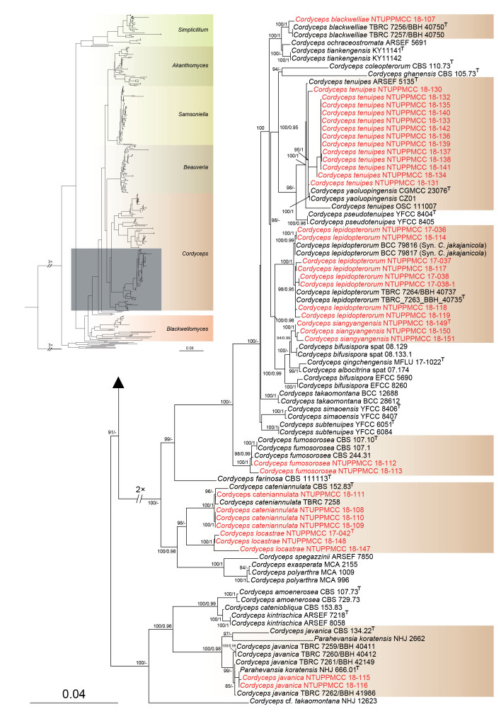
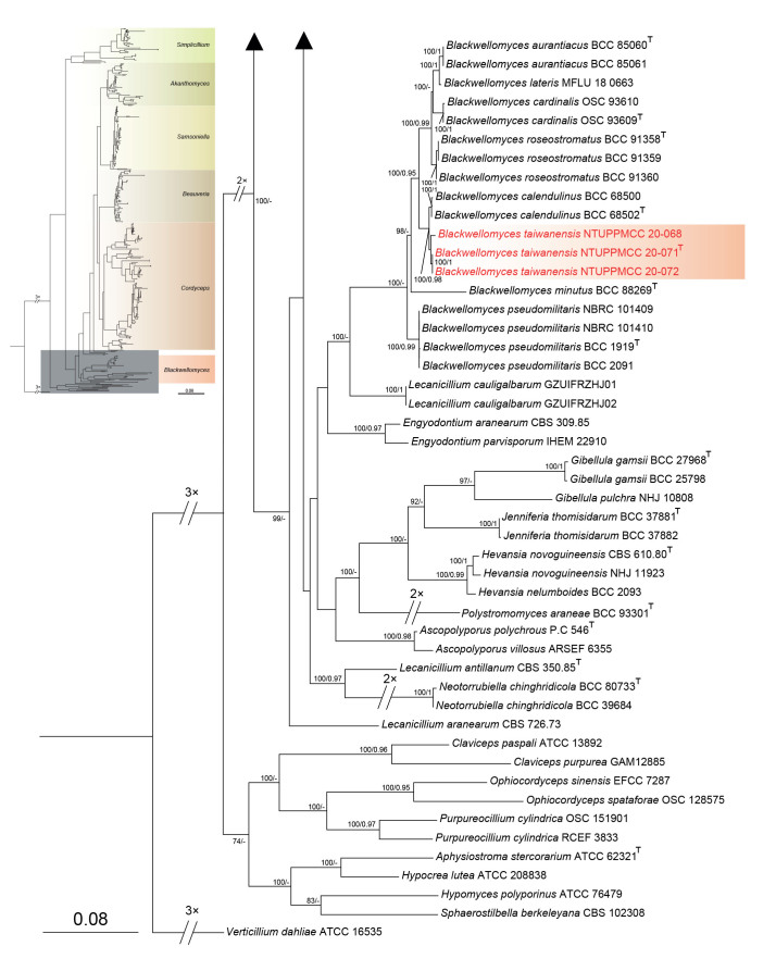
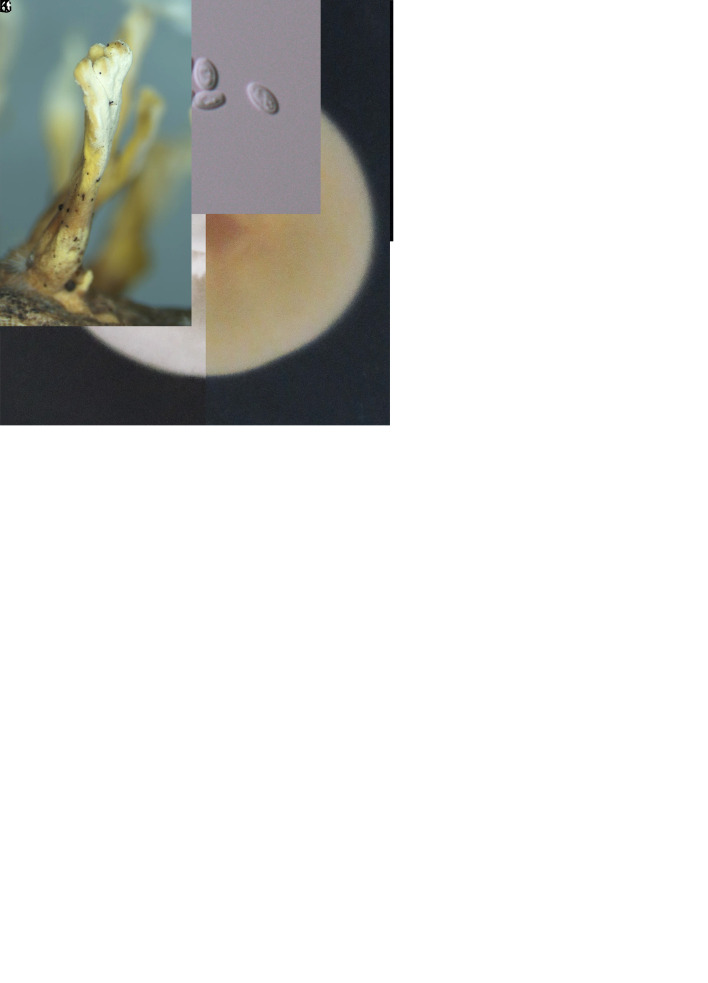
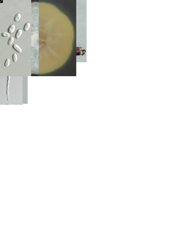
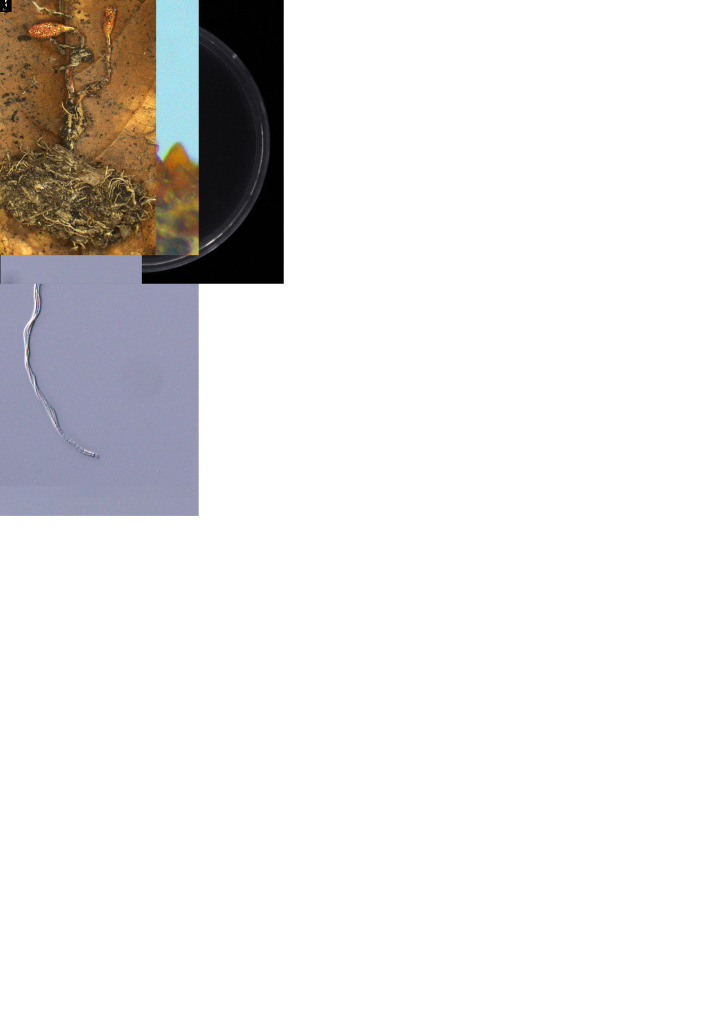
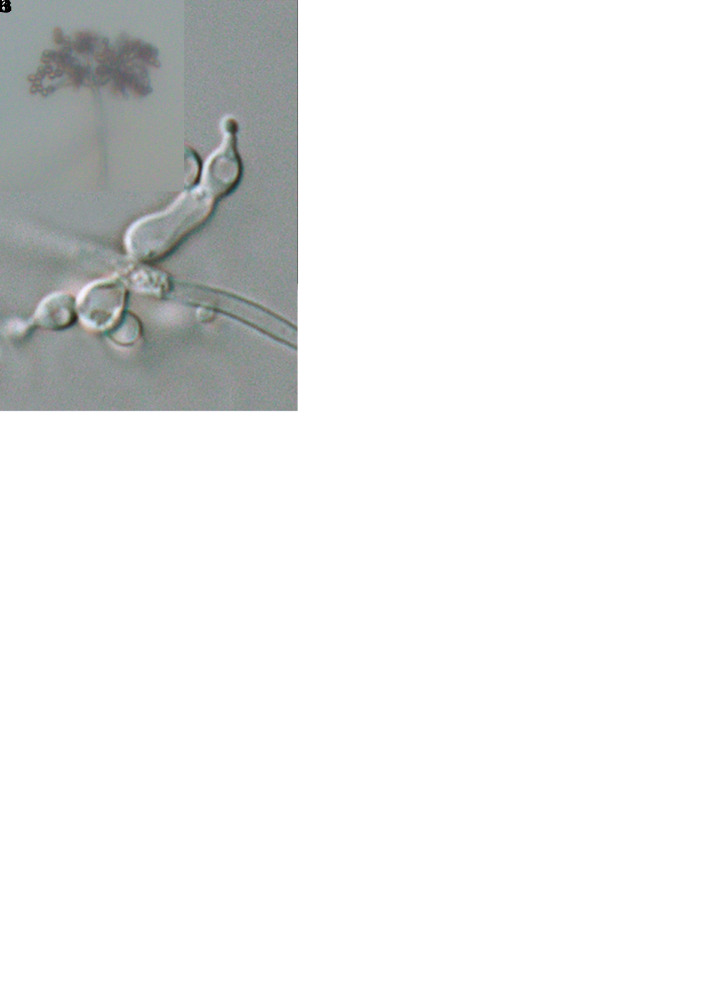
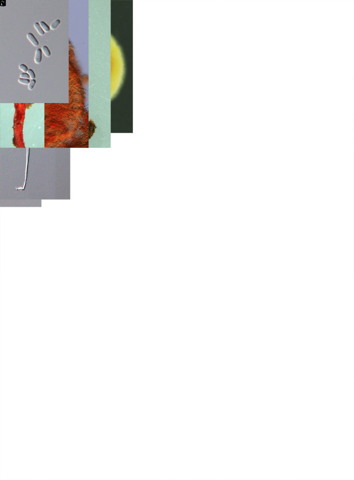
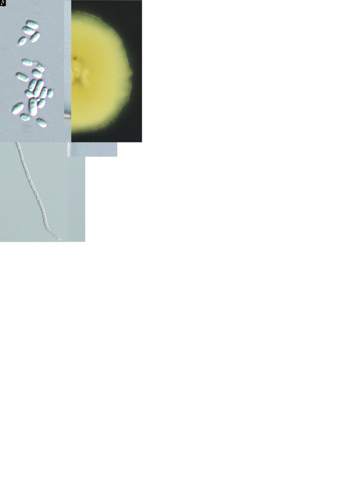
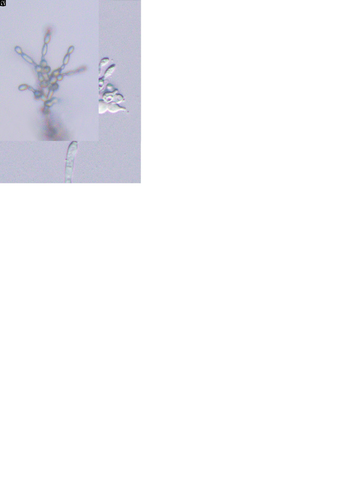
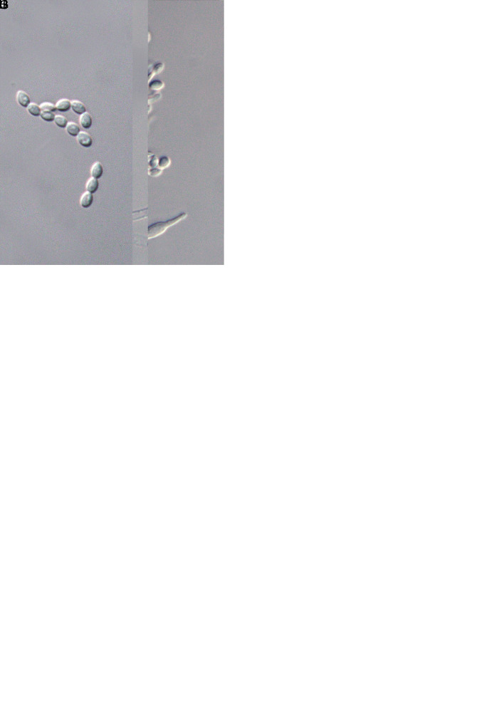
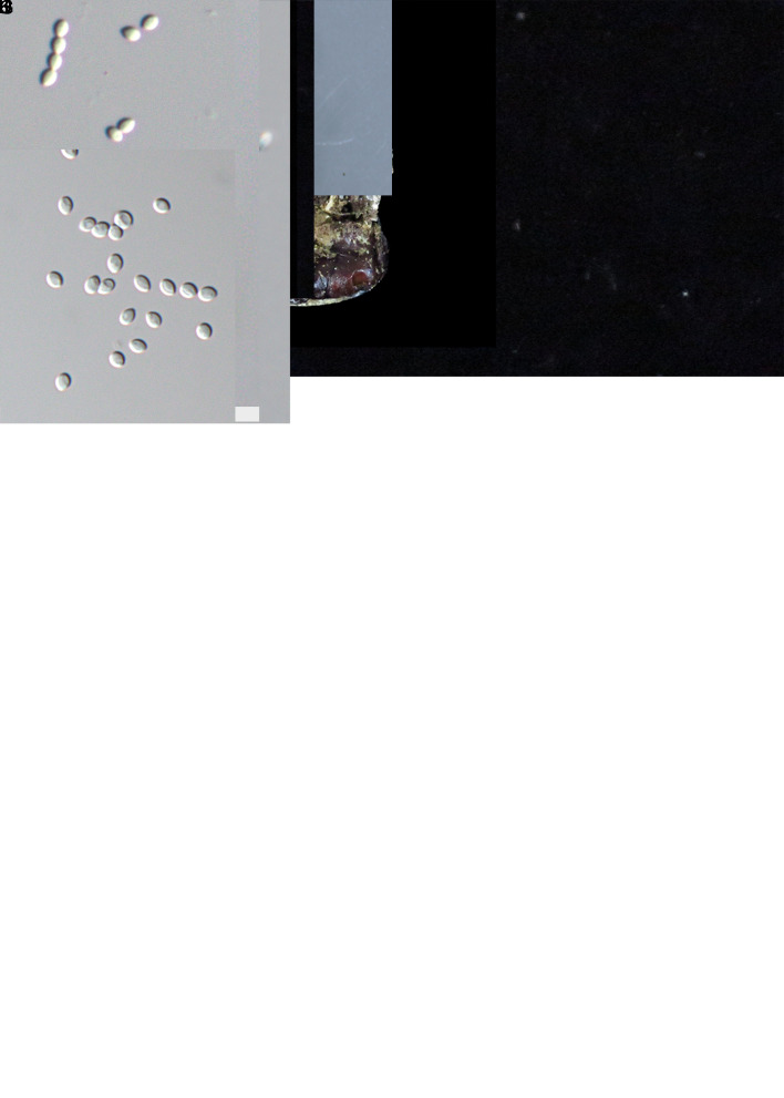
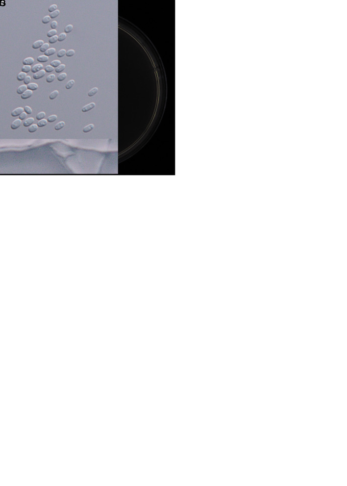
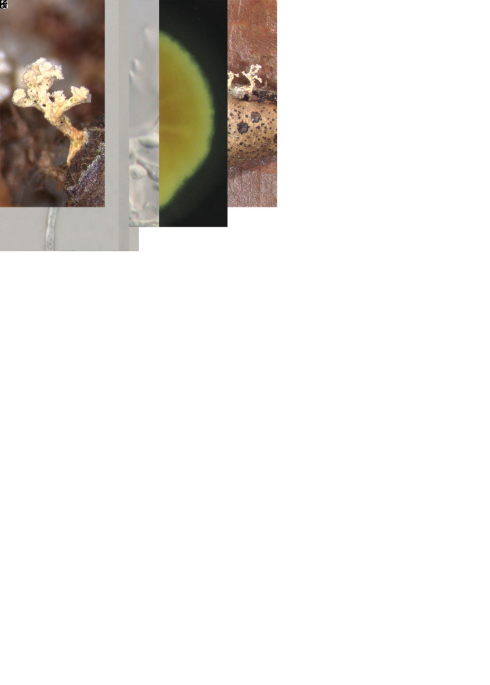
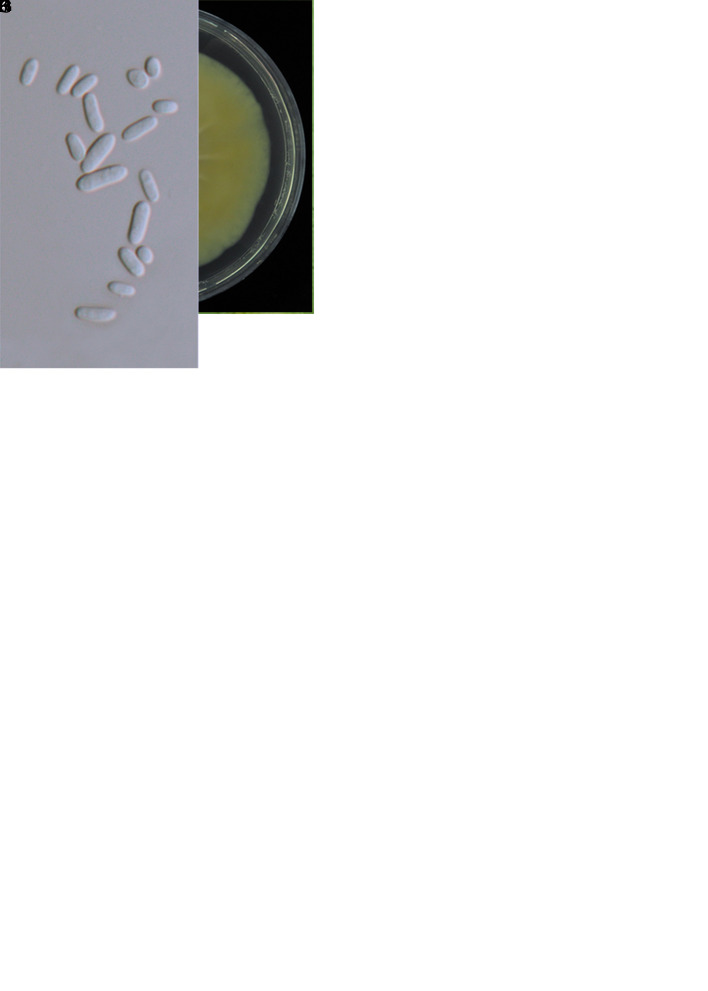
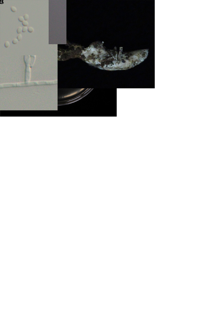
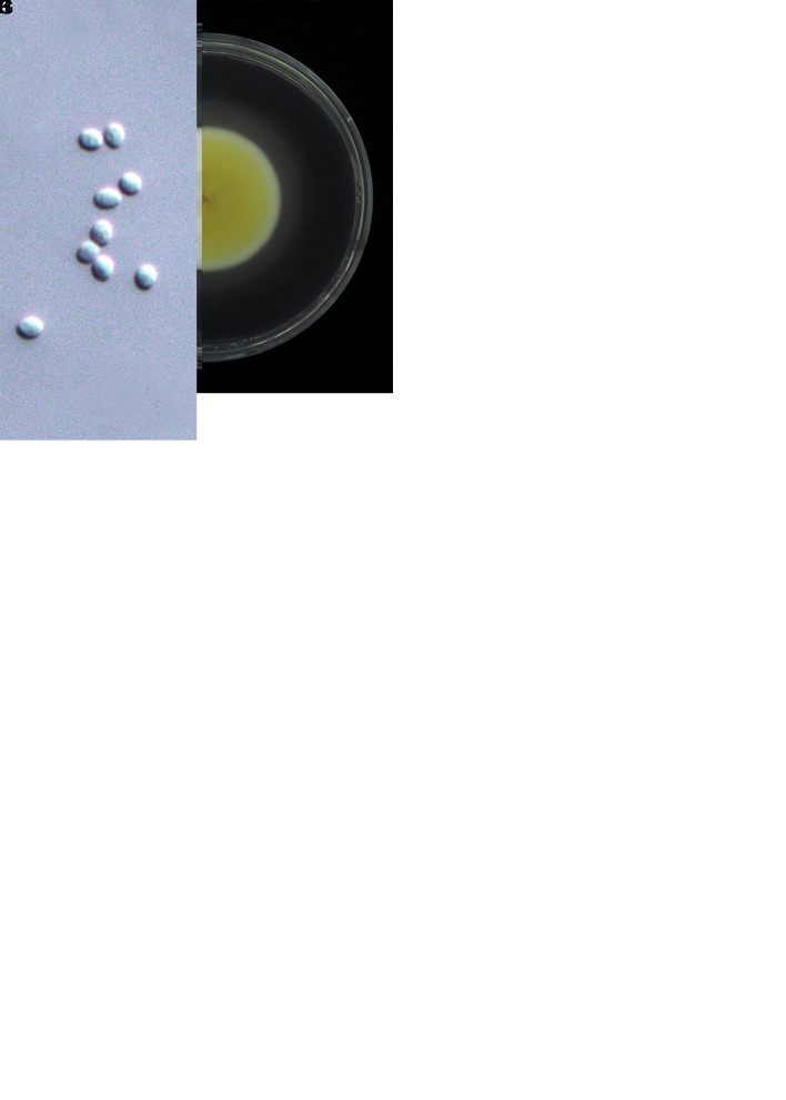
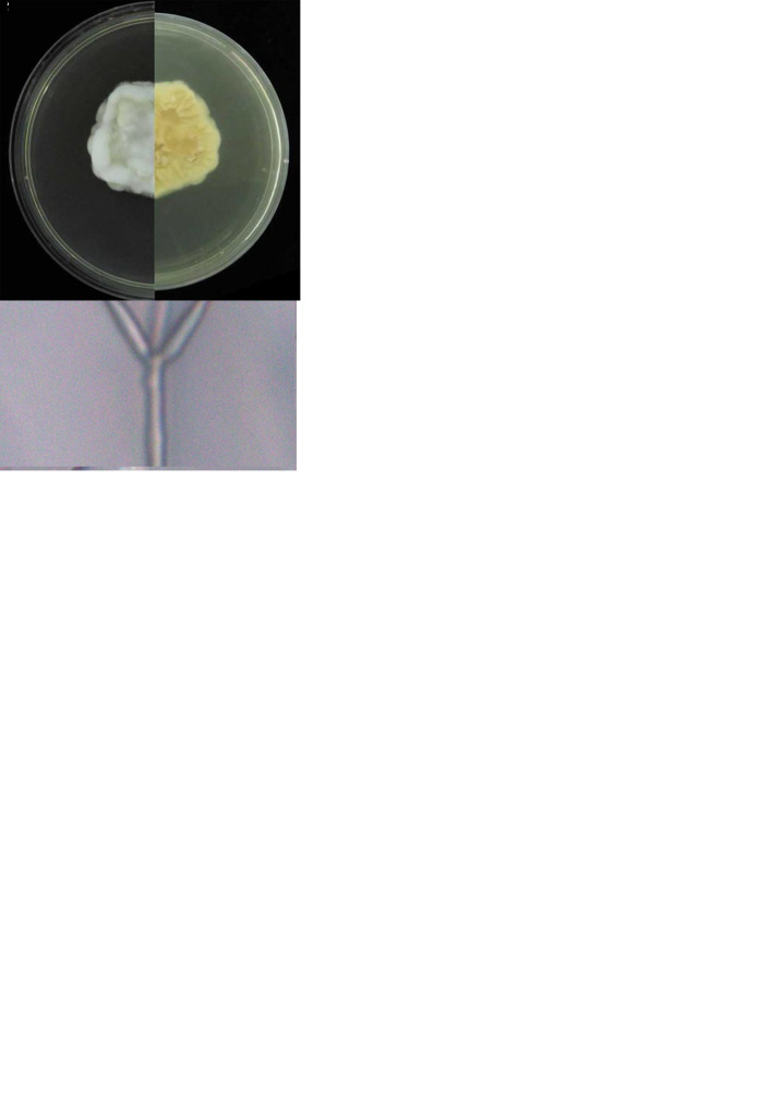
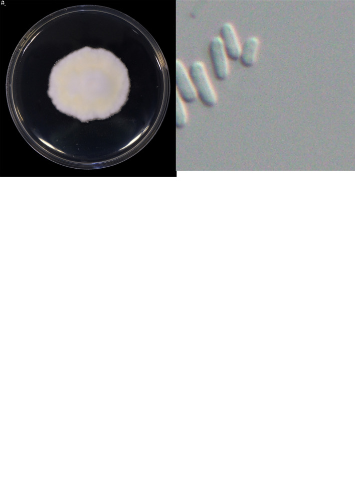
Species classified in Cordycipitaceae (Hypocreales) include multiple entomopathogenic fungi. Numerous changes have recently occurred in the nomenclature of cordycipitaceous fungi due to the single naming system proposed for pleomorphic fungi in 2011. Species of Cordycipitaceae are widely applied as herbal medicines, especially in Asian cultures. However, the diversity of Cordycipitaceae in Taiwan is based on relatively few literature records. Here we conducted a comprehensive survey of this family throughout the island of Taiwan and provided a glimpse of the diversity and distribution patterns. In addition, the present study reassesses the generic and species boundaries of Cordycipitaceae and finally provides an updated phylogenetic overview of Cordyceps and allied genera. Phylogenetic reconstructions using combined ITS, nrLSU, tef1-α, rpb1, and rpb2 sequence data, along with morphological features, resulted in 10 novel species: Akanthomyces taiwanicus sp. nov., Blackwellomyces taiwanensis sp. nov., Cordyceps hehuanensis sp. nov., C. locastrae sp. nov., C. malleiformis sp. nov., C. pseudorosea sp. nov., C. siangyangensis sp. nov., Samsoniella lasiocampidarum sp. nov., S. yuanzuiensis sp. nov., and Simplicillium salviniae sp. nov.; and nine new records for Taiwan: A. kanyawimiae, A. muscarius, S. cardinalis, S. hepiali, B. lii, B. medogensis, C. lepidopterorum, C. neopruinosa, and Si. chinense. Furthermore, we provided DNA sequence data of the ex-type strains of C. ninchukispora for the first time and determined the species limits of the taxon. In addition, the present study proposed to synonymize B. staphylinidicola and C. jakajanicola under B. bassiana and C. lepidopterorum, respectively. Moreover, three species, C. roseostromata, C. kyushuensis, and C. shuifuensis, that clustered within the species clade of C. militaris are proposed to be synonymized under the latter taxon. To maintain the monophyly of Cordyceps, we propose to classify Parahevansia koratensis in Cordyceps, which makes the genus Parahevansia obsolete. Taxonomic novelties: New species: Akanthomyces taiwanicus W.Y. Chuang, B. Shrestha & H.A. Ariyaw., Blackwellomyces taiwanensis W.Y. Chuang & H.A. Ariyaw., Cordyceps hehuanensis W.Y. Chuang & H.A. Ariyaw., C. locastrae W.Y. Chuang & H.A. Ariyaw., C. malleiformis W.Y. Chuang & H.A. Ariyaw., C. pseudorosea W.Y. Chuang & H.A. Ariyaw., C. siangyangensis W.Y. Chuang & H.A. Ariyaw., Samsoniella lasiocampidarum W.Y. Chuang & H.A. Ariyaw., S. yuanzuiensis W.Y. Chuang & H.A. Ariyaw., Simplicillium salviniae W.Y. Chuang & H.A. Ariyaw. New combination: Cordyceps koratensis (Hywel-Jones) H.A. Ariyaw., M. Stadler & Luangsa-ard. New synonyms: Beauveria bassiana (Bals.-Criv.) Vuill., Cordyceps lepidopterorum Mongkols. et al., C. militaris (L.) Fr. Citation: Chuang WY, Lin YC, Shrestha B, Luangsa-ard JJ, Stadler M, Tzean SS, Wu S, Ko CC, Hsieh SY, Wu ML, Wang SC, Shen TL, Ariyawansa HA (2024). Phylogenetic diversity and morphological characterization of cordycipitaceous species in Taiwan. Studies in Mycology 109: 1-56. doi: 10.3114/sim.2024.109.01.

Keywords: Cordycipitaceae; entomopathogenic fungi; new species; phylogenetic analyses.


Conflict of interest statement

The authors declare that there is no conflict of interest.

Figures

Fig. 1
Fig. 1
Overall information of the 86 cordycipitaceous fungi collected in this study. A. Geographic distribution of cordycipitaceous fungi collected in this study. (Satellite imagery source: FORMOSAT-5 (FS-5) satellite, National Space Organization, Hsinchu City, Taiwan.) B. Proportions of the genera of 86 cordycipitaceous fungi collected in this study.
Fig. 2
Fig. 2
Maximum likelihood phylogenetic tree of Cordycipitaceae and related families based on a combined dataset of ITS, nrLSU, tef1-α, rpb1, and rpb2 gene regions. Verticillium dahliae (ATCC 16535) was selected as the outgroup taxon. ML bootstrap values (BS) ≥ 70 % and Bayesian posterior probabilities (PP) ≥ 0.95 are given at the nodes. Strains obtained in the present study are marked in red and type strains are indicated with a superscript T.
Fig. 2
Fig. 2
Maximum likelihood phylogenetic tree of Cordycipitaceae and related families based on a combined dataset of ITS, nrLSU, tef1-α, rpb1, and rpb2 gene regions. Verticillium dahliae (ATCC 16535) was selected as the outgroup taxon. ML bootstrap values (BS) ≥ 70 % and Bayesian posterior probabilities (PP) ≥ 0.95 are given at the nodes. Strains obtained in the present study are marked in red and type strains are indicated with a superscript T.
Fig. 3
Fig. 3
Morphology of Akanthomyces taiwanicus (holotype NTUPPMH 20-060, ex-type culture NTUPPMCC 20-060). A. Synnemata arising from an adult Agrius convolvuli. B. The host adult A. convolvuli. C. Proboscis of A. convolvuli. D, E. Magnified synnemata. F. Phialides on the host. G. Conidia on the host. H. Obverse and reverse of colonies on PDA at 14 d. I, J. Phialides on PDA. K. Conidia from PDA culture. Scale bars: F, G = 10 μm; I, J = 10 μm; K = 5 μm.
Fig. 4
Fig. 4
. Morphology of Blackwellomyces taiwanensis (holotype NTUPPMH 20-071, ex-type culture NTUPPMCC 20-071). A. Stromata on Tenebrionidae pupa. B. Stroma. C. Section of stroma. D. Section of perithecium. E–G. Immaure asci. H. Mature ascus. I. Ascus cap. J. Part-spores. K. Obverse and reverse of colonies on PDA. L, M. Phialides on PDA. N. Conidia from PDA culture. Scale bars: C = 200 μm; D = 40 μm; E–H = 20 μm; I, J = 5 μm; L–N = 10 μm.
Fig. 5
Fig. 5
Morphology of Cordyceps hehuanensis (holotype NTUPPMH 18-145, ex-type culture NTUPPMCC 18-145). A. Stromata arising from a lepidopteran adult. B, C. Enlarged fertile part of stroma showing semi-immersed perithecia. D. Section of perithecia. EG. Ascus. H. Ascus cap. I. Part-spores. J. Obverse and reverse of colonies on PDA at 14 d. K, L Phialides on PDA. M, N. Conidia from PDA culture. Scale bars: D = 200 μm; E–G = 50 μm; H, I = 10 μm; K–N = 10 μm.
Fig. 6
Fig. 6
Morphology of Cordyceps locastrae (holotype NTUPPMH 17-042, ex-type culture NTUPPMCC 17-042). A, B. Synnemata on Locastra muscosalis pupa. C. Conidiophore with phialides and conidia. D, E. Conidia on the host. F. Obverse and reverse of colonies on PDA in 14 d. G, H. Evlachovaea-like conidiogenesis. I–K Phialides on PDA. L. Conidia on PDA culture. Scale bars = 5 μm.
Fig. 7
Fig. 7
Morphology of Cordyceps malleiformis (holotype NTUPPMH 18-143, ex-type culture NTUPPMCC 18-143). A, B. Stromata arising from a lepidopteran pupa. C. Part of stroma showing semi-immersed perithecia. D. Perithecium. E. Ostiole of perithecia. F. Perithecia cell wall. G–I. Ascus. J, K. Ascus cap L. Obverse and reverse of colonies on PDA at 14 d. M. Evlachovaea-like conidiogenesis. N. Phialides on PDA. O, P. Conidia from PDA culture. Scale bars: D = 50 μm; E, F = 20 μm; G–I = 50 μm; J, K = 5 μm; M = 20 μm; N–P = 10 μm.
Fig. 8
Fig. 8
. Morphology of Cordyceps pseudorosea (holotype NTUPPMH 17-041, ex-type culture NTUPPMCC 17-041). A. Stromata arising from the ventral side of the anterior end of the erebid adult. B. Erebid adult host. C, D. Sections of perithecia. E–G. Asci. H. Ascus cap. I. Part-spores. J. Obverse and reverse of colonies on PDA. K, L. Evlachovaea-like conidiogenesis. M. Conidia from PDA culture. Scale bars: C = 200 μm; D = 40 μm; E–G = 50 μm; H, I = 10 μm; K–M = 10 μm.
Fig. 9
Fig. 9
. The morphology of Cordyceps siangyangensis (holotype NTUPPMH 18-149, ex-type culture NTUPPMCC 18-149). A, B. Fungus on host. C. Magnified synnemata. D, E. Host. F. Phialides on the host. G. Conidia on the host. H. Obverse and reverse of colonies on PDA at 14 d. I, J. Isaria-like conidiogenesis. K, L. Phialides with conidia on PDA. M. Conidia from PDA. Scale bars: F, G = 5 μm; I, J = 20 μm; K–M = 10 μm.
Fig. 10
Fig. 10
Morphology of Samsoniella lasiocampidarum (holotype NTUPPMH 20-063, ex-type culture NTUPPMCC 20-063). A. Synnemata on a lasiocampid larva. B, C. Synnemata and conidial mass. D. Phialide on lasiocampid host. E. Conidia on lasiocampid host. F. Obverse and reverse of colonies on PDA. G. Isaria-like conidiogenesis. H, I. Conidiophore on PDA. J. Conidia from PDA culture. Scale bars: D = 10 μm; E = 5 μm; G–J =10 μm.
Fig. 11
Fig. 11
The morphology of Samsoniella yuanzuiensis (holotype NTUPPMH 20-064, ex-type culture NTUPPMCC 20-064). A, B. Synnemata growing on a lepidopteran pupa. C. Conidial mass on the apex of synnemata. D. Phialides on the host. E. Conidia on the host. F. Obverse and reverse of colonies on PDA. G, H. Isaria-like conidiogenesis. I, J. Conidiophore on PDA. K. Conidia from PDA culture. Scale bars =10 μm.
Fig. 12
Fig. 12
Morphology of Simplicillium salviniae (ex-type culture NTUPPMCC 20-074/BCRC 34536). A, B. Obverse and reverse of colonies on PDA. C–E. Conidial mass on the top of a conidiophore. F–H. Conidiophore on PDA. I. Conidia from PDA culture. Scale bars = 10 μm.
Fig. 13
Fig. 13
Morphology of Akanthomyces kanyawimiae (NTUPPMH 20-058, living culture NTUPPMCC 20-058). A. Synnemata of fungus arising from host (Araneae). B, C. Magnified synnemata. D. Phialides on host (Araneae). E, F. Conidia from host (Araneae). G. Obverse and reverse of colonies on PDA at 14 d. H. Isaria-like conidiogenesis. I, J. Phialides on PDA. K. Conidia from PDA culture. Scale bars = 10 μm.
Fig. 14
Fig. 14
Morphology of Akanthomyces muscarius (NTUPPMH 20-059, living culture NTUPPMCC 20-059). A. Mycelium covering Araneae host. B. Phialides on the host. C, D. Conidia on the host. E. Obverse and reverse of colonies on PDA at 14 d. F. Conidiophores arising on aerial hyphae. G. Conidial mass on the top of a conidiophore. H–J. Phialides on PDA. K. Conidia from PDA culture. Scale bars: B = 20 μm; C, D = 10 μm; G = 20 μm; H–K = 10 μm.
Fig. 15
Fig. 15
Mycelial colony and conidiation of Beauveria lii (NTUPPMCC 20-054). A, B. Obverse and reverse of colonies on PDA at 21 d. C–E. Conidiogenesis pattern and phialides on PDA. F–H. Conidia from PDA culture. Scale bars = 10 μm.
Fig. 16
Fig. 16
Morphology of Beauveria medogensis (NTUPPMH 20-055, living culture NTUPPMCC 20-055). A. Mycelium arising from Formicidae host. B. Formicidae host. C. Phialide and conidia on the host. D. Obverse and reverse of colonies on PDA at 14 d. E. Conidiogenesis pattern. F–H. Phialides on PDA. I–K. Conidia from PDA culture. Scale bars: C = 10 μm, E–H = 10 μm, I–K = 5 μm.
Fig. 17
Fig. 17
Morphology of Cordyceps lepidopterorum (NTUPPMH 18-118 and living culture NTUPPMCC 18-118). A, B. Synnemata arising from a Cicadidae nymph. C. Synnemata. D. Phialides on the host. E. Conidia on the host. F. Obverse and reverse of colonies on PDA at 14 d. G, H. Isaria-like conidiogenesis. I. Phialides on PDA. J. Conidia from PDA culture. Scale bars: D–E = 10 μm; G = 50 μm; H–I = 10 μm; J = 5 μm.
Fig. 18
Fig. 18
Morphology of Cordyceps neopruinosa (NTUPPMH 18-128, living culture NTUPPMCC 18-128). A. Stromata growing on a limacodid cocoon. B. Ascomata of specimen. C. Section of perithecium. D. Cell wall of perithecium. E, F. Ascus. G. Obverse and reverse of colonies on PDA. H. Evlachovaea-like conidiogenesis. I, J. Phialides on PDA. K. Conidia from PDA culture. Scale bars: C = 50 μm, D = 20 μm, E = 40 μm, F = 30 μm, H = 30 μm. I, J = 10 μm, K, L = 5 μm.
Fig. 19
Fig. 19
Mycelium of S. cardinalis (DI) growing on fruiting body of cordyceps-like taxon (A–C) (NTUPPMH 20-066, living culture NTUPPMCC 20-066). A. Cordyceps sp. on cicada. B, C. Conidia of Cordyceps sp. D. Obverse of colonies on PDA at 21 d. E. Reverse of colonies on PDA at 21 d. F–H. Conidiogenesis pattern and phialides on PDA. I, J. Conidia from PDA culture. Scale bars: B, C = 5 μm; F–J = 10 μm.
Fig. 20
Fig. 20
Morphology of Samsoniella hepiali (NTUPPMH 18-159, living culture, NTUPPMCC 18-159). A. Synnemata arising from lepidopteran insect. B. Phialide on the host. C. Conidia on the host. D. Obverse and reverse of colonies on PDA at 14 d. E–G. Isaria-like conidiogenesis. HJ. Phialides on PDA. K. Conidia from PDA culture. Scale bars: B, C = 10 μm; E–K = 10 μm.
Fig. 21
Fig. 21
Morphology of Beauveria brongniartii (A–J) and Simplicillium chinense (K–O) (NTUPPMH 20-073, living culture NTUPPMCC 20-073). A. Beauveria brongniartii on Locastra muscosalis cocoon. B. Locastra muscosalis. C. Stromata of cordycipitaceous fungi. D. Ascomata of cordycipitaceous fungi. E, F. Section of perithecium. G, H. Ascus. I. Ascus cap. J. Part-spores. K. Obverse and reverse of colonies on PDA. L. Conidiogenesis pattern. M, N. Phialide on PDA. O. Conidia from PDA culture. Scale bars: E = 100 μm; F–H = 50 μm; I, J = 10 μm; L = 20 μm; M–O = 10 μm.
Fig. 22
Fig. 22
Morphology of C. javanica (NTUPPMH 18-115, living culture NTUPPMCC 18-115). A. Fungus on unknown insect host. B. Obverse and reverse of colonies on PDA at 14 d. C. Conidia on the host. D–F. Isaria-like conidiogenesis. G, H. Phialides on PDA. I, J. Conidia from PDA culture. Scale bars: C–E = 20 μm; F–H = 10 μm; I–J = 5 μm.
Fig. 23
Fig. 23
Morphology of C. ninchukispora (ex-type culture NTUPPMCC 20-077/BCRC 31900). A, B. Obverse and reverse of colonies on PDA at 21 d. CE. Phialides on PDA. F. Conidia from PDA. Scale bars: C–F = 10 μm.
Fig. 24
Fig. 24
Morphology of Cordyceps militaris (NTUPPMH 18-120, living culture NTUPPMCC 18-120). A. Fungus on Notodontidae pupa. B. Ascomata of specimen. C. Notodontidae pupa. D. Section of perithecium. E. Cell wall of perithecium. F, G. Asci. H. Ascus cap. I. Part-spores. J. Obverse and reverse of colonies on PDA (NTUPPMCC 18-120). K. Isaria-like conidiogenesis. L, M. Phialides on PDA. N. Conidia from PDA culture. Scale bars: D–G = 50 μm; H, I = 10 μm; K–N = 10 μm.

Similar articles

Cited by

References

    1. Aini AN, Mongkolsamrit S, Wijanarka W. et al. (2020). Diversity of Akanthomyces on moths (Lepidoptera) in Thailand. MycoKeys 71: 1. - PMC - PubMed
    1. Araújo JP, Hughes DP. (2016). Diversity of entomopathogenic fungi: which groups conquered the insect body? Advances in Genetics 94: 1–39. - PubMed
    1. Ariyawansa HA, Hyde KD, Jayasiri SC. et al. (2015). Fungal diversity notes 111–252—taxonomic and phylogenetic contributions to fungal taxa. Fungal Diversity 75: 27–274.
    1. Ariyawansa HA, Phillips AJ, Chuang WY. et al. (2018). Tzeananiaceae, a new pleosporalean family associated with Ophiocordyceps macroacicularis fruiting bodies in Taiwan. MycoKeys 37: 1. - PMC - PubMed
    1. Chen CH, Hsieh SY, Yeh YH. et al. (2020). Cladocillium musae, a new genus and species of cercosporoid fungi (Mycosphaerellaceae) on wild banana in Taiwan. Mycological Progress 19: 837–843.

LinkOut - more resources