. 2024 Dec:109:403-450.
doi: 10.3114/sim.2024.109.06. Epub 2024 Sep 26.

Known from trees and the tropics: new insights into the Fusarium lateritium species complex

Affiliations

Known from trees and the tropics: new insights into the Fusarium lateritium species complex

M M Costa et al. Stud Mycol. 2024 Dec.

Abstract

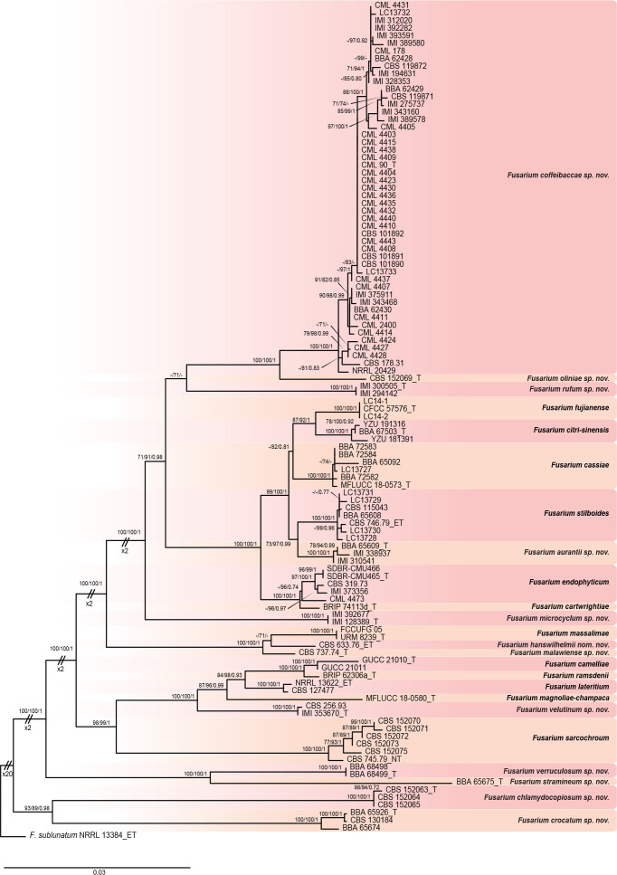
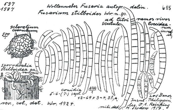
The Fusarium lateritium species complex (FLSC) currently comprises 11 phylogenetic species, including accepted names such as F. lateritium, F. sarcochroum, and F. stilboides, which have mostly been reported in association with citrus and coffee. Many varieties were documented by Wollenweber & Reinking (1935), which is indicative of a wider diversity of species within this group. The lack of type material in some cases, especially for the older names, means that definition by molecular phylogeny is very difficult. In the present study, we examined 179 strains related to F. lateritium from different countries and substrates. Historic reference material, including representative strains from the Wollenweber & Reinking (1935) varieties were included in this study, DNA sequences were generated for comparison, and the morphology correlated with original descriptions to enable the correct application of older names. Strains were characterized by multi-gene phylogenetic analyses based on fragments of the β-tubulin (tub2), calmodulin (CaM), RNA polymerase II second largest subunit (rpb2), and translation elongation factor 1-alpha (tef1) genes, evaluation of morphological characters and host-substrate preferences. The biological species concept was tested by crossings in vitro. Strains previously identified as F. lateritium, F. stilboides, or one of their varieties based on morphology, were found to belong to 16 species in the FLSC, but also to species from six other species complexes (SC), including the F. citricola SC, F. heterosporum SC, F. incarnatum-equiseti SC, F. redolens SC, F. sambucinum SC, and the F. tricinctum SC. Eleven new phylogenetic and two biological species are described in the FLSC, and emended descriptions are provided for four previously described species. An epitype is designated for F. lateritium, and F. lateritium var. longum, a former variety within the FLSC, is lecto- and epitypified, and elevated to species level with a replacement name. Taxonomic novelties: New species: F. aurantii M.M. Costa, Sand.-Den. & Crous, F. chlamydocopiosum M.M. Costa, Sand.-Den. & Crous, F. citri-sinensis L. Zhao & J.X. Deng, F. coffeibaccae M.M. Costa, L.H. Pfenning, Sand.-Den. & Crous, F. crocatum M.M. Costa, Sand.-Den. & Crous, F. malawiense M.M. Costa, Sand.-Den. & Crous, F. microcyclum M.M. Costa, Sand.-Den. & Crous, F. oliniae M.M. Costa, Sand.-Den. & Crous; F. rufum M.M. Costa, Sand.-Den. & Crous, F. stramineum M.M. Costa, Sand.-Den. & Crous, F. velutinum M.M. Costa, Sand.-Den. & Crous, F. verruculosum M.M. Costa, Sand.-Den. & Crous; Replacement name: F. hanswilhelmii M.M. Costa, Sand.-Den. & Crous; Epitype (basionym): F. lateritium Nees, F. lateritium var. longum Wollenw.; Lectotype (basionym): F. lateritium var. longum Wollenw. Citation: Costa MM, Sandoval-Denis M, Moreira GM, Kandemir H, Kermode A, Buddie AG, Ryan MJ, Becker Y, Yurkov A, Maier W, Groenewald JZ, Pfenning LH, Crous PW (2024). Known from trees and the tropics: new insights into the Fusarium lateritium species complex. Studies in Mycology 109: 403-450. doi: 10.3114/sim.2024.109.06.

Keywords: Biological species concept; fungal taxonomy; multigene phylogeny; new taxa.


Conflict of interest statement

The authors declare that there is no conflict of interest.

Figures

Fig. 1
Fig. 1
Phylogenetic tree inferred from a Maximum Likelihood (RAxML) analysis based on concatenated tef1, tub2, rpb2, and CaM sequences of species from the FLSC. Ex-type, ex-epitype, and ex-neotype strains are indicated with T, ET, and NT respectively. Numbers at branches indicate support values (RAxML-BS / IQ-TREE-BS / BI-PP) above 70 % / 70 % / 0.7. The tree was rooted to Fusarium sublunatum (CBS 189.34 = NRRL 13384). Scale bar represents expected number of changes per site.
Fig. 2
Fig. 2
Fusarium aurantii sp. nov. (ex-type culture BBA 65609). A, B. Colonies on PDA and OA, respectively, after 14 d at 24 °C in the dark. C–F. Sporodochia formed on the surface of carnation leaves and agar surface. G, H, J–L, N, O. Aerial conidiophores, conidiogenous cells and conidia. I. Projections on the hyphae. M. Aerial conidia. P. Sporodochial conidiophores and conidiogenous cells. Q. Sporodochial conidia. Scale bars: C, D = 200 μm; E, F = 100 μm; G = 50 μm; H, J–L, P = 20 μm; I, M, N, Q = 10 μm; O = 5 μm.
Fig. 3
Fig. 3
Fusarium cassiae (BBA 65092). A, B. Colonies on PDA and OA, respectively, after 14 d at 24 °C in the dark. C, D. Sporodochia formed on the surface of carnation leaves. E–N. Aerial conidiophores, conidiogenous cells and conidia. O–Q. Chlamydospores. R. Aerial conidia. S–U. Sporodochial conidiophores, conidiogenous cells and conidia. V. Sporodochial conidia. Scale bars: C, D = 100 μm; E, H–V = 10 μm; F, G = 20 μm.
Fig. 4
Fig. 4
Fusarium chlamydocopiosum sp. nov. (ex-type culture CBS 152063). A, B. Colonies on PDA and OA, respectively, after 14 d at 24 °C in the dark. C, D. Sporodochia formed on the surface of carnation leaves (C), and agar surface (D). E–M. Aerial conidiophores, conidiogenous cells and conidia. N, O. Sterile hyphal projections. P. Aerial conidia. Q–T. Chlamydospores. U, V. Sporodochial conidiophores, conidiogenous cells and conidia. W. Sporodochial conidia. Scale bars: C = 100 μm; D = 20 μm; E, F–M, P, S, U–W = 10 μm; N, O, Q, R, T = 5 μm.
Fig. 5
Fig. 5
Fusarium citri-sinensis (ex-type culture BBA 67503). A, B. Colonies on PDA and OA, respectively, after 14 d at 24 °C in the dark. C–E. Sporodochia formed on the surface of carnation leaves and agar surface. F–J, M–Q. Aerial conidiophores, conidiogenous cells and conidia. K, L. Chlamydospores. R. Aerial conidia. S–U. Sporodochial conidiophores, conidiogenous cells and conidia. V. Sporodochial conidia. Scale bars: C, D = 100 μm; E, F, U = 20 μm; G, H, K, L, O–T, V = 10 μm; I, J, M, N = 5 μm.
Fig. 6
Fig. 6
Fusarium coffeibaccae sp. nov. (ex-type culture CML 90). A, B. Colonies on PDA and OA, respectively, after 14 d at 24 °C in the dark. C, D. Sporodochia formed on the surface of carnation leaves. E–Q. Aerial conidiophores, conidiogenous cells and conidia. R. Aerial conidia, including mesoconidia. S, T. Chlamydospores. U, V. Sporodochial conidiophores, conidiogenous cells and conidia. W. Sporodochial conidia. Scale bars: C, E = 100 μm; D = 200 μm; F–T, W = 10 μm; U = 20 μm; V = 50 μm.
Fig. 7
Fig. 7
Sexual morph of Fusarium coffeibaccae sp. nov. (CML 4411 × CML 101892). A–D. Perithecia. C, D. Perithecia oozing ascospores. E, F. Section through perithecium. G–K. Asci and ascospores. L, M. Ascospores. M. Germinating ascospore. Scale bars: A–D = 100 μm; E–G = 50 μm; H–M = 10 μm.
Fig. 8
Fig. 8
Fusarium crocatum sp. nov. (ex-type culture BBA 65926). A, B. Colonies on PDA and OA, respectively, after 14 d at 24 °C in the dark. C, D. Sporodochia formed on the surface of carnation leaves. E–N. Aerial conidiophores, conidiogenous cells and conidia. O. Aerial conidia. P, Q. Sporodochial conidiophores, conidiogenous cells and conidia. R. Sporodochial conidia. Scale bars: C, D = 200 μm; E = 50 μm; F = 20 μm; G–R = 10 μm.
Fig. 9
Fig. 9
Sexual morph of Fusarium crocatum sp. nov. (BBA 65926 × BBA 65674). A–C. Perithecia. C. Perithecia oozing ascospores. D–G. Section through perithecium. H–K. Asci and ascospores. L, M. Ascospores. Scale bars: C, D = 200 μm; E = 50 μm; F, H, I = 20 μm; G, J, M = 10 μm; K, L = 5 μm.
Fig. 10
Fig. 10
Fusarium endophyticum (CBS 319.73). A, B. Colonies on PDA and OA, respectively, after 14 d at 24 °C in the dark. C, D. Sporodochia formed on the surface of carnation leaves. E–M. Aerial conidiophores, conidiogenous cells and conidia. N. Aerial conidia. O, P. Chlamydospores. Q–S. Sporodochial conidiophores, conidiogenous cells and conidia. T. Sporodochial conidia. Scale bars: C, D = 100 μm; E, F = 50 μm; G, I–K, M–T = 10 μm; H = 20 μm; L = 5 μm.
Fig. 11
Fig. 11
Fusarium hanswilhelmii nom. nov. (ex-epitype culture CBS 633.76). A, B. Colonies on PDA and OA, respectively, after 14 d at 24 °C in the dark. C, D. Sporodochia formed on the surface of carnation leaves. E–O. Aerial conidiophores, conidiogenous cells and conidia. P, Q. Chlamydospores. R. Aerial conidia. S, T. Sporodochial conidiophores, conidiogenous cells and conidia. U. Sporodochial conidia. Scale bars: C, D = 100 μm; E–H, L–P, S, T = 20 μm; I–K, Q, R, U = 10 μm.
Fig. 12
Fig. 12
Fusarium hanswilhelmii nom. nov. Lectotype of F. lateritium var. longum as illustrated in protologue by Wollenweber (1930).
Fig. 13
Fig. 13
Fusarium lateritium (NRRL 13622). A, B. Colonies on PDA and OA, respectively, after 14 d at 24 °C in the dark. C–E. Sporodochia formed on the surface of carnation leaves and agar surface. F–N, Q. Aerial conidiophores, conidiogenous cells and conidia. O. Chlamydospores. P. Aerial conidia. R–U. Sporodochial conidiophores, conidiogenous cells and conidia. V. Sporodochial conidia. Scale bars: C = 200 μm; D = 100 μm; E = 40 μm; F, G = 50 μm; H = 30 μm; I, K, M–P, R, U, V = 10 μm; J = 5 μm; L = 2 μm; Q, S, T = 20 μm.
Fig. 14
Fig. 14
Lectotype of Fusarium lateritium as illustrate and described in Nees (1817). A. On a twig of beech in natural size. B. Detached, enlarged. +. Vertical section mounted on water.
Fig. 15
Fig. 15
Fusarium malawiense sp. nov. (ex-type culture CBS 737.74). A, B. Colonies on PDA and OA, respectively, after 14 d at 24 °C in the dark. C, D. Sporodochia formed on the surface of carnation leaves. E–J. Aerial conidiophores, conidiogenous cells and conidia. K–M. Chlamydospores. N, O. Aerial conidia. P. Sporodochial conidiophores, conidiogenous cells and conidia. Q. Sporodochial conidia. Scale bars: C, D = 100 μm; E–G = 20 μm; H, I, K, N–Q = 10 μm; J, L, M = 5 μm.
Fig. 16
Fig. 16
Fusarium microcyclum sp. nov. (ex-type culture IMI 128389). A, B. Colonies on PDA and OA, respectively, after 14 d at 24 °C in the dark. C–E. Sporodochia formed on the surface of carnation leaves and agar surface. F–P. Aerial conidiophores, conidiogenous cells and conidia. Q. Aerial conidia. R–T. Aerial conidiophores, conidiogenous cells and conidia. U, V. Sporodochial conidiophores and conidiogenous cells. W. Sporodochial conidia. Scale bars: C–E = 50 μm; F, O–T, W = 10 μm; G–N = 5 μm; U, V = 20 μm.
Fig. 17
Fig. 17
Fusarium oliniae sp. nov. (ex-type culture CBS 152069). A, B. Colonies on PDA and OA, respectively, after 14 d at 24 °C in the dark. C, D. Sporodochia formed on the surface of carnation leaves. E–J. Aerial conidiophores, conidiogenous cells and conidia. K. Aerial conidia. L, M. Sporodochial conidiophores, conidiogenous cells and conidia. N. Sporodochial conidia. Scale bars: C, D = 200 μm, E = 100 μm; F, H–K, N = 10 μm; G = 5 μm; L, M = 20 μm.
Fig. 18
Fig. 18
Fusarium rufum sp. nov. (ex-type culture IMI 300505). A, B. Colonies on PDA and OA, respectively, after 14 d at 24 °C in the dark. C, D. Sporodochia formed on the surface of carnation leaves. E–M. Aerial conidiophores, conidiogenous cells and conidia. N. Aerial conidia. O–Q. Sporodochial conidiophores and conidiogenous cells. R. Sporodochial conidia. Scale bars: C, D = 200 μm; E–N, R = 10 μm; O–Q = 20 μm.
Fig. 19
Fig. 19
Fusarium sarcochroum (CBS 745.79). A, B. Colonies on PDA and OA, respectively, after 14 d at 24 °C in the dark. C–E. Sporodochia formed on the surface of carnation leaves and agar surface. F–L. Aerial conidiophores, conidiogenous cells and conidia. M–P. Chlamydospores. Q. Aerial conidia. R, S. Sporodochial conidiophores, conidiogenous cells and conidia. T. Sporodochial conidia. Scale bars: C, D = 100 μm; E–G, R, S = 20 μm; H, J–Q, T = 10 μm; I = 5 μm.
Fig. 20
Fig. 20
Fusarium stilboides (CBS 746.79). A, B. Colonies on PDA and OA, respectively, after 14 d at 24 °C in the dark. C, D. Sporodochia formed on the surface of carnation leaves. E–H, J–M, Q–S. Aerial conidiophores, conidiogenous cells and conidia. I, N. Aerial conidia. O, P. Chlamydospores. T, U. Sporodochial conidiophores, conidiogenous cells and conidia. V. Sporodochial conidia. Scale bars: C–E = 100 μm; F–J, L–P, T–V = 10 μm; K = 5 μm; Q–S = 20 μm.
Fig. 21
Fig. 21
Lectotype of Fusarium stilboides as illustrated in protologue by Wollenweber (1924).
Fig. 22
Fig. 22
Fusarium stramineum sp. nov. (ex-type culture BBA 65675). A, B. Colonies on PDA and OA, respectively, after 14 d at 24 °C in the dark. C, D. Sporodochia formed on the surface of carnation leaves. E–I, K, L. Aerial conidiophores, conidiogenous cells and conidia. J. Aerial conidia. M, N. Sporodochial conidiophores, conidiogenous cells and conidia. O. Sporodochial conidia. Scale bars: C, D = 100 μm; E–L, N, O = 10 μm; M = 20 μm.
Fig. 23
Fig. 23
Fusarium velutinum sp. nov. (ex-type culture IMI 353670). A, B. Colonies on PDA and OA, respectively, after 14 d at 24 °C in the dark. C, D. Sporodochia formed on the surface of carnation leaves. E–I, L–P. Aerial conidiophores, conidiogenous cells and conidia. J. Chlamydospores. K. Aerial conidia. Q. Sporodochial conidiophores, conidiogenous cells and conidia. R. Sporodochial conidia. Scale bars: C, D = 100 μm; E–I, K, L, N–P, R = 10 μm; J = 4; M = 5 μm; Q = 20 μm.
Fig. 24
Fig. 24
Fusarium verruculosum sp. nov. (ex-type culture BBA 68499). A, B. Colonies on PDA and OA, respectively, after 14 d at 24 °C in the dark. C–E. Sporodochia formed on the surface of carnation leaves and agar surface. F–N. Aerial conidiophores, conidiogenous cells and conidia. O–S. Chlamydospores. T. Aerial conidia. U–W. Sporodochial conidiophores, conidiogenous cells and conidia. X. Sporodochial conidia. Scale bars: C, D = 100 μm; E = 50; F–H, U, V = 20 μm; I–T, W, X = 10 μm.

Similar articles

  • An integrative re-evaluation of the Fusarium sambucinum species complex.
    Sandoval-Denis M, Costa MM, Broders K, Becker Y, Maier W, Yurkov A, Kermode A, Buddie AG, Ryan MJ, Schumacher RK, Groenewald JZ, Crous PW. Sandoval-Denis M, et al. Stud Mycol. 2025 Mar;110:1-110. doi: 10.3114/sim.2025.110.01. Epub 2024 Dec 20. Stud Mycol. 2025. PMID: 40365271 Free PMC article.
  • Fusarium: more than a node or a foot-shaped basal cell.
    Crous PW, Lombard L, Sandoval-Denis M, Seifert KA, Schroers HJ, Chaverri P, Gené J, Guarro J, Hirooka Y, Bensch K, Kema GHJ, Lamprecht SC, Cai L, Rossman AY, Stadler M, Summerbell RC, Taylor JW, Ploch S, Visagie CM, Yilmaz N, Frisvad JC, Abdel-Azeem AM, Abdollahzadeh J, Abdolrasouli A, Akulov A, Alberts JF, Araújo JPM, Ariyawansa HA, Bakhshi M, Bendiksby M, Ben Hadj Amor A, Bezerra JDP, Boekhout T, Câmara MPS, Carbia M, Cardinali G, Castañeda-Ruiz RF, Celis A, Chaturvedi V, Collemare J, Croll D, Damm U, Decock CA, de Vries RP, Ezekiel CN, Fan XL, Fernández NB, Gaya E, González CD, Gramaje D, Groenewald JZ, Grube M, Guevara-Suarez M, Gupta VK, Guarnaccia V, Haddaji A, Hagen F, Haelewaters D, Hansen K, Hashimoto A, Hernández-Restrepo M, Houbraken J, Hubka V, Hyde KD, Iturriaga T, Jeewon R, Johnston PR, Jurjević Ž, Karalti I, Korsten L, Kuramae EE, Kušan I, Labuda R, Lawrence DP, Lee HB, Lechat C, Li HY, Litovka YA, Maharachchikumbura SSN, Marin-Felix Y, Matio Kemkuignou B, Matočec N, McTaggart AR, Mlčoch P, Mugnai L, Nakashima C, Nilsson RH, Noumeur SR, Pavlov IN, Peralta MP, Phillips AJL, Pitt JI, Polizzi G, Quaedvlieg W, Rajeshkumar KC, Restrepo S, Rhaiem A, Robert J, Robert V, Ro… See abstract for full author list ➔ Crous PW, et al. Stud Mycol. 2021 Aug 17;98:100116. doi: 10.1016/j.simyco.2021.100116. eCollection 2021 Apr. Stud Mycol. 2021. PMID: 34466168 Free PMC article.
  • Redisposition of acremonium-like fungi in Hypocreales.
    Hou LW, Giraldo A, Groenewald JZ, Rämä T, Summerbell RC, Huang GZ, Cai L, Crous PW. Hou LW, et al. Stud Mycol. 2023 Jun;105:23-203. doi: 10.3114/sim.2023.105.02. Epub 2023 Jun 2. Stud Mycol. 2023. PMID: 38895703 Free PMC article.
  • Revising Clonostachys and allied genera in Bionectriaceae.
    Zhao L, Groenewald JZ, Hernández-Restrepo M, Schroers HJ, Crous PW. Zhao L, et al. Stud Mycol. 2023 Jun;105:205-266. doi: 10.3114/sim.2023.105.03. Epub 2023 Jun 12. Stud Mycol. 2023. PMID: 38895704 Free PMC article.
  • Fungicolous Fusarium Species: Ecology, Diversity, Isolation, and Identification.
    Torbati M, Arzanlou M, da Silva Santos AC. Torbati M, et al. Curr Microbiol. 2021 Aug;78(8):2850-2859. doi: 10.1007/s00284-021-02584-9. Epub 2021 Jun 28. Curr Microbiol. 2021. PMID: 34184111 Review.

Cited by

References

    1. Afanide B, Mabadeje SA, Naqvi SHZ. (1976). Gibberella baccata, the perfect state of Fusarium lateritium in Nigeria. Mycologia 68: 1108–1111.
    1. Bilaĭ VI. (1955). Fusarii (Biologija i sistematika). Isdatelstvo Akademii Nauk Ukraniskoi SSE, Kiev, Ukraine: (in Russian).
    1. Booth C. (1971). The genus Fusarium. Commonwealth Mycological Institute, Kew, Surrey, England, UK.
    1. Carbone I, Kohn LM. (1999). A method for designing primer sets for speciation studies in filamentous ascomycetes. Mycologia 91: 553–556.
    1. Cavalcanti AD, Santos ACS, Ferro LO. et al. (2020). Fusarium massalimae sp. nov. (F. lateritium species complex) occurs endophytically in leaves of Handroanthus chrysotrichus. Mycological Progress 19: 1133–1142.

LinkOut - more resources