. 2024 Nov 21;10(1):veae097.
doi: 10.1093/ve/veae097. eCollection 2024.

Unraveling the genomic landscape of piscine myocarditis virus: mutation frequencies, viral diversity and evolutionary dynamics in Atlantic salmon

Affiliations

Unraveling the genomic landscape of piscine myocarditis virus: mutation frequencies, viral diversity and evolutionary dynamics in Atlantic salmon

Racheal Amono et al. Virus Evol. .

Abstract

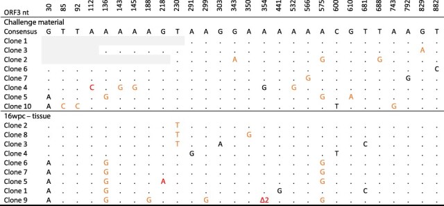
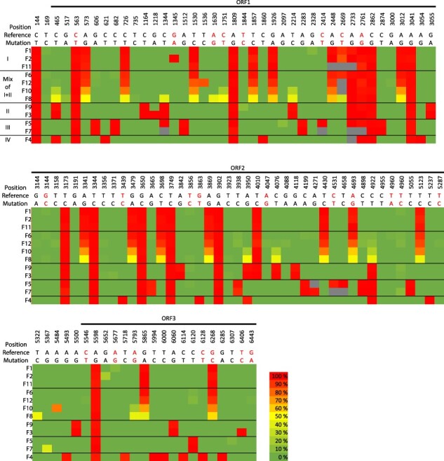
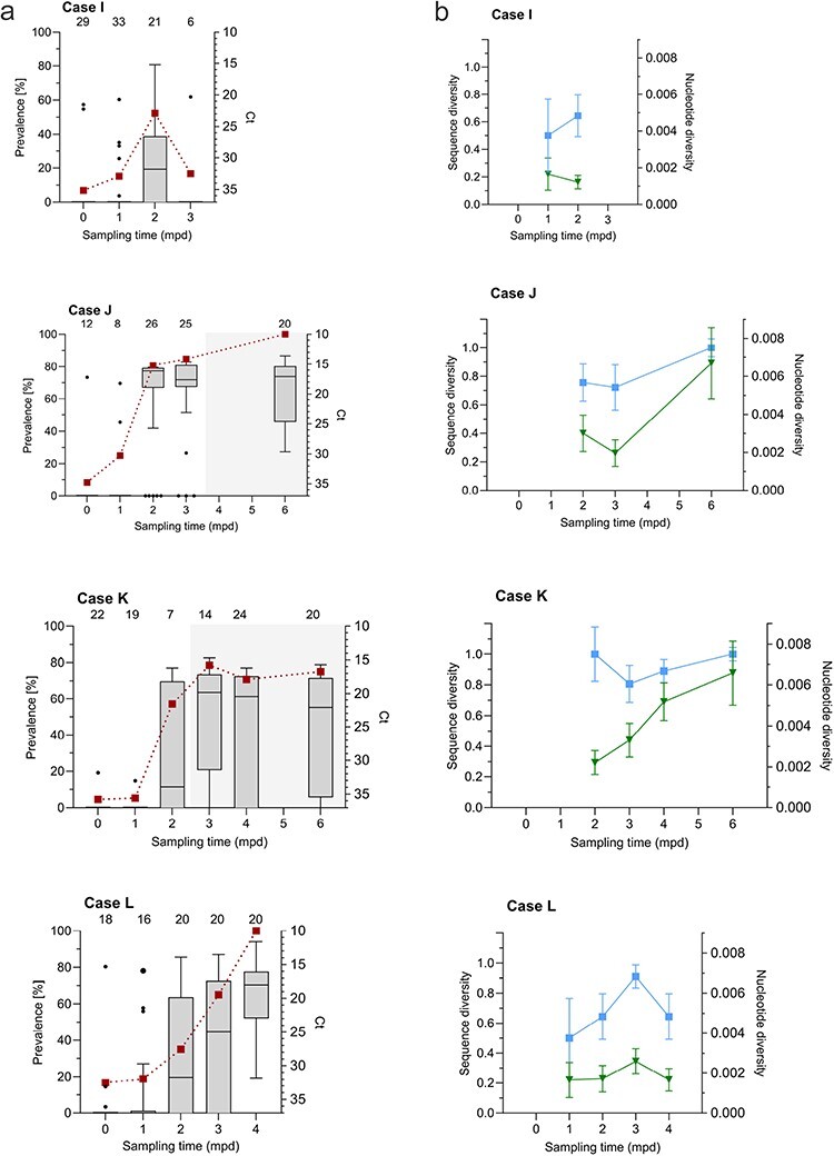
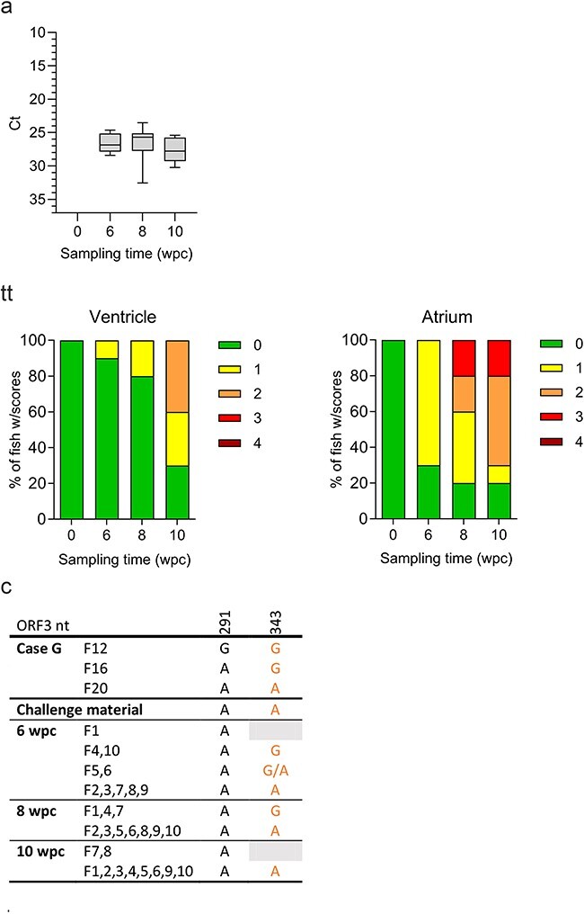
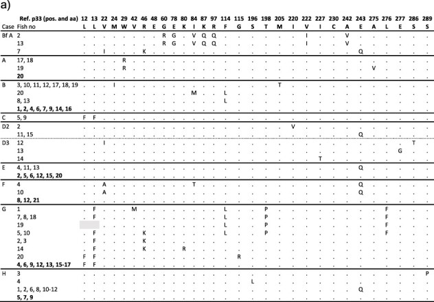
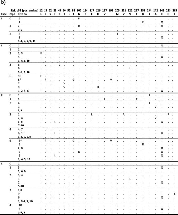
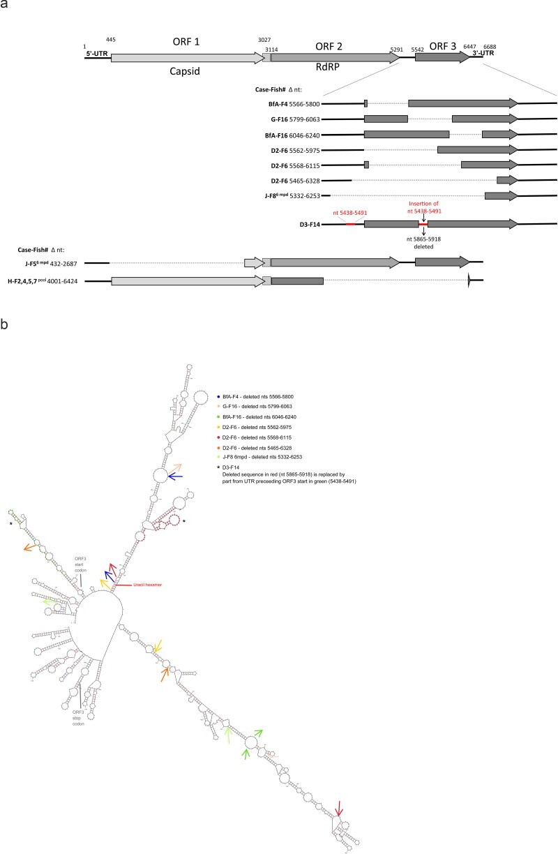
Over a decade since its discovery, piscine myocarditis virus (PMCV) remains a significant pathogen in Atlantic salmon aquaculture. Despite this significant impact, the genomic landscape, evolutionary dynamics, and virulence factors of PMCV are poorly understood. This study enhances the existing PMCV sequence dataset by adding 34 genome sequences and 202 new ORF3 sequences from clinical cardiomyopathy syndrome (CMS) cases in Norwegian aquaculture. Phylogenetic analyses, also including sequences from the Faroe Islands and Ireland revealed that PMCV sequences are highly conserved with distinct clustering by country of origin. Still, single CMS outbreaks display multiple PMCV variants, and although some clustering was seen by case origin, occasional grouping of sequences from different cases was also apparent. Temporal data from selected cases indicated increased sequence diversity in the population. We hypothesize that multiple bottlenecks and changing infection dynamics in the host population, with transfer to naïve individuals over time, represent a continuous selection pressure on the virus populations. No clear relation was found between PMCV variants and the severity of heart pathology. However, specific non-synonymous and synonymous mutations that might impact protein function and gene expression efficiency were identified. An additional factor that may impact PMCV replication is the presence of defective viral genomes, a novel finding for viruses of the order Ghabrivirales. This study provides new insights into PMCV genomic characteristics and evolutionary dynamics, highlighting the complex interplay of genetic diversity, virulence markers, and host-pathogen interactions, underscoring the epidemiological complexity of the virus. Keywords: piscine myocarditis virus; evolutionary dynamics; diversity; phylogeny; genomic sequencing; defective viral genomes.

Keywords: defective viral genomes; diversity; genomic sequencing; phylogeny; piscine myocarditis virus.


Conflict of interest statement

M.L. was affiliated by the diagnostic service company PatoGen AS, which offers PMCV screening, when the analyses were performed. F.M. is affiliated by the aquaculture production company Mowi ASA.

Figures

Figure 1.
Figure 1.
Map of Norway showing geographic location of the seawater Atlantic salmon aquaculture sites of cases included in the study. The cases (Cases A–L and Bf A) are color-coded by case origin and with year of sampling, as indicated.
Figure 2.
Figure 2.
Overview of the positions in the 34 PMCV genomes with diverging nucleotides identified by comparison to PMCV AL V-708 reference genome. Bar height represents the number of genomes with divergent nucleotides in each specific position. Red-colored bars indicate positions resulting in a non-synonymous change, leading to amino acid divergence. A few notable examples of single diverging nucleotides or combinations are marked by a–d. (a) G6268C in ORF3, resulting in E243Q in p33, is highly prevalent in the dataset and is consistently found concurrent with the synonymous C726T and A3012G in ORF1 and C3698T in ORF2 in the full-length genome dataset. (b) If the putative −1 ribosomal frameshift site at the 3ʹ end of ORF1 is translationally functional, the link1−2 is translated as a linker between the capsid and RdRp (Haugland et al. 2011, Sandlund et al. 2021), and this A3054G represents a non-synonymous mutation. (c) C563T in ORF1, resulting in an A40V substitution in the capsid, and the synonymous mutations G3650C and G3902A in ORF2 (all marked by C1) are examples of the 13 most prevalent mutations. However, a few variants concurrently lack these three mutations. Genomes lacking the C1 marked mutations include a unique combination of the synonymous A708G and A1392G in ORF1 (both marked by C2). (d) The synonymous mutations A3041G and A5598G are other examples of high-prevalence mutations. Still, four full-length genomes originating from three cases did not include these two mutations concurrently.
Figure 3.
Figure 3.
Phylogenetic analysis of 34 Norwegian and 10 Faroese PMCV complete and near-complete genome sequences from Atlantic salmon aquaculture sites experiencing CMS outbreaks or with suspected CMS. One near-complete genome sequence obtained from Faroese wild Atlantic salmon, sampled in 2012, and the Norwegian reference genome sequence AL V-708 from 2007 are also included. Norwegian sequences (Cases A–H) are color-coded by case origin and identified with the unique ID (“case” “two letter code for Norwegian county of origin” “year of sampling”-“fish number”’) followed by county of origin. Sequences from the Faroe Islands are all shown in light brown color and identified with the unique ID (FO/ “case number”-“fish number”/“year of sampling”). All bootstrap values 70 or above are shown in red color. The tree was rooted to the sequence from Faroese wild Atlantic salmon by default due to its high divergence towards the remaining 45 sequences. GenBank accession numbers are given in Supplementary Table S3.
Figure 4.
Figure 4.
Phylogenetic analysis of 251 complete or near complete PMCV ORF3 sequences, including all ORF3 sequences obtained in the present study and sequences publicly available from Norway, the Faroe Islands, and Ireland. The ORF3 sequences extracted from the full-genome sequences and additional ORF3 sequences obtained from Norwegian Cases A–H are color-coded and identified with unique IDs as in Fig. 3, with extra color coding for Cases I–L and Bf A. From the three time points available from Case D, near-complete sequences for phylogeny were only available from sampling points D2 and D3. For Cases I–L, only sequences from the latest time point of the time course are included. Previously published Norwegian sequences from 2009 to 2010 and sequences from the Faroe Islands and Ireland are given as indicated with a single color, independent of case origin. These Norwegian sequences are identified by GenBank accession number and unique ID (“case no.” “fish ID by lower case letter”- “letter code for Norwegian county of origin”/year of sampling). Sequences from cases of which only one individual is included are not identified using fish ID by lower-case letter. Faroese sequences are identified with the unique ID (FO/“case number”-“fish number”/“year of sampling”) and Irish samples by GenBank accession number and unique ID including info on case (e.g “IRE-F58” or “F-25”), followed by year of sampling and fish ID (e.g. “#13” or “1”). The Norwegian PMCV reference genome sequence AL V-708 from 2007 is highlighted in black. All sequences originate from farmed Atlantic salmon unless otherwise stated. The ORF3 sequences from wild salmon are highlighted with asterisks. All Norwegian sequences are also provided with information on the county of origin. For display purposes, the tree has been manually compressed by showing identical Norwegian ORF3 sequences originating from the same case in the present study together on the same branch. For the remaining sequences from Norway, identical sequences are shown on the same branch only if obtained from the same county, with the accession numbers followed by an ID in respective order. Identical sequences from the Faroe Islands are also shown on the same branch regardless of origin. All bootstrap values 70 or above are shown in red color with the value separating the two main groups in the tree highlighted by a larger font size. Brackets are used to group sequences encoding the IKR, IKQ, or VQQ motifs in residues 84, 87, and 97 of the ORF3-encoded protein p33 (see text for details). The tree has been rooted against the wild salmon PMCV ORF3 sequence from the Faroe Islands, also for display purposes.
Figure 5.
Figure 5.
Diverging nucleotides observed between ORF3 amplicon clones from a cell culture preparation of PMCV challenge material and ORF3 amplicon clones originating from heart tissue of one Atlantic salmon parr at 16  wpc, infected using the challenge material. For comparative presentation purposes a consensus of the most prevalent nucleotides found in the challenge material amplicon clones are given on top. Nucleotides (nts) in the clone sequences deviating from this consensus are indicated, with a dot representing no difference. Black nts—synonymous mutation, orange nts—non-synonymous mutation, red nts—mutation resulting in stop codon or codon frame shift, Δ2—deletion of two adenines within a stretch of six adenines in positions 349–354. Grey background—no information due to low-quality of sequencing chromatograms.
Figure 6.
Figure 6.
Intra-host single-nucleotide variants (iSNVs) with variant frequencies identified from PMCV sequence reads from RNA seq obtained from the 12 individuals of Case H. The nucleotide variants, and their positions in the genome with ORFs indicated, are shown relative to the PMCV reference AL V-708 reference sequence. Nucleotides colored in red indicate non-synonymous variants. Varying nucleotides in <2% of the reads are excluded. Frequencies of iSNVs are shown per individual as a colored heat map (green 0–2%, yellow 50%, red 100%). Grey color indicates no sequence information. Black lines separate the samples into four groups (I–IV) based on similarity of iSNVs. F6, F8, F10, and F12 are characterized by a mix of iSNVs described by groups I and II in varying frequencies.
Figure 7.
Figure 7.
Characteristics of PMCV infection progress and ORF3 diversity over time post first virus detection in field populations (Cases I–L). (a) Prevalence of fish positive for PMCV RNA of the total number tested (n = 6–33 as indicated on top of diagrams) following monthly real-time PCR screening of the study populations in Cases I–L from primary detection (0  mpd; red squares, left axis). PMCV RNA levels per individual are indicated by Ct values (right axis). Negative samples are included, all set to Ct 37 (the cut-off Ct defining a negative result). The Ct data are presented as box plots representing the interquartile range (middle 50% of the data) with a median line. Whiskers are drawn from minimum to maximum. Outliers are indicated by dots beyond either whisker. The larger dot symbol (Case L, 1 mpd) represents two samples differing by 0.5 Ct. Sampling time points are given as mpd. Onset and period including mortality in Cases J and K is indicated by grey shade in the diagram. (b) Sequence diversities (blue squares, left axis) and nucleotide diversities (π, green triangles, right axis) calculated for ORF3. Sequence diversity describes the uniqueness of a particular sequence in a given population, i.e. the probability that two sequences randomly sampled are different. Nucleotide diversity (π) represents the average number of pairwise nucleotide differences between two randomly chosen sequences per position. The two diversity measures are only calculated for time points where a minimum of four ORF3 sequences are available (n = 4–10). Sampling time points are given as mpd.
Figure 8.
Figure 8.
Characteristics of PMCV infection progress and diversity over time post-challenge in an experimental challenge (Ex B). (a) Levels of PMCV RNA indicated by Ct values for all sampling time points in the wpc in heart tissue. The Ct data are presented as box plots representing the interquartile range (middle 50% of the data) with a median line. Whiskers are drawn from minimum to maximum. (b) Distribution of histopathology scores of the atrium and ventricular cardiac compartments among the individuals for all sampling time points, with scoring from 0 (no lesions observed) to 4 (severe lesions). No fish with a score of 4 were found at any time point in any cardiac compartments. (c) Comparison of available PMCV ORF3 sequences from the field samples Case G F12, 16 and 20 pooled for the challenge material, the resulting challenge material, and from challenged fish from all time points with position and nucleotide present of the only two positions of a total of 909 in ORF3 with observed diversity. Two nucleotides separated by a slash indicate that they were both found in significant amounts among the reads from RNA seq. Black nts—synonymous mutation, orange nts—non-synonymous mutation, grey background—RNA seq data resulted in low or no coverage. RNA seq of the challenge material and all heart tissue samples produced partial genomic sequences covering more than ORF3.
Figure 9.
Figure 9.
PMCV p33 protein sequence variants from Cases A–L and broodfish Case Bf A. Amino acid substitutions in the PMCV ORF3-encoded p33 shown relative to the AL-V 708 reference p33 sequence with residue numbers and amino acid in reference shown on top. Fish individuals are identified by fish number and those with identical p33 sequences are shown on the same row. Fish individuals with a p33 sequence identical to the reference sequence are highlighted in bold. Grey background indicates that sequence information was not obtained. (a) Cases Bf A and A–H. (b) Cases I–L, including p33 sequences from each sampling time point (mpd). A highly divergent variant found in single fish from both Cases J and K (see Fig. 4, J Tr2019-10-F8 and K Tr2019-9-F6, uppermost part of tree) is marked by an asterisk.
Figure 9.
Figure 9.
continued
Figure 10.
Figure 10.
Deletions identified in PMCV RNA and their relation to predicted RNA secondary structure. (a) Schematic presentation of the deletions found in the PMCV genome from a selection of infected tissue samples from field cases. Deleted nucleotides (Δ nts) are indicated by positions relative to the 5ʹ end of the genome. One deletion-insertion event resulting in 54 nts of UTR2−3 replacing a deletion of equal size in ORF3 is also shown. (b) Predicted secondary structure of ORF3 RNA, including upstream UTR2−3 and downstream 3ʹUTR. Color-coded arrows indicate where each deletion is initiated (arrow facing out) and ends (arrow facing in). Hexamer of uracils where deletions are initiated are indicated by red letters. Nucleotides representing a likely deletion-insertion event are indicated in red (deleted) and green (inserted) shades.

Similar articles

Cited by

References

    1. Amato KA, Haddock LA 3rd, Braun KM. et al. Influenza A virus undergoes compartmentalized replication in vivo dominated by stochastic bottlenecks. Nat Commun 2022;13:3416. doi: 10.1038/s41467-022-31147-0 - DOI - PMC - PubMed
    1. Amin AB, Trasti J. Endomyocarditis in Atlantic salmon in Norwegian seafarms. Bull Eur Assoc Fish Pathol 1988;8:70–71.
    1. Bačnik K, Kutnjak D, Černi S. et al. Virome analysis of signal crayfish (Pacifastacus leniusculus) along its invasion range reveals diverse and divergent RNA Viruses. Viruses 2021;13:2259. doi: 10.3390/v13112259 - DOI - PMC - PubMed
    1. Bailey SF, Alonso Morales LA, Kassen R. Effects of synonymous mutations beyond codon bias: the evidence for adaptive synonymous substitutions from microbial evolution experiments. Genome Biol Evol 2021;13:evab141. doi: 10.1093/gbe/evab141 - DOI - PMC - PubMed
    1. Bancroft JD, Stephens A. Theory and Practice of Histological Techniques. London: Churchill Livingstone, 1990.

LinkOut - more resources