Review
. 2025 Mar;55(3):36.
doi: 10.3892/ijmm.2024.5477. Epub 2024 Dec 24.

Recent advances in nanomaterials for the detection of mycobacterium tuberculosis (Review)

Affiliations
Review

Recent advances in nanomaterials for the detection of mycobacterium tuberculosis (Review)

Jianmeng Zhu et al. Int J Mol Med. 2025 Mar.

Abstract

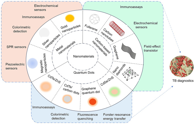
The world's leading infectious disease killer tuberculosis (TB) has >10 million new cases and ~1.5 million mortalities yearly. Effective TB control and management depends on accurate and timely diagnosis to improve treatment, curb transmission and reduce the burden on the medical system. Current clinical diagnostic methods for tuberculosis face the shortcomings of limited accuracy and sensitivity, time consumption and high cost of equipment and reagents. Nanomaterials have markedly enhanced the sensitivity, specificity and speed of TB detection in recent years, owing to their distinctive physical and chemical features. They offer several biomolecular binding sites, enabling the simultaneous identification of multiple TB biomarkers. Biosensors utilizing nanomaterials are often compact, user‑friendly and well‑suited for detecting TB on location and in settings with limited resources. The present review aimed to review the advances that have occurred during the last five years in the application of nanomaterials for TB diagnostics, focusing on their detection capabilities, structures, working principles and the significance of key nanomaterials. The current review addressed the limitations and challenges of nanomaterials‑based TB diagnostics, along with potential solutions.

Keywords: biosensors; diagnostics; nanomaterials; tuberculosis.


Conflict of interest statement

The authors declare that they have no competing interests.

Figures

Figure 1
Figure 1
Nanomaterial-based biosensing strategies for TB diagnosis. TB, tuberculosis; SPR, surface plasmon resonance.
Figure 2
Figure 2
AuNPs-based colorimetric detection of TB DNA Schematic diagram: Two PCR tubes were made using TB primers and PCR mix. The TB DNA template was placed in one tube and the other tube without the TB DNA template. After PCR, AuNPs and ethanol were added. The tube without the DNA template stayed red, while the tube with TB DNA turned purple. Reproduced from (32), Copyright (2023), with permission from Royal Society of Chemistry. AuNPs, gold nanoparticles; TB, tuberculosis.
Figure 3
Figure 3
Procedure of piezoelectric sensor based on AuNPs-mediated enzyme-assisted signal amplification. (A) SEM images of electrodes promoting the growth of AuNPs in HAuCl4 and NADH solutions containing target DNA and Exo III for (Ba) 0 min, (b) 10 min, (c) 30 min and (d) blank control. Reproduced from (37), Copyright (2022), with permission from Elsevier. AuNPs, gold nanoparticles.
Figure 4
Figure 4
Procedure for acpcPNA-induced AgNP aggregation. (A) AgNPs were initially well dispersed by the negatively charged electrostatic repulsion. Positively charged acpcPNA shielded them from electrostatic repulsion, causing silver particles to aggregate and a color reaction to occur. When complementary DNA was present, the specific PNA-DNA interaction replaced the PNA-AgNPs interaction, forming negatively charged PNA-DNA double strands that depolymerized the nanoparticles. In the case of non-complementary DNA, the nanoparticles did not depolymerize and no color change occurred. Reproduced from (54), Copyright (2017), with permission through Creative Commons public use license from Teengam P et al, American Chemical Society. (B) Schematic of CFP10-ESAT6 detection using the portable electrochemical reader. Sputum sample analysis was performed locally with the modified SPGE (circled in red) and a portable reader. Following GP/PANI modification of SPGE, the CapAb was immobilized on its surface to capture the target antigen and the Ab-loaded Fe3O4/Au particle bound to the target and amplified the detection signal. Reproduced from (41), Copyright (2021), with permission from Springer Nature. acpcPNA, PNA with a positively charged lysine modification at its C-terminus; AuNPs, gold nanoparticles; PNA, peptide nucleic acid; CFP10, culture filtrate antigen, 10 kDa; ESAT-6, early secreted antigenic target-6; SPGE, screen-printed gold electrode; GP/PANI, graphene/polyaniline; CapAb, capture antibody; MTB, Mycobacterium tuberculosis.
Figure 5
Figure 5
QD-based FRET system and colorimetric platform for TB diagnosis. (A) Schematic illustration of FRET-based MTB detection using QDs-DNA(fluorescence donor) and Cu-TCPP (fluorescence acceptor). Reproduced from (67), Copyright (2021), with permission from Elsevier. (B) Graphical representation of the QD-NB-based colorimetric platform for TB diagnosis. Reproduced from (61), Copyright (2023), with permission from American Chemical Society. FRET, fluorescence resonance energy transfer; MTB, Mycobacterium tuberculosis; QD, quantum dot; NB, nanobeacon; TB, tuberculosis.
Figure 6
Figure 6
QDs as fluorescent signal switches to detect MTB. Schematic for (A) vapor sample collection and (B) MTB methyl nicotinate detection based on CoTCPP nanosheets CdTe QDs and CoTCPP. Reproduced from (62), Copyright (2022), with permission from Springer. MTB, Mycobacterium tuberculosis; QD, quantum dot.
Figure 7
Figure 7
Schematic Illustration for preparing nanoCoTPyPs with different morphology and the simultaneous detection of rpoB531 and katG315 based on double QDs-ssDNA and nanoCoTPyP. The spherical nano-CoTPyP performed the best quenching and sensing properties. Reproduced from (63), Copyright (2022), with permission from American Chemical Society. nanoCoTPyP, nanocobalt 5,10,15,20-tetra(4-pyridyl)-21H,23H porphine; QD, quantum dot; ssDNA, single-stranded probe DNA.
Figure 8
Figure 8
Schematic diagram of rGO/PNE/Au nanocomposites synthesis and detection procedure for MTB DNA. Reproduced from (81), Copyright (2022), with permission from Elsevier. rGO/PNE MTB, Mycobacterium tuberculosis; rGO, reduced graphene oxide; PNE, polynorepinephrine.
Figure 9
Figure 9
Graphene functionalization for the TB biosensor. (A) Illustration of surface modification of graphene-based biosensor and the coupling process of MPT64 with 1,5-DAN and glutaraldehyde. AFM photos of graphene (B) before surface modification, (C) following 1,5-DAN treatment and (D) following MTP64 Ab conjugation. (E) Raman spectroscopy and (F) X-ray photoelectron spectroscopy characterized the surface of graphene. Reproduced with permission through Creative Commons Attribution License (CC BY) from Seo G et al (82), Frontiers in Bioengineering and Biotechnology; published by Frontiers, 2023. TB, tuberculosis; MPT64, MTB 64 protein; 1,5-DAN, 1,5-diaminonaphthalene.
Figure 10
Figure 10
SWCNT-based FET device and FND-based immunosensor for MTB detection. (A) Schematic representation of SWCNTs modified by EDC/NHS and connected to Ab85B. Reproduced with permission through Creative Commons Attribution 4.0 International License from Wang J et al (79), ACS Sensors; published by American Chemical Society, 2024. (B) Schematic representation of ESAT6 (MTB critical virulence factor) detection by competitive spin-enhanced lateral flow immunoassay. Magnetically modulated fluorescence allows background-free detection. ESAT6 in the sample and test strip compete for the few Ab binding sites on the FND to accomplish competitive detection. Movement of the strip is indicated by a black arrow. Reproduced from (80), Copyright (2024), with permission through Creative Commons Attribution-NonCommercial 3.0 Unported Licence, Royal Society of Chemistry. SWCNTs, single-walled carbon nanotubes; EDC/NHS, 1-Ethyl-3-(3-dimethylaminopropyl)carbodiimide/N-Hydroxysuccinimide; ESAT6, early secreted antigenic target-6; FNDs, fluorescent nanodiamonds; TB, tuberculosis.

Similar articles

Cited by

References

    1. Bagcchi S. WHO's global tuberculosis report 2022. Lancet Microbe. 2023;4:e20. doi: 10.1016/S2666-5247(22)00359-7. - DOI - PubMed
    1. Asadi L, Croxen M, Heffernan C, Dhillon M, Paulsen C, Egedahl ML, Tyrrell G, Doroshenko A, Long R. How much do smear-negative patients really contribute to tuberculosis transmissions? Re-examining an old question with new tools. EClinicalMedicine. 2022;43:101250. doi: 10.1016/j.eclinm.2021.101250. - DOI - PMC - PubMed
    1. Meriki HD, Wung NH, Tufon KA, Tony NJ, Ane-Anyangwe I, Cho-Ngwa F. Evaluation of the performance of an in-house duplex PCR assay targeting the IS6110 and rpoB genes for tuberculosis diagnosis in Cameroon. BMC Infect Dis. 2020;20:791. doi: 10.1186/s12879-020-05523-4. - DOI - PMC - PubMed
    1. Natarajan S, Ranganathan M, Hanna LE, Tripathy S. Transcriptional profiling and deriving a seven-gene signature that discriminates active and latent tuberculosis: An integrative bioinformatics approach. Genes (Basel) 2022;13:616. doi: 10.3390/genes13040616. - DOI - PMC - PubMed
    1. Molloy A, Harrison J, McGrath JS, Owen Z, Smith C, Liu X, Li X, Cox JAG. Microfluidics as a novel technique for tuberculosis: From diagnostics to drug discovery. Microorganisms. 2021;9:2330. doi: 10.3390/microorganisms9112330. - DOI - PMC - PubMed

MeSH terms

LinkOut - more resources