. 2024 Dec 24:12:RP89638.
doi: 10.7554/eLife.89638.

Improving PD-1 blockade plus chemotherapy for complete remission of lung cancer by nanoPDLIM2

Affiliations

Improving PD-1 blockade plus chemotherapy for complete remission of lung cancer by nanoPDLIM2

Fan Sun et al. Elife. .

Abstract

Immune checkpoint inhibitors (ICIs) and their combination with other therapies such as chemotherapy, fail in most cancer patients. We previously identified the PDZ-LIM domain-containing protein 2 (PDLIM2) as a bona fide tumor suppressor that is repressed in lung cancer to drive cancer and its chemo and immunotherapy resistance, suggesting a new target for lung cancer therapy improvement. In this study, human clinical samples and data were used to investigate PDLIM2 genetic and epigenetic changes in lung cancer. Using an endogenous mouse lung cancer model faithfully recapitulating refractory human lung cancer and a clinically feasible nano-delivery system, we investigated the therapeutic efficacy, action mechanism, and safety of systemically administrated PDLIM2 expression plasmids encapsulated in nanoparticles (nanoPDLIM2) and its combination with PD-1 antibody and chemotherapeutic drugs. Our analysis indicate that PDLIM2 repression in human lung cancer involves both genetic deletion and epigenetic alteration. NanoPDLIM2 showed low toxicity, high tumor specificity, antitumor activity, and greatly improved the efficacy of anti-PD-1 and chemotherapeutic drugs, with complete tumor remission in most mice and substantial tumor reduction in the remaining mice by their triple combination. Mechanistically, nanoPDLIM2 increased major histocompatibility complex class I (MHC-I) expression, suppressed multi-drug resistance 1 (MDR1) induction and survival genes and other tumor-related genes expression in tumor cells, and enhanced lymphocyte tumor infiltration, turning the cold tumors hot and sensitive to ICIs and rendering them vulnerable to chemotherapeutic drugs and activated tumor-infiltrating lymphocytes (TILs) including those unleashed by ICIs. These studies established a clinically applicable PDLIM2-based combination therapy with great efficacy for lung cancer and possibly other cold cancers.

Keywords: PD-1; PDLIM2; cancer biology; combination therapy; human; immune checkpoint; lung cancer; mouse; nanotherapy.

Plain language summary

Lung cancer remains the leading cause of all cancer-related deaths. Treatment options are limited because drug-based therapies including chemotherapy and immune checkpoint inhibitors (or ICIs, for short) are ineffective in most patients. PDLIM2 is a protein that normally prevents tumors from forming by regulating the activities of other genes. However, lung cancer cells generally have lower levels of this protein than healthy cells and this appears to be linked to the ability of the cancer cells to become resistant to chemotherapy and ICIs. Cells make proteins using templates encoded in our DNA. It remains unclear how PDLIM2 production is repressed in lung cancer: it is possible that cancer cells may acquire genetic alterations that affect PDLIM2 production, or there may be other changes to the structure of the DNA known as epigenetic changes. Sun et al. investigated the production of PDLIM2 in samples from human lung cancer patients. The experiments found that in over 90% of the patients, the levels of PDLIM2 were lower than in cells from healthy individuals. This was due to genetic alterations or epigenetic changes, or a combination of the two. Further experiments in a mouse model of lung cancer demonstrated that it is possible to use nanotechnology to deliver PDLIM2 to cancer cells for effective cancer therapy with low toxicity. Combining this nanotechnology (known as nanoPDLIM2) with both ICIs and chemotherapy drugs was able to completely eradicate all tumors in most of the mice. The findings provide a firm basis for further studies of the potential of nanoPDLIM2 as a safe and effective therapy for human lung cancer. PDLIM2 production is also repressed in numerous other types of cancer, so it is possible that nanoPDLIM2 may have broader uses in cancer treatment.


Conflict of interest statement

FS, PY, YX, HZ, SS, GX, ZQ No competing interests declared

Figures

Figure 1.
Figure 1.. PDLIM2 repression in human lung cancer involves both epigenetic alteration and genetic deletion, and Pdlim2 genetic deletion in mice leads to development of spontaneous tumors, majorly lung tumors.
(A) TCGA data showing PDLIM2 repression in over 90% of lung tumors if using 50% of the expression level in normal lung tissues as the cut-off (NL, n=110; Tumor, n=1019). (B) TCGA data showing PDLIM2 promoter hypermethylation and expression repression (dashed box) in over 70% of lung tumors when using 125% of the methylation level in normal lung tissues as the cut-off (n=827). (C) TCGA data showing positive associations between PDLIM2 expression and its gene copy numbers as well as PDLIM2 genetic deletion and expression repression (dashed box) in about 58% of lung tumors using the copy number variation of –0.1 as the cut-off (n=1010). (D) TCGA data showing simultaneous promoter hypermethylation and genomic deletion of PDLIM2 (dashed box) in about 44% of lung tumors (n=816). (E) Microsatellite-PCR showing PDLIM2 loss in human lung tumors (n=21). (F) qPCR showing PDLIM2 loss in human lung cancer cell lines with known copy number of the PDLIM2 gene (n=25). (G) Kaplan-Meier tumor-free survival curve showing increased spontaneous tumors in Pdlim2-/- and Pdlim2+/- mice compared to WT mice. Gehan-Breslow-Wilcoxon test was performed. *<0.05. (H) Percentage of tumor types spontaneously developed in Pdlim2-/- and Pdlim2+/- mice showing a majority of lung tumors.
Figure 1—figure supplement 1.
Figure 1—figure supplement 1.. Mice with PDLIM2 deletion develop spontaneous tumors, majorly lung tumors.
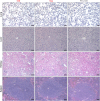
(A) IHC assays showing decreased and complete loss of PDLIM2 protein expression in the lungs of PDLIM2+/- and PDLIM2-/- mice, respectively. Scale bar, 20 µm. (B) H&E staining of the lung tissues showing spontaneous lung tumors in PDLIM2+/- and PDLIM2-/- mice. Scale bar, 20 µm.
Figure 2.
Figure 2.. Systemic administration of PDLIM2 plasmid nanoparticles shows efficacy in mouse model of refractory lung cancer.
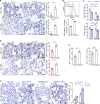
(A) Schedule of lung cancer induction and treatment. (B) Urethane model showing efficacy of intravenous administration of PDLIM2-expression plasmid nanoparticles for refractory lung cancer (n≥6). Nanoparticles with an empty vector plasmid (Vec) that was employed to express PDLIM2 were used as a control. (C) IHC staining showing decreased nuclear expression of STAT3 and RelA in lung tumors by PDLIM2 nanotherapy (n=6). (D) IHC staining showing decreased Bcl-xL and increased apoptosis marker cleaved caspase –3 in lung tumors by PDLIM2 nanotherapy (n=6). (E) IHC staining showing decreased Cyclin D1 and proliferation (BrdU incorporation) in lung tumors by PDLIM2 nanotherapy (n=6). Scale bar in (C–E), 20 μm. Student’s t test was performed (two tailed, unpaired) and data represent means ± SEM in (B–E). **p<0.01; ns, not statistically significant.
Figure 3.
Figure 3.. PDLIM2 nanotherapy shows high tumor specificity and low toxicity.
(A) IHC staining showing high PDLIM2 re-expression in lung tumors after PDLIM2 nanotherapy. (B) PCR and IB assays showing lung tumor-specific plasmids delivery and PDLIM2 expression by PDLIM2 nanotherapy (n=3). (C) IHC staining showing comparable expression of PDLIM2 in the indicated tissues of mice treated with PDLIM2 expression plasmid or empty vector plasmid nanoparticles. (D) No significant changes in animal body weight by nanoPDLIM2 (n=5). (E) H&E staining showing no noticeable changes in major organs by nanoPDLIM2. Scale bar: (A and C) 20 μm; (E) 50 μm.
Figure 4.
Figure 4.. PDLIM2 nanotherapy renders lung cancers more vulnerable to chemotherapy.
(A) Urethane model showing synergy of PDLIM2 nanotherapy and chemotherapy in lung cancer treatment (n≥5). (B) IHC staining showing increased lung tumor cell apoptosis by PDLIM2 nanotherapy, chemotherapy, and further increase by their combination (n=6). (C) IHC staining showing RelA activation by chemotherapy and blockage of chemo activation of RelA by PDLIM2 nanotherapy (n=6). (D) IHC staining showing strong MDR1 induction by chemotherapy and blockage of MDR1 induction by PDLIM2 nanotherapy (n=6). Scale bar in (B–D), 20 μm. Student’s t test was performed (two tailed, unpaired) and data represent means ± SEM. *p<0.05; **p<0.01; ns, not statistically significant.
Figure 5.
Figure 5.. PDLIM2 nanotherapy increases the efficacy of PD-1 blockade immunotherapy for refractory lung cancer.
(A) IHC staining showing increased TILs in lung tumors by PDLIM2 nanotherapy (n=6). (B) FACS showing increased MHC-I expression in lung tumor cells by PDLIM2 nanotherapy (n=4). (C) Urethane model showing PDLIM2 nanotherapy enhancing PD-1 immunotherapy efficacy in lung cancer treatment (n≥5). (D) IHC staining showing increased TILs by PDLIM2 nanotherapy in the context of immunotherapy (n=6). (E) FACS showing increased activation of CD8+ T cells by PDLIM2 nanotherapy in the context of immunotherapy (n=3). (F) IHC staining showing increased lung tumor cell apoptosis by PDLIM2 nanotherapy, immunotherapy, and further increase by their combination (n=6). Scale bar in (A, D, F), 20 μm. Student’s t test was performed (two tailed, unpaired) and data represent means ± SEM. **p<0.01; ns, not statistically significant.
Figure 6.
Figure 6.. Combination of PDLIM2 nanotherapy, chemotherapy and immunotherapy shows great efficacy in lung cancer treatment.
(A) IHC staining showing PD-L1 induction by chemotherapy but not PDLIM2 nanotherapy. (B) Schedule of lung cancer induction and treatment. (C) Urethane model showing high resistance of lung tumors to the chemo and αPD-1 combination therapy in lung epithelial specific PDLIM2 deletion mice (ΔSPC) (n≥4). (D) IHC staining showing no MHC-I induction by chemotherapy, PD-1 immunotherapy or their combination (n=6). (E) Tumor examination showing complete remission of all lung tumors in 60% of mice by combination of the three therapies (n≥5). (F) IHC staining showing increased TILs by PDLIM2 nanotherapy in mice treated with anti-PD-1 and chemotherapeutic drugs (n=6). (G) FACS analysis showing increased lung CD8+ T-cell activation by PDLIM2 nanotherapy in mice treated with anti-PD-1 and chemotherapeutic drugs (n=5). (H) No significant effect of PDLIM2 nanotherapy on the body weight of mice treated with anti-PD-1 and chemotherapeutic drugs (n=5). Scale bar in (A and F), 20 μm. Student’s t test was performed (two tailed, unpaired) and data represent means ± SEM in (c–g). *p<0.05; **p<0.01; ns, not statistically significant.
Figure 6—figure supplement 1.
Figure 6—figure supplement 1.. PDLIM2 nanotherapy causes no obvious toxicity in major organs.
(H & E) staining showing comparable toxicity in lung, liver, kidney, and spleen between Vec and PDLIM2 group in the context of combinational chemotherapy and PD-1 blockade immunotherapy (n=5). Scale bar: 50 µm.
Figure 6—figure supplement 2.
Figure 6—figure supplement 2.. Epigenetic drugs cause body weight loss in mice with lung cancer.
Mice were i.p. injected with urethane (1 g/kg) for 6 weeks to induce lung tumors, and then treated for 6 weeks with 5-aza-dC and MS-275 (Epi, i.p., 1 mg/kg each, twice per week), carboplatin and paclitaxel (Chemo, i.p., 30 mg/kg and 15 mg/kg, respectively, once per week), or their combinations. (A) Body weight change between prior to the first treatment and the sacrifice endpoint. (B) Percentage of mice with peritoneal effusion (n ≥ 4). Excel file for the data shown in Figure 6—figure supplement 3.
Figure 6—figure supplement 3.
Figure 6—figure supplement 3.. Epigenetic drugs show better efficacy in lung cancer treatment.
Mice were i.p. injected with urethane (1 g/kg) for 6 weeks to induce lung tumors, and then treated with 5-aza-dC and MS-275 (Epi, i.p., 1 mg/kg each, twice per week), carboplatin and paclitaxel (Chemo, i.p., 30 mg/kg and 15 mg/kg, respectively, once per week), anti-PD-1 antibody (aPD-1, i.p., 200 µg/mouse, three times per week), PDLIM2-expression plasmids containing nanoparticles (i.v., 25 µg plasmid/mouse, once per week), or their combinations for 6 weeks before they were sacrificed for tumor examination (n≥4).

Update of

Similar articles

References

    1. Baxevanos P, Mountzios G. Novel chemotherapy regimens for advanced lung cancer: have we reached a plateau? Annals of Translational Medicine. 2018;6:139. doi: 10.21037/atm.2018.04.04. - DOI - PMC - PubMed
    1. Bonnet ME, Erbacher P, Bolcato-Bellemin AL. Systemic delivery of DNA or siRNA mediated by linear polyethylenimine (L-PEI) does not induce an inflammatory response. Pharmaceutical Research. 2008;25:2972–2982. doi: 10.1007/s11095-008-9693-1. - DOI - PubMed
    1. Buscail L, Bournet B, Vernejoul F, Cambois G, Lulka H, Hanoun N, Dufresne M, Meulle A, Vignolle-Vidoni A, Ligat L, Saint-Laurent N, Pont F, Dejean S, Gayral M, Martins F, Torrisani J, Barbey O, Gross F, Guimbaud R, Otal P, Lopez F, Tiraby G, Cordelier P. First-in-man phase 1 clinical trial of gene therapy for advanced pancreatic cancer: safety, biodistribution, and preliminary clinical findings. Molecular Therapy. 2015;23:779–789. doi: 10.1038/mt.2015.1. - DOI - PMC - PubMed
    1. Commisso C, Davidson SM, Soydaner-Azeloglu RG, Parker SJ, Kamphorst JJ, Hackett S, Grabocka E, Nofal M, Drebin JA, Thompson CB, Rabinowitz JD, Metallo CM, Vander Heiden MG, Bar-Sagi D. Macropinocytosis of protein is an amino acid supply route in Ras-transformed cells. Nature. 2013;497:633–637. doi: 10.1038/nature12138. - DOI - PMC - PubMed
    1. Doroshow DB, Sanmamed MF, Hastings K, Politi K, Rimm DL, Chen L, Melero I, Schalper KA, Herbst RS. Immunotherapy in non-small cell lung cancer: facts and hopes. Clinical Cancer Research. 2019;25:4592–4602. doi: 10.1158/1078-0432.CCR-18-1538. - DOI - PMC - PubMed

MeSH terms