. 2025 Jan 14;97(1):86-95.
doi: 10.1021/acs.analchem.4c02421. Epub 2024 Dec 24.

Microarray-Based Methodology for Lipid Profiling, Enzymatic Activity, And Binding Assays in Printed Lipid Raft Membranes from Astrocytes and Neurons

Affiliations

Microarray-Based Methodology for Lipid Profiling, Enzymatic Activity, And Binding Assays in Printed Lipid Raft Membranes from Astrocytes and Neurons

Laura Sánchez-Sánchez et al. Anal Chem. .

Abstract

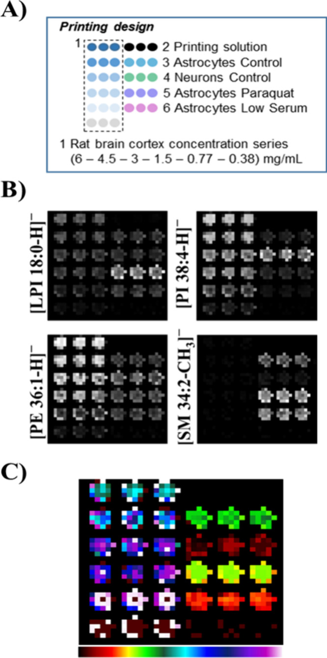
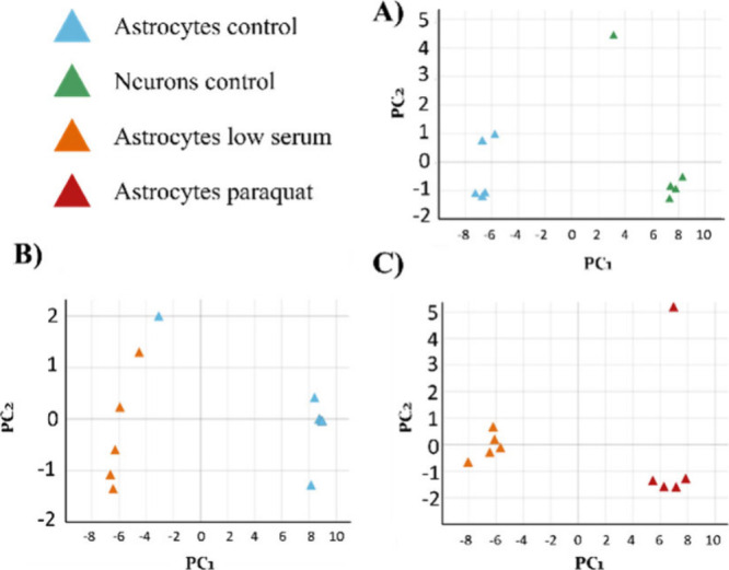
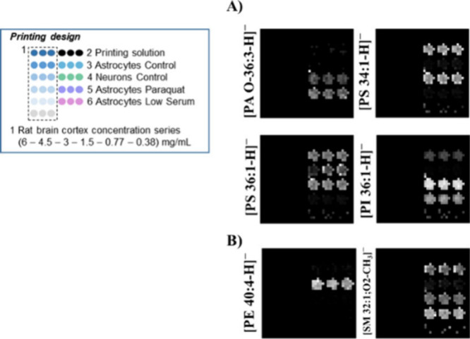
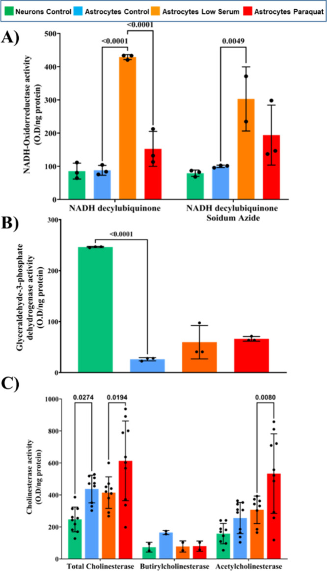
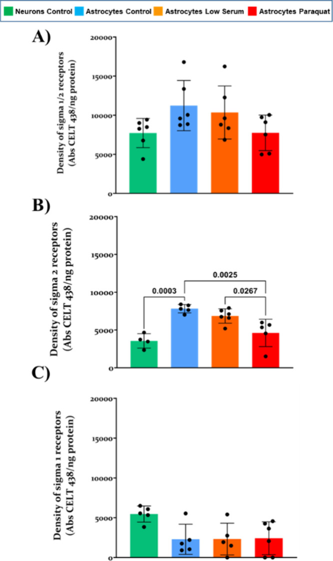
Lipid rafts are liquid-ordered domains in which specific enzymes and receptors are located. These membrane platforms play crucial roles in a variety of signaling pathways. Alterations in the lipid environment, such as those elicited by oxidative stress, can lead to important functional disruptions in membrane proteins. Cell membrane microarrays have emerged in the past decade as a powerful methodology for the study of both lipids and membrane proteins at large scales. Based on that technology and the importance of liquid-ordered subdomains, we have developed a new printed lipid raft technology with a preserved native protein structure and lipid environment. To validate this technology and evaluate its potential for different aims, raft membrane microarrays (RMMAs) containing two different cell types (astrocytes and neurons) and three different conditions (astrocytes in control situation, metabolic stress, and oxidative stress) were developed. To study differences in lipid profiles between raft domains, the MALDI-MS assay was performed on RMMAs. To evaluate the preservation of native protein activities (enzymatic activity and ligand binding) in the printed raft domains, differences in NADH oxidoreductase, GAPDH, cholinesterase activities, and sigma-1 and sigma-2 binding assays were performed. We demonstrate the performance of this new microarray technology, adapted to membrane subdomains, as valid to explore changes in lipid composition and protein activities in raft domains from brain cell lines under different stress conditions relevant for neuropathology.


Conflict of interest statement

The authors declare no competing financial interest.

Figures

Figure 1
Figure 1
Western blot assay with markers for both raft and nonraft membrane subdomains. Example for raft preparation of 1321N1 cells in a control situation. Note that part of the strong ApoD signal was still present after stripping and is visible above the Cav1 signal.
Figure 2
Figure 2
Lipid fingerprint analysis in RMMAs from astrocytic and neuronal cell lines using mass spectrometry in MS–. (A) Printing design for microarrays. Standard curves and samples were printed in triplicate. (B) Example images showing the relative abundance of selected lipids in raft samples or standards using gray scale. (C) Segmentation analysis using our modified RankCompete algorithm. Colors were assigned by using the rainbow color scale.
Figure 3
Figure 3
Principal component analysis (PCA) of lipid raft samples from neuronal and astrocytic cell lines under different conditions using MS– data. (A) PCA of astrocytic and neuronal rafts in control situations (92% variability explained by the first 2 principal components PC1 and PC2). (B) PCA of astrocytic rafts in metabolic stress and control situation (90% variance explained by 4 principal components PC1–PC4). (C) PCA of astrocytic rafts in metabolic and oxidative stress (98% variability explained by PC1 and PC2).
Figure 4
Figure 4
Relative abundance of selected example lipids in printed raft samples using the gray scale. Shown on the left is the design for the standard curve and samples, printed in triplicate. (A) Relative abundance of selected lipids that presented differences due to the treatment. (B) Relative abundance of selected lipids that presented differences between both cell lines in the control situation. No signal over the set threshold (0.1% of higher peak relative intensity) was detected in the rat cortex controls for these lipids.
Figure 5
Figure 5
Enzymatic activity of selected enzymes in printed raft domains. (A) NADH oxidoreductase enzymatic activity in the presence or absence of a selective mitochondrial complex IV inhibitor. (B) Glyceraldehyde-3-phosphate dehydrogenase activity. (C) Total cholinesterase, butyrilcholinesterase, and acetylcholinesterase activity assay. Two-way ANOVA two-tailed test was performed in panels A and C. One-way ANOVA two-tailed test was used in panel B. In all cases, α was set at 0.05. Obtained p-values are shown in graphs. Data are expressed as mean ± SD.
Figure 6
Figure 6
Fluorescent ligand binding assay for sigma-1 and sigma-2 receptors in printed lipid raft domains in different conditions. (A) Density of active total sigma-1 and sigma-2 receptors. (B) Density of sigma-2 receptors using a sigma-1 masking agent. (C) Density of sigma-1 receptors calculated as de difference between panels A and B. One-way ANOVA two-tailed wtest ith Tukey’s posthoc was carried out with α set at 0.05, obtained p-values are shown in graphs. Data are expressed as mean ± SD.

Similar articles

References

    1. Lu S. M.; Fairn G. D. Mesoscale Organization of Domains in the Plasma Membrane - beyond the Lipid Raft. Crit Rev. Biochem Mol. Biol. 2018, 53 (2), 192–207. 10.1080/10409238.2018.1436515. - DOI - PubMed
    1. Kusumi A.; Fujiwara T. K.; Tsunoyama T. A.; Kasai R. S.; Liu A.-A.; Hirosawa K. M.; Kinoshita M.; Matsumori N.; Komura N.; Ando H.; Suzuki K. G. N. Defining Raft Domains in the Plasma Membrane. Traffic 2020, 21 (1), 106–137. 10.1111/tra.12718. - DOI - PubMed
    1. George K. S.; Wu S. Lipid Raft: A Floating Island of Death or Survival. Toxicol. Appl. Pharmacol. 2012, 259 (3), 311–319. 10.1016/j.taap.2012.01.007. - DOI - PMC - PubMed
    1. Simons K.; Ikonen E. Functional Rafts in Cell Membranes. Nature 1997, 387 (6633), 569–572. 10.1038/42408. - DOI - PubMed
    1. Levental I.; Grzybek M.; Simons K. Greasing Their Way: Lipid Modifications Determine Protein Association with Membrane Rafts. Biochemistry 2010, 49 (30), 6305–6316. 10.1021/bi100882y. - DOI - PubMed

MeSH terms