. 2025 Mar;66(3):929-941.
doi: 10.1111/epi.18239. Epub 2024 Dec 24.

Preferential superficial cortical layer activation during seizure propagation

Affiliations

Preferential superficial cortical layer activation during seizure propagation

Anastasia Brodovskaya et al. Epilepsia. 2025 Mar.

Abstract

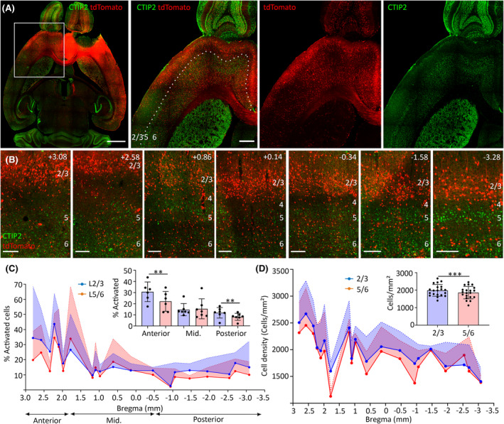
Objective: Focal cortical seizures travel long distances from the onset zone, but the long-distance propagation pathways are uncertain. In vitro and in vivo imaging techniques have investigated the local spread of seizures but did not elucidate long-distance spread. Furthermore, classical studies in slices suggested seizure spread locally along deep cortical layers, whereas more recent in vivo imaging studies posit a role for superficial cortical layers in local spread.

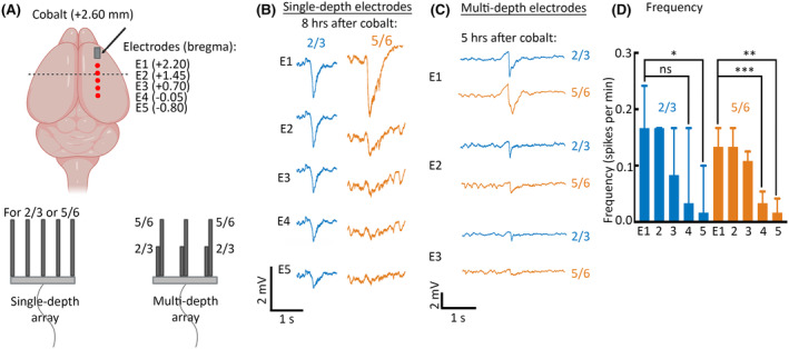
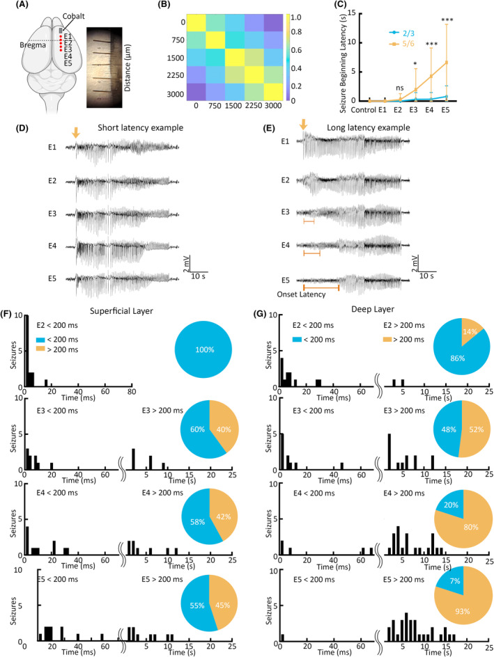
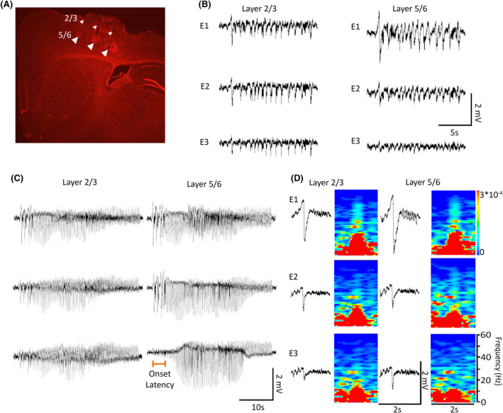
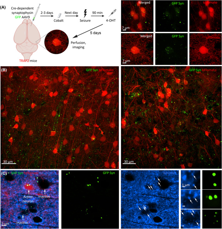
Methods: We imaged seizure-activated neurons using activity reporter mice and measured local field potentials (LFPs) using microelectrode arrays to map cortical seizure propagation in awake mice.

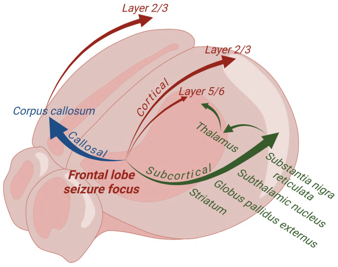
Results: Frontal lobe onset seizures activate more neurons in superficial layers 2-3 than deep layers 5-6 throughout the cortex. LFP recordings demonstrate that seizures spread faster through the superficial than deep layers over long cortical distances of 3.5 mm. We also show that monosynaptically connected long-distance neurons are in the seizure circuit.

Significance: We propose that long-distance cortical seizure spread occurs preferentially via synaptically connected superficial cortical neurons.

Keywords: Layer 2/3; Layer 5/6; deep layers; epilepsy; excitatory synaptic connectivity; local field potentials; seizures; superficial layers.


Conflict of interest statement

J.K. is a consultant for Ovid therapeutics and Marinus Pharmaceuticals. There are no other conflict of interest. We confirm that we have read the Journal's position on issues involved in ethical publication and affirm that this report is consistent with those guidelines.

Figures

FIGURE 1
FIGURE 1
Frontal lobe seizures activated layers 2 and 3 (L2/3) stronger than layers 5 and 6 (L5/6) from the anterior to posterior cortex. (A) tdTomato expression (red) in the horizontal slice immunolabeled for CTIP2 (L5/6, green). Scale bars = 1500, 100 μm. (B) Superficial L2/3 expressed more tdTomato than did deep L5/6 during a seizure from the anterior to posterior cortex. The white number at the top right indicates bregma. Scale bars = 150 μm. (C) Superficial L2/3 (blue) were more activated than deep L5/6 (pink) during a seizure in the anterior and posterior cortex contralateral to the seizure focus (shaded regions indicate error bars, each point represents average for that bregma region across mice). (D) Neuronal cell density (cells/mm2) in L2/3 (blue) versus L5/6 (pink) from anterior to posterior contralateral cortex (each point represents average for that bregma region across mice). Data are mean ± SEM. **p < .01, ***p < .001.
FIGURE 2
FIGURE 2
(A) Schematic of local field potential microelectrode placement (red dots) behind cobalt (gray rectangle). In one group of mice, E1–E5 electrodes targeted superficial layers (200 μm below the dura) and in another group deep layers (700 μm below the dura); electrode schematic is on the bottom left. The image on the bottom right depicts the custom‐made array of microelectrodes for simultaneous layers 2 and 3 (L2/3) + layers 5 and 6 (L5/6) recordings. (B) Interictal spikes propagate further away from the seizure focus through L2/3 but not through L5/6. These recordings were done with single‐depth microelectrode arrays recorded from either L2/3 or L5/6, 8 h after cobalt implantation. (C) Simultaneous multidepth array recordings (from L2/3 and L5/6 in the same mouse) 5 h after cobalt implantation, showing that interictal spikes propagate further away from the seizure focus through L2/3 but not through L5/6. (D) Interictal spike frequency (spikes per minute [60 s], median ± interquartile range, 8 h after cobalt placement) at E1–E5 from superficial (blue) and deep layers (orange). ns, nonsignificant. *p < .05, **p < .01, ***p < .001.
FIGURE 3
FIGURE 3
Long‐distance seizure spread is faster through superficial layers and slower through deep layers during frontal lobe onset seizures. (A) Schematic of local field potential microelectrode placement (red dots) behind cobalt (gray rectangle). (B) A correlation matrix plots the correlation coefficients (from 1.0 to 0) for each pair of microelectrodes. E1 corresponds to 0 μm away from cobalt, and E5 corresponds to 3000 μm away from cobalt. (C) Seizure onset latency (in seconds) in E1–E5 in layers 2–3 (blue) and 5–6 (orange) and one contralateral electrode across from E1 (control). Plot is a fit of the 3rd degree polynomial equation. (D, E) Examples of short‐latency seizures with immediate seizure onset in the posterior right cortex (E5) and long‐latency seizures with onset delay in E5 with the corresponding power spectrums. (F, G) Seizure onset times (in milliseconds or seconds) in superficial (F) and deep layers (G) from the anterior right premotor cortex (E1) to the posterior right parietal cortex (E5). Pie charts indicate percentage of seizures that arrived at the corresponding electrodes in <200 ms (blue) or >200 ms (orange). Data are mean ± SD. ns, nonsignificant. *p < .05, ***p < .001.
FIGURE 4
FIGURE 4
Double depth simultaneous local field potential (LFP) recordings confirm a faster spread of seizures through layers 2 and 3 (L2/3) and slower through layers 5 and 6 (L5/6). (A) Lesions done after the recordings mark the locations of the microelectrode tips that targeted L2/3 and L5/6 (small and big white arrows). (B) Representative simultaneous LFP recording from L2/3 and L5/6. (C) Representative simultaneous LFP recording from L2/3 and L5/6 during focal to bilateral seizure with the corresponding power spectrums. The orange bar for the posterior E3 electrode in L5/6 indicates seizure onset delay. (D) Spike–wave discharges from L2/3 and L5/6 with gamma oscillation bursts and power spectrums.
FIGURE 5
FIGURE 5
Seizure spread is mediated by excitatory synaptic connectivity. (A) Cre‐dependent green fluorescent protein (GFP) synaptophysin (Syn) adeno‐associated virus serotype 9 (AAV9) virus was injected at the seizure focus in TRAP2 mice that express Cre in the activated neurons. Cobalt implantation caused frontal lobe focal to bilateral tonic–clonic seizures the next day. The injection of 4‐hydroxytamoxifen (4‐OHT) within 90 min of a seizure caused GFP expression in the activated synapses and tdTomato expression in the activated neurons. (B) GFP AAV9 synaptophysin expression in the activated synapses (green) in the right posterior superficial cortex of TRAP2 mice after focal to bilateral seizure. (C) Synaptophysin (green) colocalized with presynaptic marker bassoon (blue) in activated tdTomato‐positive neurons (red).
FIGURE 6
FIGURE 6
Circuit map of frontal lobe focal to bilateral tonic–clonic seizures. (1) Subcortical seizure spread (green) is through the striatum, globus pallidus externus, subthalamic nucleus, and substantia nigra reticulata. (2) Contralateral seizure spread is in blue, where thalamocortical connections amplify seizures, whereas the corpus callosum (genu and body) spread them bilaterally. (3) Horizontal intracortical seizure spread is in red, where superficial layers 2 and 3 allow faster seizure spread posteriorly.

Similar articles

Cited by

References

    1. Weiler N, Wood L, Yu J, Solla SA, Shepherd GMG. Top‐down laminar organization of the excitatory network in motor cortex. Nat Neurosci. 2008;11:360–366. - PMC - PubMed
    1. Wenzel M, Hamm JP, Peterka DS, Yuste R. Reliable and elastic propagation of cortical seizures in vivo. Cell Rep. 2017;19:2681–2693. - PMC - PubMed
    1. Rossi LF, Wykes RC, Kullmann DM, Carandini M. Focal cortical seizures start as standing waves and propagate respecting homotopic connectivity. Nat Commun. 2017;8:1–11. - PMC - PubMed
    1. Wenzel M, Hamm JP, Peterka DS, Yuste R. Acute focal seizures start as local synchronizations of neuronal ensembles. J Neurosci. 2019;39:8562–8575. - PMC - PubMed
    1. Schevon CA, Weiss SA, McKhann G Jr, Goodman RR, Yuste R, Emerson RG, et al. Evidence of an inhibitory restraint of seizure activity in humans. Nat Commun. 2012;3:1060. - PMC - PubMed

LinkOut - more resources