. 2024 Dec 24;135(6):e185443.
doi: 10.1172/JCI185443.

A conserved human CD4+ T cell subset recognizing the mycobacterial adjuvant trehalose monomycolate

Affiliations

A conserved human CD4+ T cell subset recognizing the mycobacterial adjuvant trehalose monomycolate

Yuki Sakai et al. J Clin Invest. .

Abstract

Mycobacterium tuberculosis causes human tuberculosis (TB). As mycobacteria are protected by a thick lipid cell wall, humans have developed immune responses against diverse mycobacterial lipids. Most of these immunostimulatory lipids are known as adjuvants acting through innate immune receptors, such as C-type lectin receptors. Although a few mycobacterial lipid antigens activate unconventional T cells, the antigenicity of most adjuvantic lipids is unknown. Here, we identified that trehalose monomycolate (TMM), an abundant mycobacterial adjuvant, activated human T cells bearing a unique αβ T cell receptor (αβTCR). This recognition was restricted by CD1b, a monomorphic antigen-presenting molecule conserved in primates but not mice. Single-cell TCR-RNA-Seq using newly established CD1b-TMM tetramers revealed that TMM-specific T cells were present as CD4+ effector memory T cells in the periphery of uninfected donors but expressed IFN-γ, TNF, and anti-mycobacterial effectors upon TMM stimulation. TMM-specific T cells were detected in cord blood and PBMCs of donors without bacillus Calmette-Guérin vaccination but were expanded in patients with active TB. A cryo-electron microscopy study of CD1b-TMM-TCR complexes revealed unique antigen recognition by conserved features of TCRs, positively charged CDR3α, and long CDR3β regions. These results indicate that humans have a commonly shared and preformed CD4+ T cell subset recognizing a typical mycobacterial adjuvant as an antigen. Furthermore, the dual role of TMM justifies reconsideration of the mechanism of action of adjuvants.

Keywords: Immunology; Infectious disease; Structural biology; T cell receptor; Tuberculosis.


Figures

Figure 1
Figure 1. Identification of mycobacterial lipid–reactive T cells.
(A) Schematic representation of the experimental procedure. Human PBMCs were cultured with plate-coated crude lipids extracted from M. tuberculosis. The expanded T cells were sorted and analyzed by single-cell TCR-RNA-Seq. Highly expanded CTVlo TCR clonotypes were reconstituted into NFAT-GFP reporter cells to examine the reactivity to M. tuberculosis lipids. (B) Forty-four TCR clonotypes were reconstituted into reporter cells and analyzed for their surface expression using anti-CD3 antibody. (C) NFAT-GFP reporter cells (44 clonotypes) expressing each different TCR were stimulated with M. tuberculosis (Mtb) crude lipids in the presence of PBMCs or cytokine-differentiated monocytes as APCs and, after a 20-hour incubation, analyzed for GFP and CD69 expression. Representative results from 2 independent experiments are shown. (D) UMAP plots of T cells expanded in response to M. tuberculosis lipids (left panel). T cell clones expressing Y-50 clonotype are highlighted by red dots (right panel). CTL, cytotoxic T lymphocytes. (E) Heatmap of the gene expression signature of Y-50 cells, with expression of characteristic genes in each cell expressing Y-50 TCR clonotype shown.
Figure 2
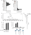
Figure 2. Identification of TMM as a T cell antigen.
(A and B) M. tuberculosis H37Rv crude lipids were fractionated by HPTLC using chloroform/methanol/water (C/M/W, 65:25:4; vol/vol/vol) (A) and 90:10:1; vol/vol/vol (B) and stained with copper(II) acetate-phosphoric acid. Y-50 reporter cells were stimulated with each fraction in the presence of APCs and analyzed for GFP and CD69 expression. White and black arrowheads denote the origin and the solvent front, respectively. (C) MALDI-TOF MS spectrum of lipid fraction 2 (Fr2). (D) The chemical structure of TMM of α-mycolate is shown, and methoxy-mycolate and keto-mycolate are the other major subclasses of mycolate found in M. tuberculosis TMM. (E) Y-50 reporter cells were cocultured with cytokine-differentiated human monocytes preincubated with whole bacteria (heat-killed M. tuberculosis H37Rv or living BCG) and analyzed for GFP and CD69 expression. (F) Y-50 reporter cells were stimulated with the indicated concentration of TMM, TDM, or GMM. Expression of GFP and CD69 is shown in the bar graphs. Schematic ligand structures are shown below. Data are shown as the mean ± SD of triplicate assays (E and F) and representative results from 2 independent experiments are shown (A, B, E, and F).
Figure 3
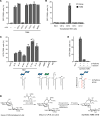
Figure 3. CD1b restricts TMM recognition by Y-50 T cells.
(A) Y-50 reporter cells were cocultured with cytokine-differentiated human monocytes and TMM (0.3 nmol /well) in the presence of 5 μg/mL anti-CD1a, -CD1b, -CD1c, -CD1d or isotype control antibodies (IgG1 and IgG2b) and analyzed for GFP and CD69 expression. (B) The reporter cells expressing Y-50 TCR were stimulated with TMM (1 nmol/well) in the presence of HEK293T cells transfected with human CD1a, CD1b, CD1c, or CD1d. (C) Y-50 reporter cells were stimulated with TMM (1 nmol/well) purified from M. tuberculosis CDC1551, M. bovis BCG, M. intracellulare, M. smegmatis, Rhodococcus equi, and R. sp 4306. Also, GMM, MMM, GroMM, and MA were tested in the presence of human CD1b-expressing DC2.4 cells (CD1b-DC2.4). (D) Scheme for synthetic TMM. (E) Y-50 reporter cells were stimulated with synthetic TMM harboring a β-hydroxy group and α-branched alkyl chains or synthetic analogs lacking hydroxy (–, +) or both moieties (–, –) in the presence of CD1b-DC2.4 cells as APCs. Data are shown as the mean ± SD of triplicate assays, and representative results from 2 independent experiments are shown (AC and E).
Figure 4
Figure 4. Mutagenesis and structural analysis of TMM-reactive TCR.
(A) The amino acid sequences of Y-50 CDR3α. Arginine residues which were mutated to alanine are shown in red. TMM reactivities of each mutant are shown as a percentage of the maximum (max) response induced by plate-coated anti-CD3 Ab. The number of amino acids is shown in accordance with the ImMunoGeneTics (IMGT) definition (https://imgt.org/IMGTScientificChart/). (B) Nucleotide and amino acid sequences of the Y-50 TCR CDR3β region and its junction deletion mutants (Δ). D region and N or P nucleotide sequences that constitute junctional sequences are unshaded. Cells were stimulated as indicated in A. (C) Crystal structure of the Y-50 TCRαβ heterodimer (PDB: 8XUB). The main chains of TCRα and β are shown in violet and brown, respectively. CDR3αβ regions are boxed. (D and E) A 2Fo-Fc map contoured at 2.0 σ (D) and B-factor diagram (E) of CDR3αβ are shown as gray mesh and color gradient, respectively. Junction regions of CDR3β are boxed. Data are shown as the mean ± SD of triplicate assays (A and B), and representative results from 2 independent experiments are shown.
Figure 5
Figure 5. Ternary complex structure of Y-50 TCR-TMM-CD1b.
(A) Overall structure of the Y-50 TCR-TMM-CD1b complex. The main chains of TCRα, TCRβ, and CD1b are shown. TMM is presented as yellow spheres. (B) Upper panel: Superimposition of the structure of Y-50 TCR alone (PDB: 8XUB) (pink) and Y-50 TCR bound to TMM-CD1b (PDB: 8ZOX) (blue) . Lower panels: CDR3β regions (boxed area in the upper panel) magnified. (C) Close-up view of TMM (R,R) and the side chain of R114 within CDR3α. The β-hydroxy group of TMM is shown in red. (D) Y-50 reporter cells were stimulated with the natural configuration of synthetic TMM (R,R) or non-natural stereoisomers (S,S) or (S,R+R,S) in the presence of CD1b-DC2.4 and analyzed for GFP and CD69 expression. The stereoisomer structures are shown below (R, red; S, black). Data are shown as the mean ± SD of triplicate experiments, and a representative result from 2 independent experiments is shown. (E) Close-up view of TMM (R,R) and the side chain of R107 (CDR3α) and D114 (CDR3β). Hydroxy groups of TMM that formed hydrogen bonds to the TCR side chains are shown in red. (F) Close-up view of the side chains of R79, E80, and D83 in CD1b that interact with the side chains of R37 (CDR1β), Y58 and E63 (CDR2β), and G110 (CDR3β). (G) Multi-bonded interaction of the CD1b RExxD motif and TRBV4-1 residues. Individual interaction is shown by dotted lines. (H) Conservation of the RExxD motif in human CD1b and CD1c. The amino acid sequences of CD1a (NP_001307581), CD1b (NP_001755.1), CD1c (NP_001756.2), and CD1d (NP_001306074) are aligned. Numbers indicate the amino acid position of the mature peptide (excluding signal peptide).
Figure 6
Figure 6. Functional maturation of TMM-reactive T cells upon TMM stimulation.
(A and B) Cluster shift of the Y-50 clonotype before and after TMM stimulation. T cells expressing the Y-50 clonotype defined by scTCR-RNA-Seq are overlaid (A) on a UMAP plot of PBMCs from donors including the donor sample used in Figure 1 (B), as described in Methods. Tem, effector memory T cells; CTL, cytotoxic T lymphocytes. (CE) Differentially expressed genes in Y-50 T cells upon TMM stimulation. Violin plots show the expression of representative genes encoding cytotoxic effector molecules (C), proinflammatory cytokines and chemokines (D), and stemness-related molecules (E). Unstim., unstimulated.
Figure 7
Figure 7. TMM-specific T cells with similar characteristics are shared among individuals.
(A and B) Frequent TMM-specific clonotypes identified by TMM-CD1b-tetramer sorting and scTCR-RNA-Seq are overlaid (A) on a UMAP plot of all TMM-tetramer–sorted T cells and unsorted CD3+ T cells from 13 healthy donors (B). Three clones were detected from different individual donors; 2 clones (clones 17 and 439) were from another donor. CD4+ Tem, CD4+ effector memory T cells; CD4 Tcm, CD4+ central memory T cells. Naive T cells were rare within TMM-tetramer+ cells and were not clustered on the UMAP. (C) TCR usages, CDR3 sequences, and length of the CDR3β region of the clonotypes detected in A. (D) Each clonotype was reconstituted into reporter cells and analyzed for TMM, TDM, and GMM reactivity using CD1b-DC2.4 as APCs. Data are shown as the mean ± SD of triplicate assays, and representative results from 2 independent experiments are shown. Reporter cells were stained with PE-conjugated endo-CD1b (Cont.) or TMM-CD1b (TMM) tetramers and anti-CD3 antibodies. Y-50 TCR is shown as a control. (E and F) Frequency of TCRVβ usage (D) and length of the CDR3β region (E) of unsorted or the top 27 TMM-CD1b tetramer+ T cell clonotypes. (G) PBMCs from Japanese donors (uninfected donors, n = 7; patients with TB, n = 13) or PBMCs (n = 10) and cord blood cells (n = 10) from uninfected US donors were stained with PE-conjugated TMM-loaded CD1b tetramer, APC-conjugated CD1b-endo tetramer and anti-CD3 antibody. The percentages of TMM-CD1b tetramer+ and endo-CD1b tetramer populations in CD3+ T cells are shown (TMM-CD1b-tet+). Medians are indicated with horizontal bars. *P < 0.05, by unpaired, 2-tailed Welch’s t test.

Comment in

  • Sensing mycobacteria through unconventional pathways

References

    1. Dulberger CL, et al. The mycobacterial cell envelope — a moving target. Nat Rev Microbiol. 2020;18(1):47–59. doi: 10.1038/s41579-019-0273-7. - DOI - PubMed
    1. Freund J, et al. Sensitization and antibody formation after injection of tubercle bacilli and paraffin oil. Proc Soc Exp Biol Med. 1937;37(3):509. doi: 10.3181/00379727-37-9625. - DOI
    1. Dube JY, et al. Synthetic mycobacterial molecular patterns partially complete Freund’s adjuvant. Sci Rep. 2020;10(1):5874. doi: 10.1038/s41598-020-62543-5. - DOI - PMC - PubMed
    1. Ishikawa E, et al. Recognition of mycobacterial lipids by immune receptors. Trends Immunol. 2017;38(1):66–76. doi: 10.1016/j.it.2016.10.009. - DOI - PubMed
    1. Brown GD, et al. C-type lectins in immunity and homeostasis. Nat Rev Immunol. 2018;18(6):374–389. doi: 10.1038/s41577-018-0004-8. - DOI - PubMed

MeSH terms