. 2025 Feb;35(2):216-224.
doi: 10.1089/thy.2024.0393. Epub 2024 Dec 24.

Linsitinib Decreases Thyrotropin-Induced Thyroid Hormone Synthesis by Inhibiting Crosstalk Between Thyroid-Stimulating Hormone and Insulin-Like Growth Factor 1 Receptors in Human Thyrocytes In Vitro and In Vivo in Mice

Affiliations

Linsitinib Decreases Thyrotropin-Induced Thyroid Hormone Synthesis by Inhibiting Crosstalk Between Thyroid-Stimulating Hormone and Insulin-Like Growth Factor 1 Receptors in Human Thyrocytes In Vitro and In Vivo in Mice

Alisa Boutin et al. Thyroid. 2025 Feb.

Abstract

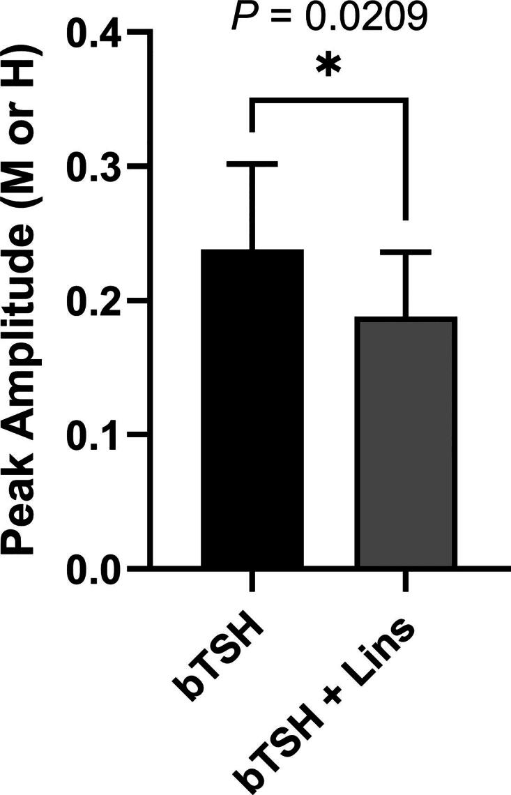
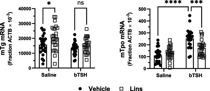
Background: Thyrotropin receptor (TSHR) and insulin-like growth factor 1 receptor (IGF-1R) have been shown to crosstalk in primary cultures of human thyrocytes (hThyros) and Graves' orbital fibroblasts. The phenomenon of TSHR/IGF-1R crosstalk has been largely studied in the pathogenesis of thyroid eye disease (TED) in human orbital fibroblasts. Here, we investigated the effects of inhibiting the IGF-1R-mediated contribution to crosstalk by linsitinib (Lins), a small-molecule IGF-1R kinase inhibitor, on TSH-induced regulation of thyroperoxidase (TPO) and thyroglobulin (TG) mRNAs and proteins in hThyros in vitro, and on TPO and TG mRNAs and free thyroxine (fT4) levels in vivo in mice. Methods: Steady-state levels of mRNAs of TPO and TG in hThyros in vitro and mouse thyroid glands were measured by RT-qPCR. Human TG (hTG) and human TPO (hTPO) proteins in human thyroid cell cultures were measured by Western blot or ELISA. Translation rates of hTG were quantified by stable isotope labeling by amino acids method (SILAC). Thyroidal mouse Tpo (mTpo) and Tg (mTg) mRNAs and fT4 in mice were assessed after Lins administration on 3 consecutive days followed by an intraperitoneal dose of bovine TSH (bTSH) 3 hours prior to drawing blood. Results: In primary cultures of hThyros, Lins inhibited bTSH-induced upregulation of hTPO mRNA by 61.5%, and hTPO protein was inhibited by 42.4%. There was no effect of Lins on hTG mRNA, but Lins inhibited the upregulation of secreted and cell-associated hTG protein by 50.1% and 42.2%, respectively, by inhibiting hTG mRNA translation. mTpo mRNA measured in thyroid glands after treatment with Lins was reduced by 31.5%. There was no effect of Lins on mTg mRNA, however, Lins decreased fT4 levels in mice under basal (endogenous mTSH levels) and bTSH-treated conditions. Conclusions: The IGF-1R antagonist Lins inhibited bTSH-stimulated hTG and hTPO protein expression in primary cultures of hThyros and fT4 levels in mice. We suggest that thyroid function studies be monitored when Lins is administered to humans, for example, if it is used to treat TED.

Keywords: IGF-1R; TSHR; crosstalk; linsitinib; thyroid hormone synthetic genes.


Figures

FIG. 1.
FIG. 1.
Lins inhibits bTSH-induced mRNA upregulation of hTPO but not of hTG in hThyros. hThyros were seeded at 1 × 10⁵ cells per well, allowed to attach overnight, then stimulated in arresting medium with 10 µM Lins alone or in combination with bTSH dose response. After 5 days of treatment, cells were lysed and analyzed for hTG, and hTPO mRNA levels using quantitative RT-PCR. Each experiment contained biological duplicates. Data are expressed as mean ± SEM; n = 3 patient donors. TSH, thyrotropin; TG, thyroglobulin; TPO, thyroperoxidase; h, human.
FIG. 2.
FIG. 2.
Lins inhibits bTSH-stimulated hTPO protein levels. hThyros were seeded at a density of 1 × 10⁵ cells per well, allowed to adhere overnight, and then stimulated in arresting medium, with 10 µM Lins alone or in combination with 1 mU/mL bTSH. After 5 days, cells were lysed, and hTPO expression was analyzed by Western blot. A representative blot from three independent experiments is shown (A). Protein levels were quantified and normalized to β-tubulin (B). Data are presented as the mean ± SEM, expressed as a percentage of the bTSH-only response; n = 3 patient donors. Group differences were assessed using an unpaired Student’s t-test, with a highly significant difference observed between the bTSH-treated and bTSH + Lins-treated wells (p = 0.0007). Lins, linsitinib.
FIG. 3.
FIG. 3.
Lins decreases bTSH-induced cell-associated hTG protein and hTG secretion but has no effect on hTG mRNA. hThyros were plated at a density of 1 × 10⁵ cells per well, allowed to adhere overnight, and then stimulated in arresting medium, with 10 µM Lins either alone or in combination with 1 mU/mL bTSH. After 5 days, hTG mRNA expression was quantified using RT-PCR (A), secreted hTG was measured in the conditioned media via ELISA (B), and hTG expression in cell lysates was analyzed by Western blot (C–D). A representative blot from three independent experiments is shown (C). Protein expression levels were quantified and normalized to β-tubulin (D). Data are presented as the mean ± SEM, expressed as a percentage of the bTSH-only response; n = 3 patient donors. Differences between groups were assessed using an unpaired Student’s t-test. Significant differences were observed between the bTSH-treated and bTSH + Lins-treated wells for hTG secretion (B, p < 0.0001) and cell-associated hTG (D, p = 0.0012), but not for mRNA expression (A).
FIG. 4.
FIG. 4.
Lins effect on hTG translation measured by pSILAC. hThyros were seeded at 1 × 10⁵ cells/well in growth media in 12-well plates and allowed to attach overnight. Next day, growth media was replaced with arresting media for 24 hours. The following day, cells were subjected to a 30 minutes starvation in pSILAC arresting media lacking lysine and arginine with either DMSO or 10 µM Lins pretreatment. Then pretreatment medium was aspirated and replaced with arresting medium in DMEM for pSILAC supplemented with labeled amino acids (lysine and arginine) under the following conditions: 1 mU/mL bTSH with isotopically medium amino acids (M); 1 mU/mL bTSH with 10 µM Lins with heavy amino acids (H) Arg and Lys; master control samples: 1 mU/mL bTSH with normal (light) amino acids. After 3 hours, monolayers were washed 3 times with ice cold PBS on ice and cells scraped with 200 µL PBS. Data are shown as mean ± SEM of the amplitudes of the spikes of the bTSH only and bTSH+Lins responses; n = 7 patient donors. The difference between groups was analyzed using paired Student’s t-test. The difference between wells treated with bTSH and bTSH + Lins was significant (p = 0.0209). PBS, phosphate-buffered saline.
FIG. 5.
FIG. 5.
Lins inhibits TSH-stimulated mTpo but not mTg mRNA expression in vivo. Lins (15 mg/kg) was administered i.p. once daily on 3 consecutive days. On the third day, bTSH was administered i.p. 1 hour after the last dose of Lins, and 3 hours later the mice were sacrificed for sample collection. Thyroid genes were measured in mice treated with vehicle/saline, vehicle/bTSH, Lins/saline, and Lins/bTSH. The increase of mTpo mRNA induced by bTSH was significantly inhibited with Lins treatment. In contrast, Lins had no effect on mTg mRNA. Two-way ANOVA statistical analysis was used for significance testing. The data represent the summary of three independent experiments.
FIG. 6.
FIG. 6.
Lins decreases fT4 levels in vivo. Lins (15 mg/kg) was administered i.p. once daily on 3 consecutive days. On the third day, bTSH was administered i.p. 1 hour after the last dose of Lins, and 3 hours later the mice were sacrificed for sample collection. fT4 was measured in mice treated with vehicle/saline, vehicle/bTSH, Lins/saline, and Lins/bTSH. Basal fT4 was significantly lower in Lins-treated mice (p = 0.0013). bTSH caused a significant increase in fT4 overall (bTSH main effect p < 0.0001). The increase of fT4 induced by bTSH was significantly inhibited with Lins treatment (p < 0.0001). Two-way ANOVA statistical analysis was used for significance testing. The data represent the summary of three independent experiments. fT4, free thyroxine.

Similar articles

Cited by

References

    1. Eggo MC, Bachrach LK, Burrow GN. Interaction of TSH, insulin and insulin-like growth factors in regulating thyroid growth and function. Growth Factors 1990;2(2–3):99–109; doi: 10.3109/08977199009071497 - DOI - PubMed
    1. Tramontano D, Cushing GW, Moses AC, et al. . Insulin-like growth factor-I stimulates the growth of rat thyroid cells in culture and synergizes the stimulation of DNA synthesis induced by TSH and Graves’-IgG. Endocrinology 1986;119(2):940–942; doi: 10.1210/endo-119-2-940 - DOI - PubMed
    1. Santisteban P, Kohn LD, Di Lauro R. Thyroglobulin gene expression is regulated by insulin and insulin-like growth factor I, as well as thyrotropin, in FRTL-5 thyroid cells. J Biol Chem 1987;262(9):4048–4052. - PubMed
    1. Kimura T, Van Keymeulen A, Golstein J, et al. . Regulation of thyroid cell proliferation by TSH and other factors: A critical evaluation of in vitro models. Endocr Rev 2001;22(5):631–656; doi: 10.1210/edrv.22.5.0444 - DOI - PubMed
    1. Morgan SJ, Neumann S, Marcus-Samuels B, et al. . Thyrotropin and insulin-like growth Factor 1 receptor crosstalk upregulates Sodium-Iodide symporter expression in primary cultures of human thyrocytes. Thyroid 2016;26(12):1794–1803; doi: 10.1089/thy.2016.0323 - DOI - PMC - PubMed

MeSH terms

LinkOut - more resources