. 2025 Feb;28(2):293-307.
doi: 10.1038/s41593-024-01829-7. Epub 2024 Dec 24.

In vivo hyperphosphorylation of tau is associated with synaptic loss and behavioral abnormalities in the absence of tau seeds

Affiliations

In vivo hyperphosphorylation of tau is associated with synaptic loss and behavioral abnormalities in the absence of tau seeds

Naoto Watamura et al. Nat Neurosci. 2025 Feb.

Abstract

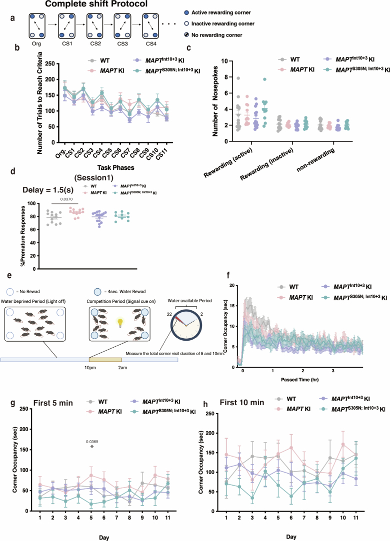
Tau pathology is a hallmark of several neurodegenerative diseases, including frontotemporal dementia and Alzheimer's disease. However, the sequence of events and the form of tau that confers toxicity are still unclear, due in large part to the lack of physiological models of tauopathy initiation and progression in which to test hypotheses. We have developed a series of targeted mice expressing frontotemporal-dementia-causing mutations in the humanized MAPT gene to investigate the earliest stages of tauopathy. MAPTInt10+3G>A and MAPTS305N;Int10+3G>A lines show abundant hyperphosphorylated tau in the hippocampus and entorhinal cortex, but they do not develop seed-competent fibrillar structures. Accumulation of hyperphosphorylated tau was accompanied by neurite degeneration, loss of viable synapses and indicators of behavioral abnormalities. Our results demonstrate that neuronal toxicity can occur in the absence of fibrillar, higher-order structures and that tau hyperphosphorylation is probably involved in the earliest etiological events in tauopathies showing isoform ratio imbalance.


Conflict of interest statement

Competing interests: T.E. serves as a Representative for Phenovance LLC, which provides commercial service using IntelliCage. T.C.S. serves as an Executive Consultant for RIKEN BIO Co. Ltd., which sublicenses App knock-in mice to for-profit organizations. K.E.D. is a board member of Ceracuity LLC. H.Z. has served at scientific advisory boards and/or as a consultant for Abbvie, Acumen, Alector, Alzinova, ALZPath, Annexon, Apellis, Artery Therapeutics, AZTherapies, CogRx, Denali, Eisai, Nervgen, Novo Nordisk, Optoceutics, Passage Bio, Pinteon Therapeutics, Prothena, Red Abbey Labs, reMYND, Roche, Samumed, Siemens Healthineers, Triplet Therapeutics and Wave, has given lectures in symposia sponsored by Cellectricon, Fujirebio, Alzecure, Biogen and Roche, and is a cofounder of Brain Biomarker Solutions in Gothenburg AB (BBS), which is a part of the GU Ventures Incubator Program (outside submitted work). The other authors declare no competing interests.

Figures

Fig. 1
Fig. 1. Generation of humanized mutant MAPT KI mice with BEs.
a, Schematic image of BE-mediated genome editing of the MAPT gene. b, Schematic illustration of BE-mediated genome editing in MAPT KI mouse zygotes by microinjection. c, Sanger sequencing results determined MAPTInt10+3 (upper panel) and MAPTS305N;Int10+3 (lower panel) KI mice. Mutation loci are indicated with arrowheads in black and substituted amino acids highlighted in red. d, Regional, genetic and annotated information regarding off-target sites identified in MAPTInt10+3 and MAPTS305N;Int10+3 KI mice. Alt., alternative; chr, chromosome; ref., reference. Schematic in a created using BioRender.com.
Fig. 2
Fig. 2. Shift from 3R- to 4R-tau expression induced by Int10+3G>A and S305N-Int10+3G>A mutations in MAPT KI mice.
a, Real-time PCR results of 3R-tau, 4R-tau and total tau levels in MAPT KI, MAPTInt10+3 KI and MAPTS305N;Int10+3 KI mice (n = 5 for each group; n = 3 female and n = 2 male) using specific primers validated in Mapt KO mice. b, Immunoblotting of tau detected by tau13 antibody in MAPT KI, MAPTInt10+3 KI and MAPTS305N;Int10+3 KI mice (n = 3 for each group, n = 1 female, n = 2 male) after alkaline phosphatase treatment. These blots were derived from separate membranes, with an equal amount of protein loaded. c,d, Quantification of 3R (c) and 4R (d) tau levels as shown in b. e,f, Relative quantification of tau peptides from MAPT KI, MAPTInt10+3 KI and MAPTS305N;Int10+3 KI mice by LC–MS/MS analysis shown for individual tau peptides (e) and 3R and 4R tau levels (f). Red outline in e describes 3R- and 4R-tau-specific tau peptides, respectively (n = 5–9 for each group; for MAPT KI: n = 6 sex matched; for MAPTInt10+3 KI: n = 5 of which n = 3 female and n = 2 male; for MAPTS305N;Int10+3 KI: n = 9 of which n = 4 female and n = 5 male). g,h, Immunostaining of 3R- and 4R-tau in the entorhinal cortex (g) and hippocampal CA3 region (h) of 3-month-old Mapt KO, MAPT KI, MAPTInt10+3 KI and MAPTS305N;Int10+3 KI mice. Scale bars, 100 µm (inset, 50 µm), with four biological replicates with similar observations (Extended Data Fig. 3c–e). In a, c and d, data represent mean ± s.e.m. (two-way ANOVA with Tukey’s multiple comparison test). Source data
Fig. 3
Fig. 3. Map of pathological tau detected by AT8 antibody staining in MAPTInt10+3 and MAPTS305N;Int10+3 mice.
a, Immunostaining of phosphorylated tau detected by AT8 antibody in the brains of 15-month-old WT, MAPT KI, MAPTInt10+3 KI and MAPTS305N;Int10+3 KI mice; n = 6 mice were used for each strain, n = 3 male and n = 3 female. Scale bars, 200 µm (upper panel), 50 µm (lower panel). b, Heatmap showing AT8 signal intensity from a in each region. c, Immunostaining of phosphorylated tau detected by AT8 antibody in the frontal cortex of patients with FTLD with the Int10+3 mutation on the MAPT gene, with three biological replicates with similar observations. Scale bar, 50 µm. d, Immunostaining of phosphorylated tau detected by AT8 antibody in the frontal cortex of patients with FTLD with the S305N mutation on the MAPT gene, with at least three technical replicates with similar observations. Scale bar, 50 µm. ACAd, anterior cingulate area, dorsal part; AHNa, anterior hypothalamic nucleus, anterior part; AI, agranular insular area; AON, anterior olfactory nucleus; AUD, auditory area; BMAa, basomedial amygdalar nucleus, anterior part; CA1, hippocampal CA1; cing, cingulum bundle; CA2, hippocampal CA2; CA3, hippocampal CA3; COA, cortical amygdalar area; COApl3, cortical amygdalar area, posterior part, lateral zone, layer3; COApm, cortical amygdalar area, posterior part, medial zone; CP; caudoputamen; DG-po, dentate gyrus, polymorph layer; DP, dorsal peduncular area; DTN, dorsal tegmental nucleus; ECT, ectorhinal area; ENTI2, entorhinal area, lateral part, layer II; ENTI3, entorhinal area, lateral part, layer3; ENTI4, entorhinal area, lateral part, layer IV; ENTI5, entorhinal area, lateral part, layer5; ENTm2, entorhinal area, medial part, layer2; FRP, frontal pole; GRN, gigantocellular reticular nucleus; IC, inferior colliculus; LM, lateral mammillary nucleus; LS, lateral septal nucleus; MO, somatomotor area; NDB, diagonal band nucleus; NTS, nucleus of the solitary tract; ORB, orbital area; ORB1, orbital area, layer 1; PAR, parasubiculum; PCG, pontine central gray; PER, perirhinal area; PERI, perirhinal area; PH, posterior hypothalamic nucleus; PL, prelimbic area; PPN, pedunculopontine nucleus; PR, pontine nucleus; PVT, paraventricular nucleus of the thalamus (not shown in map); RE, reuniens nucleus; RSP, retrosplenial area; SNr, substantia nigra, reticular part; SS, somatosensory area; SSs; supplementary somatosensory area; SSp, primary somatosensory area; SSP-ul, primary somatosensory area, upper limb; SUB, subiculum; SUBd, subiculum, dorsal part; Tea, temporal association area; TH, thalamus; VIS1, visual area layer 1. Illustrations in b created using BioRender.com.
Fig. 4
Fig. 4. Hyperphosphorylation of tau in MAPTInt10+3 and MAPTS305N;Int10+3 mice.
a, Immunostaining of phosphorylated tau detected by CP13, PHF-1, AT180 and AT270 antibodies in the brains of 15-month-old WT, MAPT KI, MAPTInt10+3 KI and MAPTS305N;Int10+3 KI mice and patients with FTLD with Int10+3 and S305N mutations on the MAPT gene, respectively, with at least three biological replicates with similar observations. Scale bars, 50 µm (mouse data) and 25 µm (data from patients with FTLD). b,c, Immunoblotting of phosphorylated and total tau detected by CP13, AT8, PHF-1, K9JA, Tau13 and HT7 antibodies in the Tris-soluble fraction of brain lysates from MAPT KI, MAPTInt10+3 KI and MAPTS305N;Int10+3 KI mice at 12 months of age (n = 6 for each group, n = 3 female and n = 3 males) and in PS19 mice at 9 months of age (b) and quantification (c). d, Relative amount of phospho-tau peptides by LC–MS analysis in the brains of 10- to 18-month-old MAPT KI (n = 6; sex matched), MAPTInt10+3 KI (n = 5; n = 3 female and n = 2 males), and MAPTS305N;Int10+3 KI mice (n = 9; n = 4 females and n = 5 males). e, Summary of histological visual quantification analyses of tau phosphorylation detected by several phospho-tau antibodies. In c and d, the data represent the mean ± s.e.m. (two-way ANOVA Tukey’s multiple comparison test). Source data
Fig. 5
Fig. 5. Insolubility and seeding activity of tau in MAPTInt10+3 and MAPTS305N;Int10+3 KI mice.
a, Immunostaining of tau detected by TOC1, T22 and MC1 antibodies in the brains of 15-month-old MAPT KI, MAPTInt10+3 KI and MAPTS305N;Int10+3 KI mice, and in patients with FTLDs with Int10+3 and S305N mutations in the MAPT gene, respectively, with at least three biological replicates with similar observations. Scale bars, 50 µm (mouse data) and 25 µm (data from patients with FTLD). b, Summary of histological visual quantification analyses of tau accumulation detected by several tau antibodies. c, Immunoblotting of tau detected by CP13 antibody in separated fractions (Top fraction to 20% range) of brain lysates from MAPT KI and MAPTS305N;Int10+3 KI mice at 6 months and from PS19 mice at 9 months. d, Immunoblotting of phosphorylated tau detected by CP13 antibody in separated fractions (Top fraction to 20% range) of brain lysates from MAPT KI and MAPTS305N;Int10+3 KI mice at 18 months of age and PS19 mouse at 9 months of age. e, Tau seeding activity in brain lysates from WT, MAPT KI, MAPTInt10+3 KI and MAPTS305N;Int10+3 KI mice at 15 months (n = 4–5 for each group, n = 2 females and n = 2 or 3 males) and PS19 mice at 9 months using S305N biosensor cells. Scale bar, 20 µm. f, Tau seeding activity from AD and patients with FTLD with Int10+3 and S305N mutations using S305N biosensor cells (n = 3–4 per group, for AD: n = 3 females and for Int10+3: n = 3 males; n = 1 male for FTLD-S305N). Scale bar, 20 µm. g, Raw RT-QuIC ThT reactions, 16 replicates, from posterior cortex of mice at 24 months, MAPT KI (n = 5) and MAPTS305N;Int10+3 KI (n = 5) and positive control PSP motor cortex human brain homogenates (n = 3). h, Endpoint reaction s.d. for MAPT KI (n = 5) and MAPTS305N;Int10+3 KI (n = 5) and PSP (n = 3). P values were calculated as the statistical difference comparing the expected distribution of each group to the observed. In e and f, data represent mean ± s.e.m. (one-way ANOVA Tukey’s multiple comparison test). In h, the box limits show the interquartile range, and the center lines show the median values. Source data
Fig. 6
Fig. 6. Synaptic loss and neurodegeneration in MAPTS305N;Int10+3 KI mice.
a, Representative super-resolution images of Homer1 and Synaptotagmin puncta colocalization in the entorhinal cortex layer II (EC-Lyll) of 16-month-old WT, MAPT KI, MAPTInt10+3 and MAPTS305N;Int10+3 KI mice. Scale bar, 5 µm. b, Quantification of Synaptotagmin/Homer1 colocalization density in the polymorph layer of the dentate gyrus (PoDG), CA3 stratum radiatum (CA3-Rad), CA1 lacunosum molecular layer (CA1-Lmol) and EC-LyII regions of 16-month-old WT, MAPT KI, MAPTInt10+3 and MAPTS305N;Int10+3 KI mice (n = 5–6 animals per genotype; n = 3 females and n = 2 or 3 males). c, Amino-cupric silver (AmCuAg) staining in the entorhinal cortices of 15-month-old MAPT KI, MAPTInt10+3 and MAPTS305N;Int10+3 KI mice. Scale bar, 50 µm. Black arrows, structures resembling neurites. d, Quantification of AmCuAg staining (MAPT KI, n = 4; MAPTInt10+ KI, n = 6; and MAPTS305N;Int10+3 KI, n = 8; sex matched per genotype). In b, data represent mean ± s.e.m. (two-way ANOVA with Tukey’s multiple comparison test). In d, data represent mean ± s.e.m. (Kruskal–Wallis one-way ANOVA test with Dunn’s multiple comparison test). Source data
Fig. 7
Fig. 7. Memory deficits indicated in MAPTS305N;Int10+3 KI mice.
a,b, Cartoon representation of the test performed (a) and percentage of spontaneous alternation of WT (n = 17), MAPT KI (n = 15), MAPTInt10+3 KI (n = 14) and MAPTS305N;Int10+3 KI (n = 16) mice at 15–16 months of age on the Y-maze test (b). c,d, Cartoon representation of the test performed (c) and total distance traveled demonstrated by WT (n = 16), MAPT KI (n = 15), MAPTInt10+3 KI (n = 14) and MAPTS305N;Int10+3 KI (n = 16) mice at 15–16 months in the open-field test (d). c,e, Cartoon representation of the test performed (c) and percentage of time spent in the center region demonstrated by WT (n = 17), MAPT KI (n = 15), MAPTInt10+3 KI (n = 14) and MAPTS305N;Int10+3 KI (n = 16) mice at 15–16 months in the open-field test (e). f–h, Cartoon representation of the test performed (f) and discrimination index for the NOL test obtained from WT (n = 17), MAPT KI (n = 15), MAPTInt10+3 KI (n = 14) and MAPTS305N;Int10+3 KI (n = 14) mice at 15–16 months at Day1 (g) and Day2 (h). ik, Cartoon representation of the test performed (i) and latency from start position to access the escape box on Day1 to Day4 (j) and time spent around the target hole at probe test by WT (n = 17), MAPT KI (n = 15), MAPTInt10+3 KI (n = 14) and MAPTS305N;Int10+3 KI (n = 16) mice at 15–16 months of age for the Barnes maze test (k). l, Values of PC1 scores (individual projection along the main variance axis, PC1) for each experimental group. m, PCA was applied on behavioral variables measured using various behavioral tests. Each dot represents a single mouse and larger dots represent the average per genotype. Colored ellipses highlight 1.5 s.d. around the mean per genotype (~87% confidence interval). In d, h and l, data represent mean ± s.e.m. (one-way ANOVA with Tukey’s multiple comparison test). In b, e and k, data represent mean ± s.e.m. (Kruskal–Wallis one-way ANOVA test (Dunn’s multiple comparison test)). In j, data represent mean ± s.e.m. (two-way ANOVA with Tukey’s multiple comparison test). Panels a, c, f and i were created using BioRender.com. Source data
Fig. 8
Fig. 8. Perseverative, disinhibition behaviors and apathy indicated in MAPTS305N;Int10+3 KI mice.
ac, Number of trials to reach criteria and nosepokes of 14- to 16-month-old WT, MAPT KI, MAPTInt10+3 KI and MAPTS305N;Int10+3 KI mice in the CS/PS-shuffled session of SP-FLEX (WT, n = 14; MAPT KI, n = 11; MAPTInt10+3 KI, n = 15; MAPTS305N;Int10+3 KI, n = 10). df, Percentage of premature responses displayed by 14- to 16-month-old WT, MAPT KI, MAPTInt10+3 KI and MAPTS305N;Int10+3 KI mice in sessions 2–3 of the behavioral inhibition test (WT, n = 12; MAPT KI, n = 11; MAPTInt10+3 KI, n = 15; MAPTS305N;Int10+3 KI, n = 9). gi, Percentages of saccharin choice made by 14- to 16-month-old WT, MAPT KI, MAPTInt10+3 KI and MAPTS305N;Int10+3 KI mice in the taste preference test and the subsequent effort-based choice test (WT, n = 11; MAPT KI, n = 11; MAPTInt10+3 KI, n = 14; MAPTS305N;Int10+3 KI, n = 9). In c, e, f and i, data represent mean ± s.e.m. (two-way ANOVA with Tukey’s multiple comparison test). For i, The P value shown is the one comparing WT with MAPTS305N;Int10+3 KI (see source data for results on all comparisons). Illustrations in d and g created using BioRender.com. Source data
Extended Data Fig. 1
Extended Data Fig. 1. Sanger sequencing of other mutant MAPT KI mice.
Sanger sequencing results determined MAPTP301L KI, MAPTP301S KI, MAPTP301V KI, MAPTP301L; Int10+3 KI, and MAPTP301S; Int10+3 KI mice. Mutation loci are indicated by arrowheads in black and the substituted amino acids are highlighted in red.
Extended Data Fig. 2
Extended Data Fig. 2. Removal of off-target mutations in MAPTInt10+3 KI and MAPTS305N; Int10+3 KI mice by backcrossing with C57B6/J mice.
a-d, Off-target mutations identified in founders (highlighted in red boxes) were removed by backcrossing with C57B6/J mice more than four times.
Extended Data Fig. 3
Extended Data Fig. 3. RT-PCR, LC-MS/MS analysis and immunostaining of tau in MAPT KI, MAPTInt10+3 KI and MAPTS305N; Int10+3 KI mice.
a, The relative expression of 3 R and 4 R tau were calculated by semi-quantitative RT-PCR using the brain from Mapt KO, MAPT KI, MAPTInt10+3 KI, and MAPTS305N; Int10+3 KI, and PS19 (n = 3 for each group). b, Amount of tau peptides based on the isoform resulting from alternative splicing by LC-MS/MS analysis in the brains of 12-15 month-old MAPT KI (n = 6; sex-matched), MAPTInt10+3 KI (n = 5; n = 3 female and n = 2 males), and MAPTS305N; Int10+3 KI mice(n = 9; n = 4 females and n = 5 male). 4R-tau (299-317 aa) in MAPTS305N; Int10+3 KI line was not detected because serine (S) at position 305 was converted to asparagine (N). c-e, Immunostaining of 3 R and 4 R tau in the brains of 3-month-old MAPT KO, MAPT KI, MAPTInt10+3 KI, and MAPTS305N; Int10+3 KI mice (n = 4 for each group, n = 2 females and n = 2 males). Scale bar represents 1 mm. Data in (b), (d), and (e) represent the mean ± SEM. (two-way ANOVA followed by Tukey’s multiple comparison test). Source data
Extended Data Fig. 4
Extended Data Fig. 4. Immunostaining of tau in MAPT KI, MAPTInt10+3 KI and MAPTS305N; Int10+3 KI mice.
a, Immunostaining of phosphorylated tau detected by AT8 antibody in the brains of 12-month-old MAPT KI, MAPTInt10+3 KI, and MAPTS305N; Int10+3 KI mice. Scale bar represents 1 mm in upper panel (hemibrain), 500 µm in the middle panel (hippocampus), 100 µm in the lower panel (piriform/entorhinal (Pir/Ent) cortex) and 50 µm in the insert. b, Immunostaining of phosphorylated tau detected by AT8 antibody in the brains of 15-month-old MAPT KI, MAPTInt10+3 KI, and MAPTS305N; Int10+3 KI mice. Scale bar represents 200 µm in lest panel (hemibrain), 50 µm in the middle panel (CA3), and 100 µm in right panel (EC). c, Immunostaining of phosphorylated tau detected by AT8 antibody in the brains of 6-month-old MAPT KI, and MAPTS305N; Int10+3 KI mice. Scale bar represents 200 µm in the left panel and 100 µm in the right panel. Arrowhead indicates AT8 positive signals. d, Immunostaining of tau using several anti-tau antibodies (CP13, AT8, T22, MC1, and TOC1) in the brains of 30-month-old MAPTS305N; Int10+3 KI mice. Scale bar represents 200 µm in the left panel (hemibrain) and 100 µm in the right panel (EC). All experiments were repeated independently at least three times with similar observations.
Extended Data Fig. 5
Extended Data Fig. 5. Neurodegenerative analysis in MAPT KI, MAPTInt10+3 KI, MAPTS305N; Int10+3 KI mice, and AD patient.
Gallyas staining in the brains of 15-month-old MAPT KI, MAPTInt10+3 KI, and MAPTS305N; Int10+3 KI mice (n = 3 animals per genotype, n = 2 females and n = 1 male) and AD patient. Scale bar represents 100 µm in mouse samples and 50 µm in the AD patient.
Extended Data Fig. 6
Extended Data Fig. 6. Insolubility of tau in PS19 mice and seeding assays of tau with P301S biosensor cell line.
a, Immunoblotting of tau detected by CP13 antibody in separated fractions (Top-50% range) of brain lysates from PS19 and WT mice at 9 months of age, with at least three technical replicates with similar observations. b, The gating strategy for FRET Flow cytometry. Cell population (left) and singlet/doublet (middle) gates are drawn using standard flow cytometry methodology. A FRET gate (right) is constructed from untreated CFP /YFP cells without YFP single-positive cells. Analysis parameters include: percent FRET positivity, median fluorescence intensity (MFI) of FRET-positive events, and the Integrated FRET Density (Integrated FRET Density = Percent FRET − positive cells × MFI). c, FRET versus donor (CFP) bivariate plots showing FRET-positive and FRET-negative P301S biosensor cells incubated with brain lysates from MAPT KI, MAPTInt10+3 KI, and MAPTS305N; Int10+3 KI mice at 15 months and from PS19 mice at 9 months. d, FRET versus donor (CFP) bivariate plots showing the FRET-positive and FRET-negative P301S biosensor cells incubated with extracts from FFPE-embedded tissue from AD and FTLD patients with Intron10 + 3 and S305N mutations in the MAPT gene. e, Integrated FRET density from brain lysates from MAPT KI (n = 5), MAPTInt10+3 KI(n = 4) and MAPTS305N; Int10+3 KI (n = 5) mice at 15 months and PS19 (n = 1) mice at 9 months as a positive control. f, Integrated FRET density from AD (n = 3) and FTLD patients with Intron10 + 3 (n = 3) and S305N (n = 1; due to un availability of tissue) mutations on the MAPT gene. Data represents the mean ± SEM. (one-way ANOVA with Tukey’s multiple comparison test). g, RT-QuIC aggregates half-way curve (t1/2) from the combination of the biological replicates from 24 M posterior cortex MAPT KI (n = 5) and MAPTS305N; Int10+3 KI (n = 5) and positive control progressive supranuclear palsy (PSP; n = 3) motor cortex brain homogenates, where the points arranged in a horizontal alignment at 67 h denote reactions where ThT fluorescence levels did not surpass the threshold, suggesting the absence of fibril formation. h, RT-QuIC ThT maxima from the combination of biological replicates as in g. Source data
Extended Data Fig. 7
Extended Data Fig. 7. Synaptic loss in MAPTS305N; Int10+3 KI mice.
a, Representative super-resolution images of Homer1 and vGLUT1 puncta colocalization in the entorhinal cortex layerII (EC-LyII) regions of 16-month-old MAPT KI, MAPTInt10+3, and MAPTS305N; Int10+3 KI mice. Scale bar represents 5 µm. b, Quantification of vGUT1/ Homer1 colocalization density in the polymorph layer of the dentate gyrus (PoDG), CA3 stratum radiatum (CA3-Rad), CA1 lacunosum molecular layer (CA1-Lmol), and entorhinal cortex layerII (EC-LyII) regions of 16-month-old MAPT KI, MAPTInt10+3, and MAPTS305N; Int10+3KI mice (n = 5 animals per genotype). c,d, Immunoblotting of synaptophysin, Homer1, PSD-95, and tublin in the tris-soluble fraction of brain lysates from WT, MAPT KI and MAPTS305N; Int10+3 KI mice at 16 months of age (n = 3 for each group, n = 2 female and n = 1 males) and in PS19 (n = 1) mice at 9 months of age. These blots were derived from separate membranes, with an equal amount of protein loaded. In (b), the data represent the mean ± SEM (two-way ANOVA with Tukey’s multiple comparison test). In (d), the data represent the mean ± SEM. Source data
Extended Data Fig. 8
Extended Data Fig. 8. Locomotor performance indicated in MAPTS305N; Int10+3 G>A KI mice.
a, Total entry number of WT, MAPT KI, MAPTInt10+3 KI, and MAPTS305N; Int10+3 KI mice at 15-16 months of age on Y-maze test (WT: n = 17, MAPT KI: n = 15, MAPTInt10+3 KI: n = 14, MAPTS305N; Int10+3 KI: n = 16). b, Total distance of WT, MAPT KI, MAPTInt10+3 KI, and MAPTS305N; Int10+3 KI mice at 15-16 months of age on the 2nd day of novel object location test (WT: n = 17, MAPT KI: n = 15, MAPTInt10+3 KI: n = 14, MAPTS305N; Int10+3 KI: n = 14). c, Higher failure (defined as <55% preference) in visuospatial memory tests on the novel object location for MAPTS305N; Int10+3 KI mice compared to WT, MAPT KI, MAPTInt10+3 KI mice at 15-16 months of age (WT: n = 17, MAPT KI: n = 15, MAPTInt10+3 KI: n = 14, MAPTS305N; Int10+3 KI: n = 14). d, Total distance of WT, MAPT KI, MAPTInt10+3 KI, and MAPTS305N; Int10+3 KI mice at 15-16 months of age on the probe test of Barnes-Maze test (WT: n = 17, MAPT KI: n = 15, MAPTInt10+3 KI: n = 14, MAPTS305N; Int10+3 KI: n = 16). e,f, Latency to fall of WT, MAPT KI, MAPTInt10+3 KI, and MAPTS305N; Int10+3 KI mice at 15-16 months of age on the Rotarod test (WT: n = 15, MAPT KI: n = 15, MAPTInt10+3 KI: n = 14, MAPTS305N; Int10+3 KI: n = 15) (created with BioRender.com). g, Summary of the behavior tests. In (b) and (d), the data represent the mean ± SEM (one-way ANOVA with Tukey’s multiple comparison test). In (c), the data represent the mean ± SD. In (f), the data represent the mean ± SEM (Kruskal-Wallis one-way ANOVA test with Dunn’s multiple comparison test). Source data
Extended Data Fig. 9
Extended Data Fig. 9. Basal activity parameters calculated from 2 weeks of data in IntelliCage.
a-c, Average number of visits (a), nosepokes (b), and licks (c) in a day in WT, MAPT KI, MAPTInt10+3 KI and MAPTS305N; Int10+3 KI mice. d, Percentages of corner visit distribution for each corner displayed by WT, MAPT KI, MAPTInt10+3 KI, and MAPTS305N; Int10+3 KI mice. e, Percentages of corner visit distribution in sequence of preferred corners displayed by WT, MAPT KI, MAPTInt10+3 KI, and MAPTS305N; Int10+3 KI mice. f, Percentages of left bottle choice exhibited by WT, MAPT KI, MAPTInt10+3 KI, and MAPTS305N; Int10+3 KI mice. g, Percentages of preferred choice exhibited by WT, MAPT KI, MAPTInt10+3 KI, and MAPTS305N; Int10+3 KI mice. h, Percentages of movement pattern (Re-entry, Short side, Long side, Diagonal) displayed by WT, MAPT KI, MAPTInt10+3 KI, and MAPTS305N; Int10+3 KI mice. All experiments were performed with 14-16-month-old female mice (WT: n = 14, MAPT KI: n = 11, MAPTInt10+3 KI: n = 15, MAPTS305N; Int10+3 KI: n = 11). Data represent the mean ± SEM (two-way ANOVA with Tukey’s multiple comparison test). Source data
Extended Data Fig. 10
Extended Data Fig. 10. Results from SP-FLEX (behavioral sequence learning and behavioral flexibility task), behavioral inhibition and competitive dominance tests.
a-c, Number of trials to reach criteria and nosepokes of 14-16-month old WT, MAPT KI, MAPTInt10+3 KI, and MAPTS305N; Int10+3 KI mice in the CS (complete shift)-only session of Self-paced Learning and Behavioral Flexibility Test (SP-FLEX) (WT: n = 14, MAPT KI: n = 11, MAPTInt10+3 KI: n = 15, MAPTS305N; Int10+3 KI: n = 10). d, Percentage of premature responses displayed by 14-16-month old WT, MAPT KI, MAPTInt10+3 KI, and MAPTS305N; Int10+3 KI mice in Session 1 of the behavioral inhibition test (WT: n = 12, MAPT KI: n = 11, MAPTInt10+3 KI: n = 15, MAPTS305N; Int10+3 KI: n = 9). e,f, Average corner occupancy time displayed by WT, MAPT KI, MAPTInt10+3 KI, and MAPTS305N; Int10+3 KI mice at 0-4 h over 11 days. (WT: n = 10, MAPT KI: n = 9, MAPTInt10+3 KI: n = 14, MAPTS305N; Int10+3 KI: n = 9) (created with BioRender.com). g, Average corner occupancy time displayed by WT, MAPT KI, MAPTInt10+3 KI, and MAPTS305N; Int10+3 KI mice in the first 5 min over 11 days. h, Average corner occupancy time displayed by WT, MAPT KI, MAPTInt10+3 KI, and MAPTS305N; Int10+3 KI mice in the first 10 min over 11 days. In (d), data represent the mean ± SEM (one-way ANOVA with Tukey’s multiple comparison test). In (g), data represent the mean ± SEM. (two-way ANOVA with Tukey’s multiple comparison test). Source data

References

    1. Hutton, M. et al. Association of missense and 5′-splice-site mutations in tau with the inherited dementia FTDP-17. Nature393, 702–705 (1998). - PubMed
    1. Poorkaj, P. et al. Tau is a candidate gene for chromosome 17 frontotemporal dementia. Ann. Neurol.43, 815–825 (1998). - PubMed
    1. Spillantini, M. G. et al. Mutation in the tau gene in familial multiple system tauopathy with presenile dementia. Proc. Natl Acad. Sci. USA95, 7737–7741 (1998). - PMC - PubMed
    1. Forrest, S. L. et al. Retiring the term FTDP-17 as MAPT mutations are genetic forms of sporadic frontotemporal tauopathies. Brain141, 521–534 (2018). - PMC - PubMed
    1. Langworth-Green, C. et al. Chronic effects of inflammation on tauopathies. Lancet Neurol.22, 430–442 (2023). - PubMed

MeSH terms

LinkOut - more resources