. 2024 Dec 24;82(1):13.
doi: 10.1007/s00018-024-05501-y.

Trimethylamine-N-oxide accelerates osteoporosis by PERK activation of ATF5 unfolding

Affiliations

Trimethylamine-N-oxide accelerates osteoporosis by PERK activation of ATF5 unfolding

Yu-Han Lin et al. Cell Mol Life Sci. .

Abstract

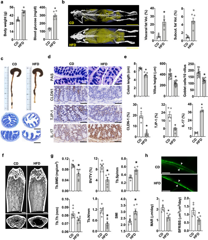
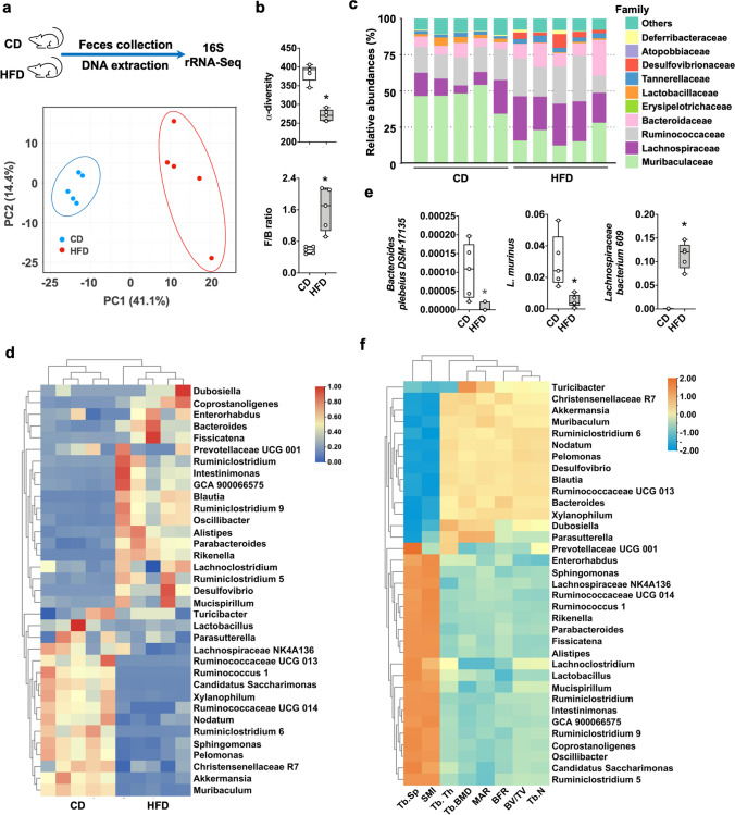
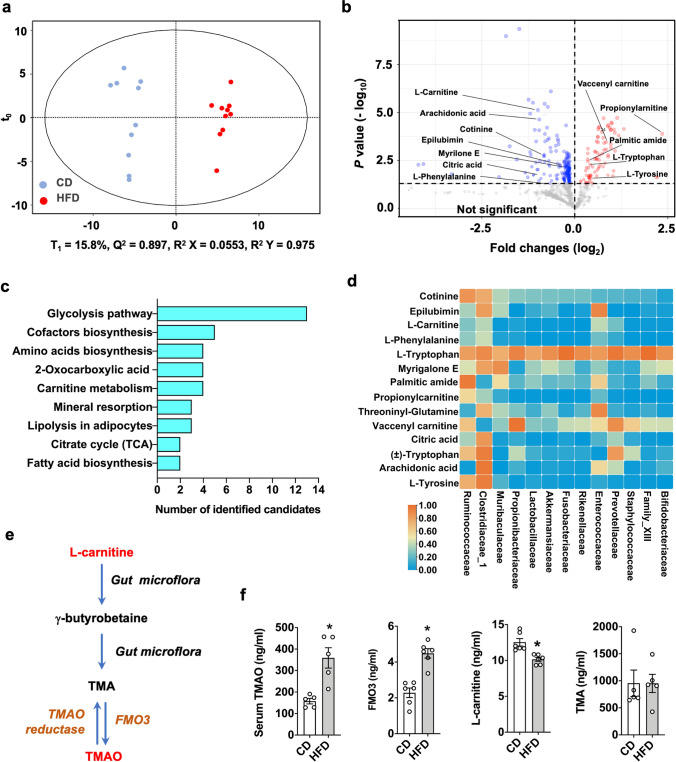
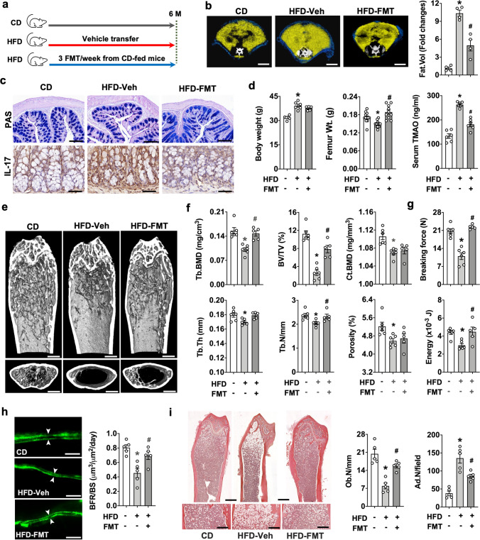
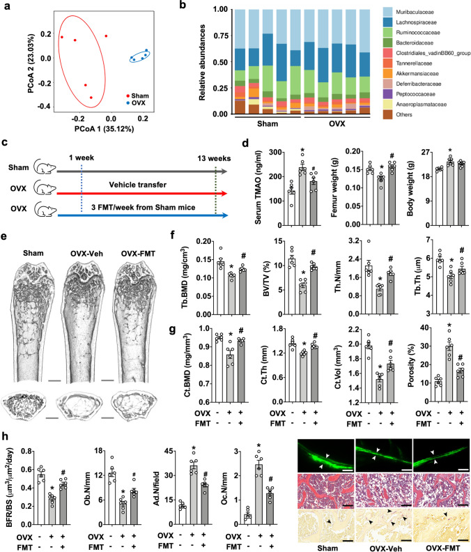
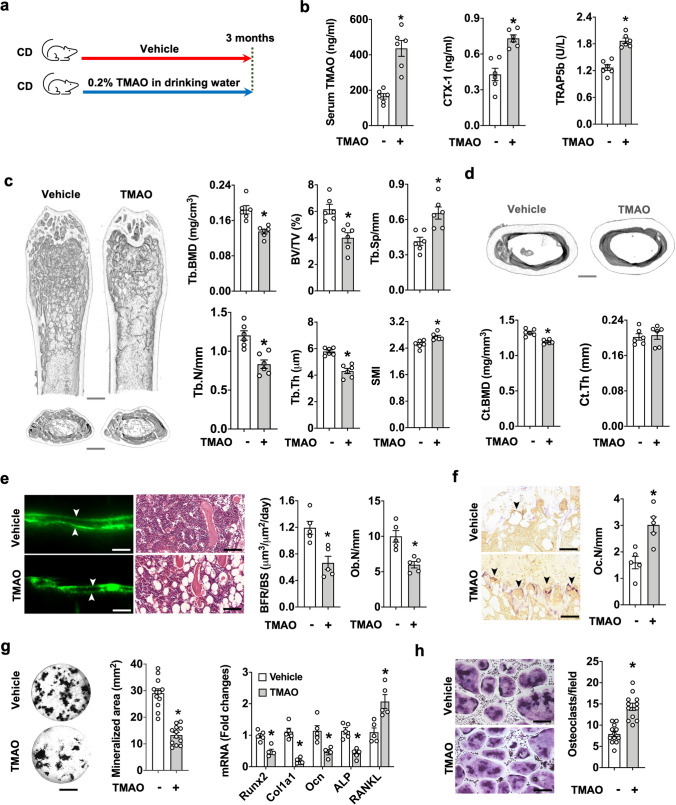
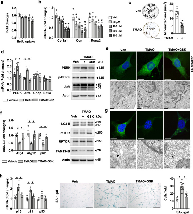
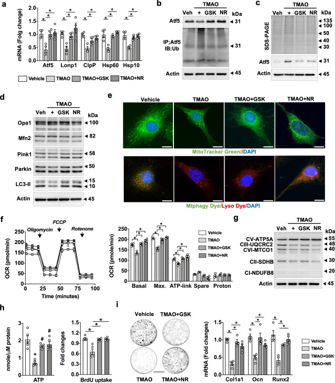
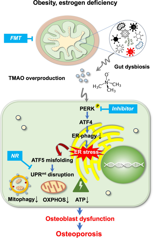
Imbalances in gut microbiota and their metabolites have been implicated in osteoporotic disorders. Trimethylamine-n-oxide (TMAO), a metabolite of L-carnitine produced by gut microorganisms and flavin-containing monooxygenase-3, is known to accelerate tissue metabolism and remodeling; however, its role in bone loss remained unexplored. This study investigates the relationship between gut microbiota dysbiosis, TMAO production, and osteoporosis development. We further demonstrate that the loss of beneficial gut microbiota is associated with the development of murine osteoporosis and alterations in the serum metabolome, particularly affecting L-carnitine metabolism. TMAO emerges as a functional metabolite detrimental to bone homeostasis. Notably, transplantation of mouse gut microbiota counteracts obesity- or estrogen deficiency-induced TMAO overproduction and mitigates key features of osteoporosis. Mechanistically, excessive TMAO intake augments bone mass loss by inhibiting bone mineral acquisition and osteogenic differentiation. TMAO activates the PERK and ATF4-dependent disruption of endoplasmic reticulum autophagy and suppresses the folding of ATF5, hindering mitochondrial unfolding protein response (UPRmt) in osteoblasts. Importantly, UPRmt activation by nicotinamide riboside mitigates TMAO-induced inhibition of mineralized matrix biosynthesis by preserving mitochondrial oxidative phosphorylation and mitophagy. Collectively, our findings revealed that gut microbiota dysbiosis leads to TMAO overproduction, impairing ER homeostasis and UPRmt, thereby aggravating osteoblast dysfunction and development of osteoporosis. Our study elucidates the catabolic role of gut microflora-derived TMAO in bone integrity and highlights the therapeutic potential of healthy donor gut microbiota transplantation to alter the progression of osteoporosis.

Keywords: ER-phagy; Gut microecosystem; Misfolding; OXPHOS; Parkin; Trimethylamine-n-oxide.


Conflict of interest statement

Declarations. Conflict of interest: Authors have no financial competing interest. Ethical approval and consent to participate: Animal protocols were reviewed and approved by the Institutional Animal Care and Use Committee of Kaohsiung Chang Gung Memorial Hospital (IACUC Affidavit #2019091103). This study did not involve human specimens or clinical data. Consent for publication: Not applicable.

Figures

Fig. 1
Fig. 1
Body adipose overdevelopment, bone mass loss, and gut atrophy in HFD-induced murine obesity. Increased body weight, blood glucose levels (a) as well as visceral fat and subcutaneous fat development in obese mice (b); scale bar, 1.5 cm. Decreased colon length and villi loss (c and e); scale bar of upper panels, 200 μm; scale bar of lower panels, 100 µm. Losses of PAS-stained mucin, CLDN-1, and TJP1 levels, but increased IL-17 levels in colon goblet cells of obese mice (d and e); scale bar, 15 μm. Sparse trabecular bone network (f); scale bar, 50 μm and decreased trabecular BMD, Tb.Th, BV/TV, Tb.N and increased Tb.Sp and SMI in obese mice (g). Decreased fluorescence calcein-labelling, MAR, and BFR/BS upon HFD consumption (h); scale bar, 20 μm. Mice were fed HFD or CD for 6 months. Data are means ± standard errors calculated from 6 mice. Asterisks (*) stand for significant difference (P < 0.05) analyzed by Kolmogorov–Smirnov test and Student t-test
Fig. 2
Fig. 2
Gut microbial dysbiosis in HFD-induced obese mice. Principal component analysis showing noticeably different gut microflora profiles (a). Decreased α-diversity and high Firmicute/Bacteroidetes ratios of the gut microflora of obese mice (b). Changes in relative abundances of gut bacterial families (c). Heatmap showing changes in the richness of gut bacterial genera and less beneficial gut microbiome variety in obese mice (d). HFD consumption affected the relative abundances of 3 gut bacterial species (e). Alterations in the gut microbiome correlated with bone mass and trabecular network loss (f). Data are calculated from 5 mice. Asterisks (*) stand for significant difference (P < 0.05) analyzed by Kolmogorov–Smirnov test and Student t-test
Fig. 3
Fig. 3
Altered serum metabolomic landscape in obese mice. Principal component analysis showing distinctive metabolomic profiles (a). Volcanic plot showing 85 and 187 metabolites elevated or reduced, respectively, in obese mice (b). KEGG pathway of metabolomic landscapes, which may contribute to plenty of cellular metabolism (c). Heatmap showing the correlation of serum metabolites and gut microorganisms (d). A drawing scheme depicting L-carnitine conversion into TMA and TMAO by gut microorganisms and FMO3 (e). Increased serum TMAO and FMO3 together with reduced L-carnitine levels in obese mice. Data are calculated from 5 mice. Asterisks (*) stand for significant difference (P < 0.05) analyzed by Kolmogorov–Smirnov test and Student t-test
Fig. 4
Fig. 4
Effects of fecal microbiota transplantation (FMT) on body fat formation, gut integrity, and bone mass in obese mice. Schematic depiction of the transplantation procedure of fecal microbiota from CD mice to HFD mice (a). FMT improves body fat volume (b); scale bar, 10 mm, mucin production and IL-17 levels in colon (c); scale bar, 10 μm, and femur weight and serum TMAO levels (d) in obese mice. FMT counteracted obesity-mediated bone loss at a trabecular microstructural level (e); scale bar, 50 μm, and Tb.BMD, Tb.Th, BV/TV, Tb.N (f), breaking force and energy (g). FMT repressed BFR/BS loss (h); scale bar, 20 μm, and osteoblast loss and marrow adiposity (i); scale bar, 20 μm. Data are means ± standard errors calculated from 5 ~ 6 mice. Asterisks (*) stand for significant difference (P < 0.05) from CD-fed group; and hashtags (#) resemble significance from HFD-Veh group analyzed by ANOVA test and post hoc Bonferroni test
Fig. 5
Fig. 5
FMT-induced bone changes in ovariectomized (OVX) mice. Distinct gut microbial profiles of sham mice and OVX mice (a). Changes in gut bacterial families in sham and OVX mice (b). Schematic illustration of the transplantation procedure of fecal microbiota from aged-matched sham mice to OVX mice (c). FMT suppressed OVX-induced femur weight loss and TMAO overproduction in serum (d). FMT suppressed the loss of trabecular bone (e), Tb.BMD, BV/TV, Tb.N, Tb.Th (f), Ct.BMD, Ct.Th, Ct.Vol and cortical porosity (g) in OVX mice. FMT preserved BFR and Ob.N and repressed marrow adipocyte formation and osteoclast overabundance in OVX mice (h); scale bar, 20 μm. Data are means ± standard errors calculated from 5 to 6 mice. Asterisks (*) stand for significant difference (P < 0.05) from sham group; and hashtags (#) resemble significance from OVX-Veh group analyzed by ANOVA test and post hoc Bonferroni test
Fig. 6
Fig. 6
Effects of TMAO intake on bone phenotypes. Schematic illustration of the TMAO feeding regime (a). Increased serum TMAO, bone resorption markers CTX-1, and TRAP5b levels in TMAO-fed mice (b). μCT images showing a sparse trabecular bone microstructure; scale bar, 50 μm. TMAO suppressed Tb.BMD, BV/TV, Tb.N, and Tb.Th and enhanced Tb.Sp. and SMI (c). Ct.BMD was reduced in TMAO-fed mice (d); scale bar, 50 μm. TMAO intake resulted in relatively low BFR/BS and Ob.N (e); scale bar, 20 μm, and high Oc.N in bone tissue (f); scale bar, 20 μm. TMAO inhibited the synthesis of mineralized matrix production as well as osteogenic marker expression, but enhanced RANKL expression of bone-marrow mesenchymal cells (g); scale bar, 5 mm. High osteoclast formation capacity of bone-marrow macrophages in TMAO-fed mice (h); scale bar, 20 μm. Data are means ± standard errors calculated from 5 mice. Asterisks (*) stand for significant difference (P < 0.05) from vehicle group analyzed by Kolmogorov–Smirnov test and Student t-test
Fig. 7
Fig. 7
TMAO regulated endoplasmic reticulum (ER) integrity and senescence in osteoblasts. TMAO dose-dependently suppressed BrdU uptake (a), expression of osteogenic makers Col1a1, Ocn, and Runx2 (b), and von Kossa-stained mineralized nodule production (c); scale bar, 5 mm. TMAO enhanced mRNA expression of PERK and ATF4 as well as protein levels of PERK and phosphorylated PERK, and ATF4 (d). Fluorescent ER tracker staining (scale bar, 10 μm) and disrupted ER ultrastructure (scale bar, 1 μm) in TMAO-treated osteoblasts (e). GSK2606144 counteracted TMAO-induced ER stress and integrity loss. Decreased mRNA expression of Atg4, Atg12, and p62 and protein levels of LC3-II, mTOR, and FAM134B (f) and autophagosome formation (scale bar, 10 μm) and ER-phagic ultrastructure (scale bar, 0.5 μm) (g); scale bar, 10 μm, upon TMAO treatment. Senescence-associated β-galactosidase staining and increased p16, and p21 expression in TMAO-treated cells (h); scale bar, 40 μm. GSK2606414 attenuated TMAO-mediated autophagy loss and senescence. Data are mean ± standard errors calculated from at least three independent experiments, including immunoblotting. Asterisks (*) stand for significant difference (P < 0.05) analyzed by ANOVA test and post hoc Bonferroni test. GSK, GSK2606414; SA-β-gal, senescence association β-galactosidase
Fig. 8
Fig. 8
TMAO suppressed UPRmt, mitophagy, mitochondrial energetics, and mineralized matrix anabolism. GSK2606144 or nicotinamide riboside counteracted TMAO-induced losses of mRNA expression of UPRmt marker, ATF5, Lonp1, ClpP, Hsp60, and Hsp10 (a) as well as suppression of protein levels of ATF5 and ubiquitinated ATF5 (b) and aggregated Aft5 (c). GSK2606144 or nicotinamide riboside preserved Pink1, Parkin, and LC3-II levels (d) and suppressed TMAO-induced losses of MitoTracker Green-staining of mitochondrial mass and Mitphay Dye/Lyso Dye-staining of mitophagic puncta formation (e); scale bar, 10 μm. GSK2606144 or nicotinamide riboside mitigated TMAO-induced suppression of mitochondrial basal, maximum, ATP-linked oxygen consumption (f), OXPHOS (g), ATP production, proliferation capacity (h), and mineralized matrix synthesis as well as osteogenic marker expression. Data are means ± standard errors calculated from a minimum 3 independent experiments, including immunoblotting. Asterisks (*) stand for significant difference (P < 0.01) analyzed by ANOVA test and post hoc Bonferroni test. GSK, GSK2606414; NR, nicotinamide riboside
Fig. 9
Fig. 9
Hypothetical model of how gut microbiota-derived TMAO changes bone integrity. Gut microorganism dysbiosis elevates TMAO production in obese or ovariectomized mice. This metabolite then activates PERK and ATF4 to suppress ER autophagy and thus elevating ER stress, which leads to mitochondrial ATF5 misfolding and UPRmt disruption, together with decreased mitophagy, OXPHOS, and ATP synthesis. This consequently accelerates osteoblast dysfunction, leading to bone loss. FMT (from healthy donors), PERK inhibitor or UPRmt activator mitigates TMAO-induced loss of osteoblastic activity loss and bone mass. FMT, fecal microbiota transplantation; TMAO, trimethylamine-n-oxide; PERK, protein kinase R-link endoplasmic reticulum kinase; ATF4, activating transcription factor 4; ER, endoplasmic reticulum; ATF5, activating transcription factor 5; NR, nicotinamide ribose; UPRmt, mitochondrial unfolding protein response; OXPHOS, oxidative phosphorylation

References

    1. Lobo RA, Gompel A (2022) Management of menopause: a view towards prevention. Lancet Diabetes Endocrinol 10:457–470. 10.1016/S2213-8587(21)00269-2 - PubMed
    1. Seimon RV, Wild-Taylor AL, Keating SE et al (2019) Effect of weight loss via severe vs moderate energy restriction on lean mass and body composition among postmenopausal women with obesity: the TEMPO diet randomized clinical trial. JAMA Netw Open 2:e1913733. 10.1001/jamanetworkopen.2019.13733 - PMC - PubMed
    1. Ali D, Figeac F, Caci A et al (2022) High-fat diet-induced obesity augments the deleterious effects of estrogen deficiency on bone: evidence from ovariectomized mice. Aging Cell 21:e13726. 10.1111/acel.13726 - PMC - PubMed
    1. Kim S, Henneicke H, Cavanagh LL et al (2021) Osteoblastic glucocorticoid signaling exacerbates high-fat-diet- induced bone loss and obesity. Bone Res 9:40. 10.1038/s41413-021-00159-9 - PMC - PubMed
    1. Behera J, Ison J, Voor MJ, Tyagi N (2022) Exercise-linked skeletal irisin ameliorates diabetes-associated osteoporosis by inhibiting the oxidative damage-dependent miR-150-FNDC5/pyroptosis axis. Diabetes 71:2777–2792. 10.2337/db21-0573 - PMC - PubMed

MeSH terms

LinkOut - more resources