. 2024 Dec 24;30(1):272.
doi: 10.1186/s10020-024-01021-4.

Doublecortin regulates the mitochondrial-dependent apoptosis in glioma via Rho-A/Net-1/p38-MAPK signaling

Affiliations

Doublecortin regulates the mitochondrial-dependent apoptosis in glioma via Rho-A/Net-1/p38-MAPK signaling

Iqra Nadeem et al. Mol Med. .

Abstract

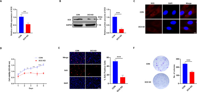
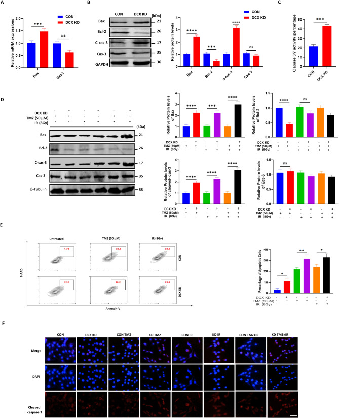
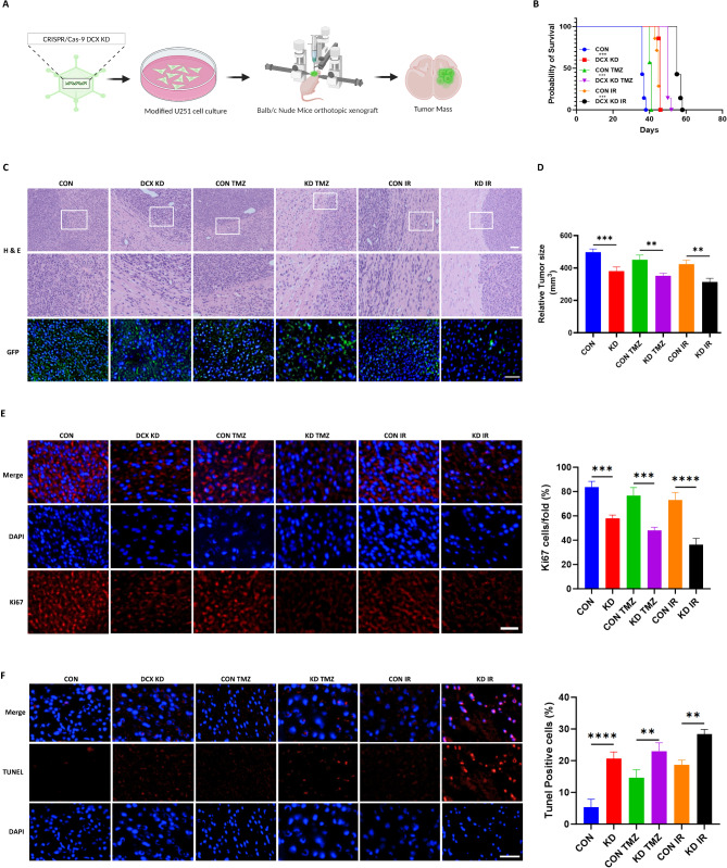
Doublecortin (DCX) is a microtubule-associated protein known to be a key regulator of neuronal migration and differentiation during brain development. However, the role of DCX, particularly in regulating the survival and growth of glioma cells, remains unclear. In this study, we utilized CRISPR/Cas9 technology to knock down DCX in the human glioma cell line (U251). DCX depletion suppressed cell proliferation and enhanced the pro-apoptotic effects of temozolomide (TMZ) and γ-radiation treatment. DCX knockdown led to the translocation of Bax to the mitochondria and mitochondria dysfunction. Furthermore, DCX deficiency-induced apoptosis took place along with the generation of reactive oxygen species (ROS), which is crucial in triggering mitochondrial membrane depolarization, the release of cytochrome c (Cyt-c), and caspase activation. Importantly, the transcriptional inhibition of DCX downregulated Rho-A, Net-1, and activated p38-MAPK cue, critical for cell survival and proliferation. Subsequent treatment with TMZ and γ-radiation further increased p38-MAPK activity through the decreased expression of Rho-A/Net-1, resulting in a significant reduction in glioma cell migration and invasion. Additionally, intracranial xenograft tumors of DCX-modified U251 cells in nude mice demonstrated inhibited tumor growth. Tumor sections treated with TMZ and γ-radiation exhibited a higher number of TUNEL-positive cells compared to the control group, indicating increased apoptosis. Our finding suggests that DCX depletion reduces glioma cell proliferation and promotes mitochondria-dependent apoptosis by enhancing the chemo and radiotherapy response. Targeting DCX represents a potential therapeutic target for glioma treatment.

Keywords: Apoptosis; CRISPR/Cas 9; Doublecortin; Glioma; Mitochondria.


Conflict of interest statement

Declarations. Ethics approval and consent to participate: This study did not include human participants, data, or tissue. All animal experiments were compliant with the Xuzhou Medical University Ethics Committee. Consent for publication: Not applicable. Competing interests: The authors declare no competing interests.

Figures

Fig. 1
Fig. 1
DCX knockdown suppresses glioma cell proliferation. (A and B) DCX knockdown by CRISPR/Cas-9 in the U251 glioma cell line was validated using Western blot and qPCR analysis (CON: Blue; DCX KD: Red). (C) Immunostaining of DCX in U251 stable cell lines using a DCX-specific antibody (Red) and nuclear counterstain DAPI (Blue). Scale bar, 25 μm. (D) MTT assay was used to examine the effect of DCX knockdown on cell viability. (E) EdU assays detected the cell proliferation after DCX knockdown. (F) Representative photographs of the colony formation assay demonstrated that the smallest number of colonies was observed in the DCX knockdown group compared with control groups: scale bar, 100 μm. All experiments were repeated three times, and data are presented as mean ± SD. *P < 0.05, **P < 0.01, ***P < 0.001, ****P < 0.0001, compared with control cells
Fig. 2
Fig. 2
DCX downregulation promotes glioma cell apoptosis. (A) The mRNA levels of Bcl-2 and Bax were detected by q-PCR. Values were normalized relative to the β-Actin mRNA levels. (B) After DCX knockdown, immunoblot analysis was performed to detect the expression of cleaved caspase-3, caspase-3, Bax, and Bcl-2 (CON: Blue; DCX KD: Red; CON TMZ: Green; KD TMZ: Purple; CON IR: Orange; KD IR: Black). Analyses of protein were performed using Image J. The protein content was normalized against the corresponding GAPDH content. (C) The caspase 3 /7 activity was analyzed using the Caspase-Glo 3/7 assay. (D) U251 cells were treated with indicated concentrations of TMZ and γ-radiation, and Western blotting was conducted to detect the expression of apoptotic proteins. β-tubulin was used as an endogenous control. (E) Cells were analyzed for apoptosis with Annexin-V/7-AAD by flow cytometry. (F) Cleaved caspase-3 immunofluorescence staining of U251 cells pre and post-treatment for 24 h, scale bar, 50 μm. For all experiments, data represent the mean ± standard error of the mean based on three independent experiments analyzed by unpaired two-tailed t-test and one-way ANOVA. *P < 0.05, **P < 0.01, ***P < 0.001, ****P < 0.0001, ns (not significant)
Fig. 3
Fig. 3
DCX depletion triggers mitochondria apoptosis in glioma cell. (A) Fluorescence microscopy images depicting the loss of MMP in DCX knockdown cells before and after treatment, as indicated by increased green fluorescence intensity of JC-1 monomers. Scale bar, 50 μm. (B) Measurement of intracellular ROS levels using fluorescent probe DCFH-DA, showing significant accumulation of ROS in DCX knockdown cells. (C) Release of cyt-C in the cytosol to mitochondria ratio measured by Western blotting. (D) Increased mitochondrial Bax expression levels in DCX knockdown cells compared to control analyzed by Western blotting. (E) Immunofluorescence analysis showing the translocation of Bax to mitochondria. (F) MitoTracker staining revealing morphological changes in mitochondria from filamentous to dot-shaped in DCX knockdown cells. (G) TEM images depicting elongated mitochondria in DCX knockdown cells and swollen mitochondria in the control group. (H) There was a significant reduction in cellular ATP levels in DCX knockdown cells compared to control. For all experiments, data represent the mean ± standard error of the mean based on three independent experiments analyzed by unpaired two-tailed t-test and one-way ANOVA. ****P < 0.0001, **P < 0.01, ***P < 0.001
Fig. 4
Fig. 4
DCX inhibits apoptosis through the Rho-A/Net-1/p38-MAPK pathway in glioma cell. (A) Western blot analysis shows increased phosphorylated p38-MAPK levels and decreased Rho-A and Net-1 expression in the DCX knockdown group compared to the control. (B) Western blotting expression analysis of Net-1, Rho-A, p-38, and phosphorylated p-38 followed by DCX knockdown with chemotherapy and radiation in U251 cells (CON TMZ: Blue; KD TMZ: Red; CON IR: Green; KD IR: Purple). Analyses of protein were performed using Image J. (C and D) Detection of Net-1 levels by mRNA and immunostaining in U251 cells with knockdown and treatment. Scale bar, 20 μm. (E) Scratch wound healing shows the migration capacity reduction in the U251 cells with DCX knockdown and TMZ/IR treatment (CON: Blue; DCX KD: Red; CON TMZ: Green; KD TMZ: Purple; CON IR: Orange; KD IR: Black). (F) DCX knockdown cells with IR treatment had the lowest average number of migrating cells among the three groups in the transwell migration assay. For all experiments, data represent the mean ± standard error of the mean based on three independent experiments analyzed by unpaired two-tailed t-test and one-way ANOVA. *P < 0.05, ****P < 0.0001, **P < 0.01, ***P < 0.001
Fig. 5
Fig. 5
DCX silencing impedes tumor growth and increases apoptosis. in vivo. (A) Construction of a glioma orthotopic model using Balb/c nude mice with DCX-modified U251 cells. (B) Mouse survival is shown by Kaplan-Meier curves. The log-rank test was used to measure survival differences in the respective groups. (C) H&E staining and GFP tracking of tumor tissue in CON and DCX KD model mice. Scale bars = 20 μm. (D) Relative tumor size before and after treatment. (E and F) Ki67 immunostaining and TUNEL measurement of apoptosis of tumor specimens. Scale bars = 20 μm. ****P < 0.0001, ***P < 0.001, **P < 0.01
Fig. 6
Fig. 6
Schematic illustration of DCX knockdown-induced mitochondrial apoptosis mechanism in glioma. This illustration depicts the molecular mechanisms by which DCX knockdown triggers apoptosis in human glioma cells. The downregulation of Rho-A and Net-1 upon loss of DCX activates p38-MAPK signaling, resulting in mitochondrial dysfunction and apoptosis induction. Additionally, the figure shows the enhanced pro-apoptotic effects observed when DCX depletion is combined with TMZ and γ-radiation treatment

References

    1. Al-Koussa H, Atat OE, Jaafar L, Tashjian H, El-Sibai M. The role of rho GTPases in Motility and Invasion of Glioblastoma Cells. Anal Cell Pathol. 2020;2020:1–9. 10.1155/2020/9274016. - PMC - PubMed
    1. Ayanlaja AA, Xiong Y, Gao Y, Ji G, Tang C, Abdullah A, Gao Z, D. Distinct features of Doublecortin as a marker of neuronal Migration and its implications in Cancer Cell mobility. Front Mol Neurosci. 2017;10:199. 10.3389/fnmol.2017.00199. - PMC - PubMed
    1. Ayanlaja AA, Ji G, Wang J, Gao Y, Cheng B, Kanwore K, Zhang L, Xiong Y, Kambey PA, Gao D. Doublecortin undergo nucleocytoplasmic transport via the RanGTPase signaling to promote glioma progression. Cell Commun Signal. 2020;18:24. 10.1186/s12964-019-0485-5. - PMC - PubMed
    1. Becker KN, Pettee KM, Sugrue A, Reinard KA, Schroeder JL, Eisenmann KM. The Cytoskeleton effectors rho-kinase (ROCK) and mammalian diaphanous-related (mDia) Formin have dynamic roles in Tumor Microtube formation in Invasive Glioblastoma cells. Cells. 2022;11:1559. 10.3390/cells11091559. - PMC - PubMed
    1. Breuzard G, Pagano A, Bastonero S, Malesinski S, Parat F, Barbier P, Peyrot V, Kovacic H. Tau regulates the microtubule-dependent migration of glioblastoma cells via the Rho-ROCK signaling pathway. J Cell Sci. 2019;132:jcs222851. 10.1242/jcs.222851. - PubMed

MeSH terms

LinkOut - more resources