

Aerobic exercise inhibits GSDME-dependent myocardial cell pyroptosis to protect ischemia-reperfusion injury
- PMID: 39719560
- PMCID: PMC11668014
- DOI: 10.1186/s10020-024-01048-7
Aerobic exercise inhibits GSDME-dependent myocardial cell pyroptosis to protect ischemia-reperfusion injury
Abstract
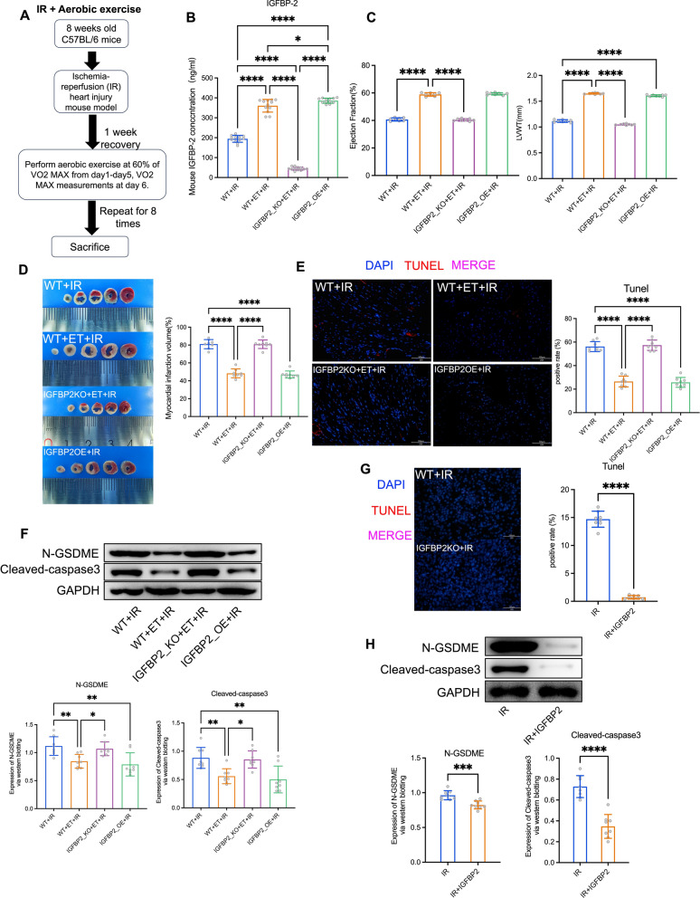
Background: Acute myocardial infarction (AMI) remains a significant cause of global mortality, exacerbated by ischemia-reperfusion (IR) injury. Myocardial cell pyroptosis has emerged as a critical pathway influencing IR injury severity.
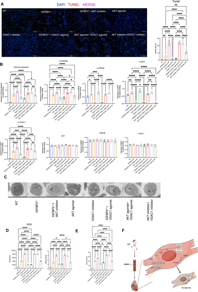
Methods: We aimed to investigate the cardioprotective effects of aerobic exercise on IR injury by examining the modulation of IGFBP2 and its impact on GSDME-dependent myocardial cell pyroptosis. Mechanistic pathways were explored using western blot analysis, ELISA, immunofluorescence, and echocardiography.
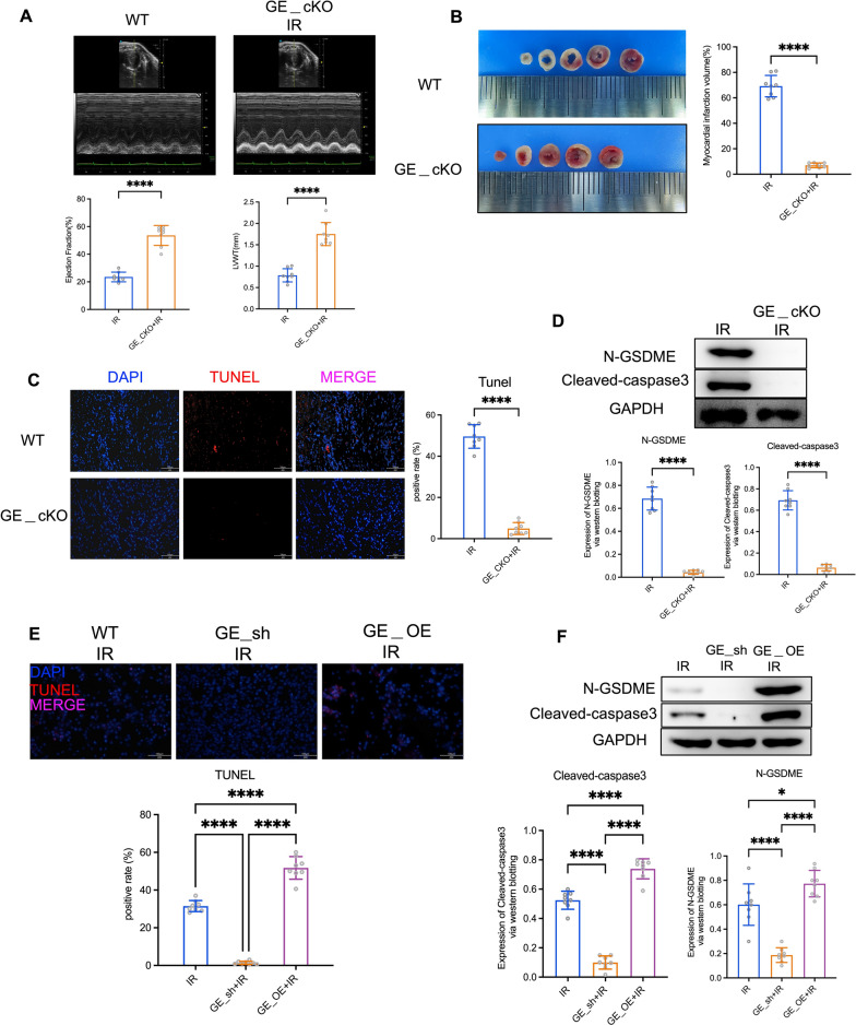
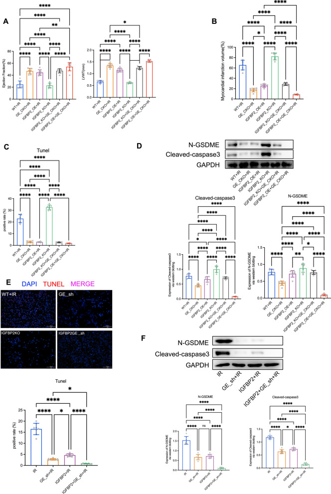
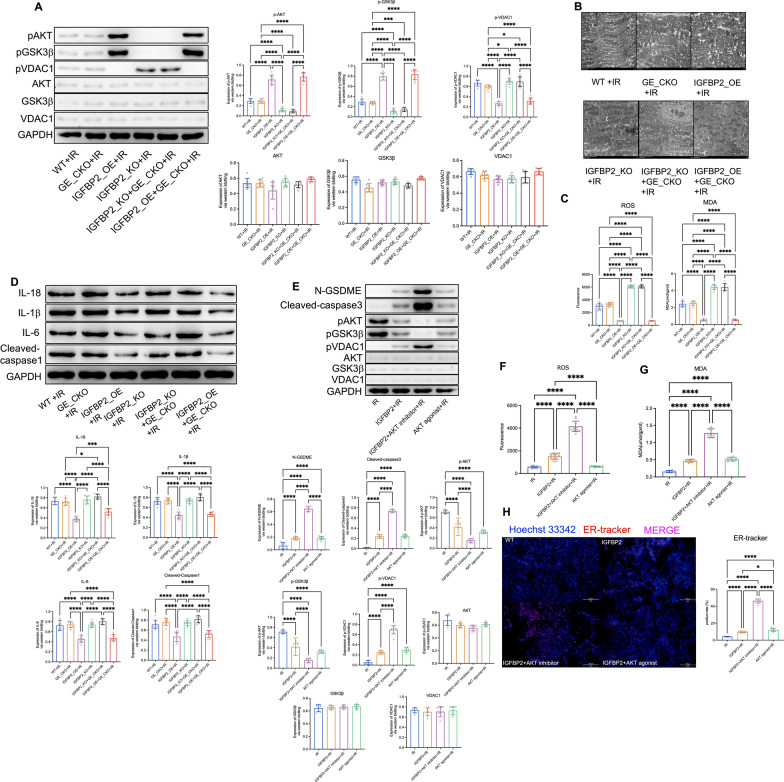
Results: Our findings demonstrate that aerobic exercise leads to increased circulating levels of IGFBP2, which effectively suppresses GSDME-dependent myocardial cell pyroptosis. This regulation occurs via the AKT-GSK3β signaling pathway, involving VDAC1 phosphorylation, thereby enhancing mitochondrial function and reducing oxidative stress.
Conclusion: In conclusion, our study highlights the role of IGFBP2 in mitigating GSDME-dependent pyroptosis as a mechanism through which aerobic exercise exerts cardioprotective effects against IR injury. These insights suggest potential therapeutic targets for managing acute myocardial infarction.
Keywords: AKT-GSK3β; Acute myocardial infarction; GSDME; IGFBP2; Ischemia-reperfusion; Pyroptosis.
© 2024. The Author(s).
Conflict of interest statement
Declarations. Ethics approval and consent to participate: All human studies were approved by the Ethical Committee of Peking Union Medical College Hospital. All animal experiments were implemented based on the Guide for the Care and Use of Laboratory Animals. Consent for publication: Not applicable. Competing interests: The authors declare no competing interests.
Figures
Similar articles
-
Kaempferol inhibits cardiomyocyte pyroptosis via promoting O-GlcNAcylation of GSDME and improved acute myocardial infarction.BMC Pharmacol Toxicol. 2025 Apr 8;26(1):76. doi: 10.1186/s40360-025-00908-0. BMC Pharmacol Toxicol. 2025. PMID: 40200275 Free PMC article.
-
Moderate-intensity aerobic exercise inhibits cell pyroptosis to improve myocardial ischemia-reperfusion injury.Mol Biol Rep. 2024 Nov 21;52(1):5. doi: 10.1007/s11033-024-10065-y. Mol Biol Rep. 2024. PMID: 39570295
-
Sevoflurane Activates PI3K/AKT Signaling Pathway by Upregulating GDF11 Expression to Attenuate Ischemia/Reperfusion Injury in Cardiomyocytes.Discov Med. 2024 Oct;36(189):2071-2078. doi: 10.24976/Discov.Med.202436189.191. Discov Med. 2024. PMID: 39463227
-
Metformin protects against myocardial ischemia-reperfusion injury and cell pyroptosis via AMPK/NLRP3 inflammasome pathway.Aging (Albany NY). 2020 Nov 24;12(23):24270-24287. doi: 10.18632/aging.202143. Epub 2020 Nov 24. Aging (Albany NY). 2020. PMID: 33232283 Free PMC article.
-
Mechanisms and Therapeutic Potential of Multiple Forms of Cell Death in Myocardial Ischemia-Reperfusion Injury.Int J Mol Sci. 2024 Dec 17;25(24):13492. doi: 10.3390/ijms252413492. Int J Mol Sci. 2024. PMID: 39769255 Free PMC article. Review.
Cited by
-
Kaempferol inhibits cardiomyocyte pyroptosis via promoting O-GlcNAcylation of GSDME and improved acute myocardial infarction.BMC Pharmacol Toxicol. 2025 Apr 8;26(1):76. doi: 10.1186/s40360-025-00908-0. BMC Pharmacol Toxicol. 2025. PMID: 40200275 Free PMC article.
References
MeSH terms
Substances
Grants and funding
LinkOut - more resources
Full Text Sources
Miscellaneous
