. 2024 Dec 24;25(1):439.
doi: 10.1186/s12931-024-03060-1.

Bronchoscopic biopsies - a novel source for primary airway epithelial cells in respiratory research

Affiliations

Bronchoscopic biopsies - a novel source for primary airway epithelial cells in respiratory research

Kimberly Barbet et al. Respir Res. .

Abstract

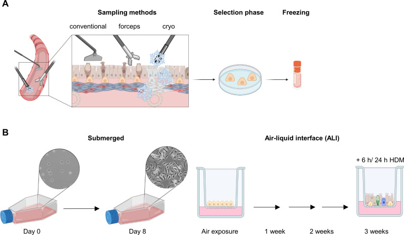
Background: Using primary airway epithelial cells (AEC) is essential to mimic more closely different types and stages of lung disease in humans while reducing or even replacing animal experiments. Access to lung tissue remains limited because these samples are generally obtained from patients who undergo lung transplantation for end-stage lung disease or thoracic surgery for (mostly) lung cancer. We investigated whether forceps or cryo biopsies are a viable alternative source of AEC compared to the conventional technique.

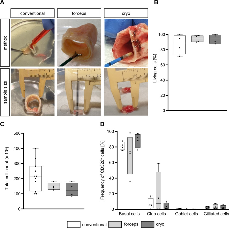
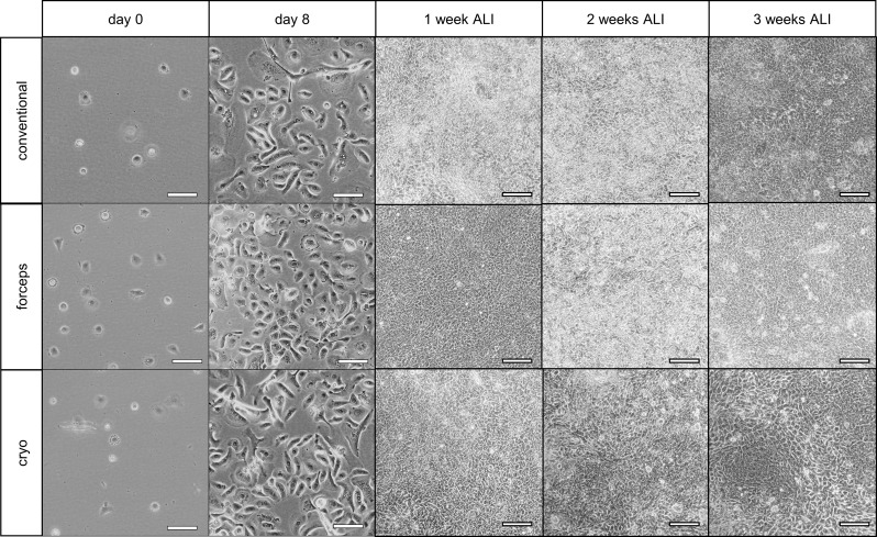
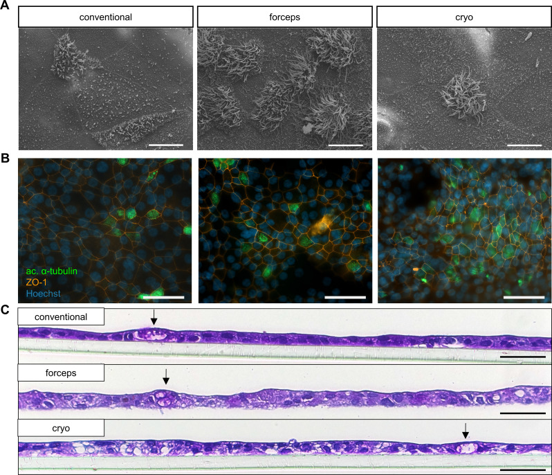
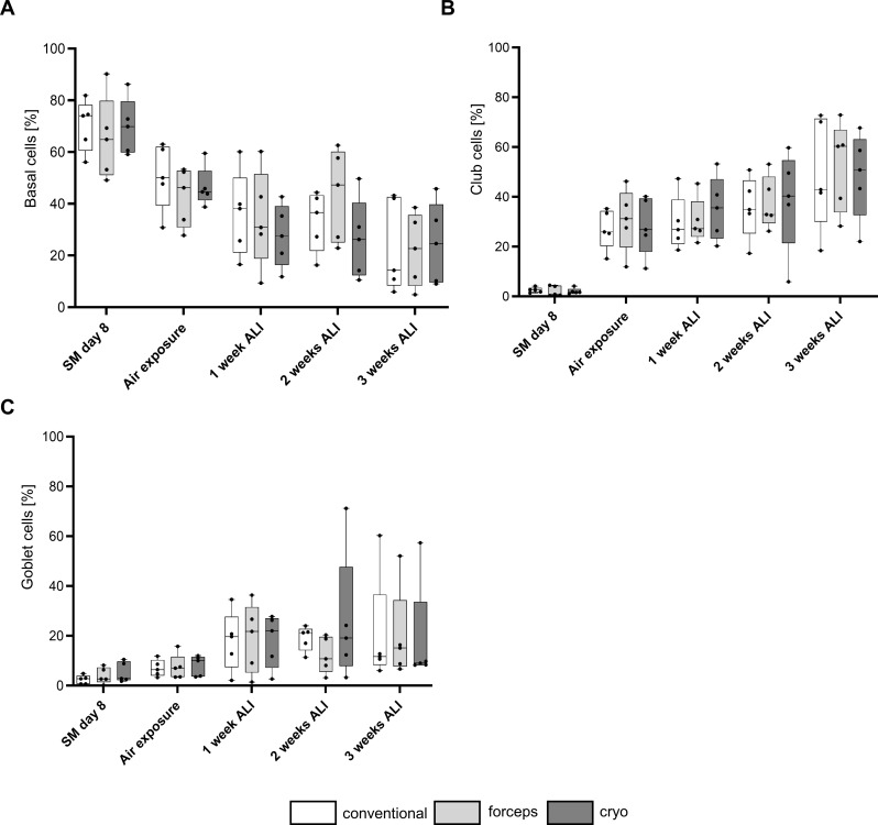
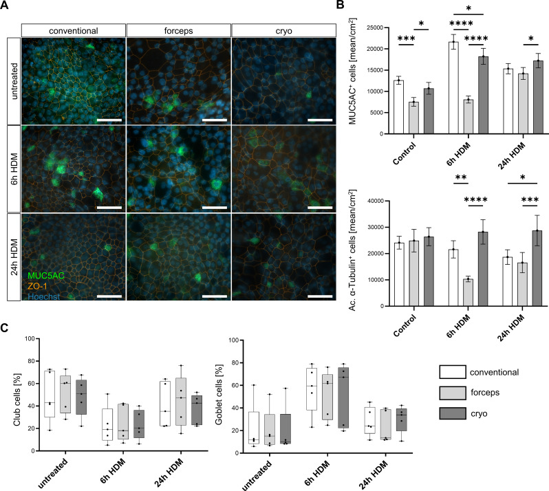
Methods: AECs were obtained ex vivo from healthy donor lung tissue using the conventional method and two biopsy procedures (forceps, cryo). The influence of the isolation method on the quality and function of AEC was investigated at different time-points during expansion and differentiation in air-liquid interface cultures. In addition, fully-differentiated AECs were stimulated with house dust mite extract (HDM) to allow functional analyses in an allergic in vitro model. Vitality or differentiation capacity were determined using flow cytometry, scanning electron microscope, periodic acid-Schiff reaction, immunofluorescence staining, and proteomics.

Results: As anticipated, no significant differences between each of the sampling methods were detected for any of the measured outcomes. The proteome composition was comparable for each isolation method, while donor-dependent effects were observed. Treatment with HDM led to minor differences in mucociliary differentiation.

Conclusions: Our findings confirmed the adequacy of these alternative approaches for attaining primary AECs, which can now expand the research for a broader range of lung diseases and for studies at an earlier stage not requiring lung surgery.

Keywords: 3R-principle; Air-liquid interface; Cryo biopsy; Forceps biopsy; In vitro disease-model; Primary airway epithelial cells.


Conflict of interest statement

Declarations. Ethics approval and consent to participate: The local ethics committee of the Medical Faculty of the University Duisburg-Essen as well as the Westdeutsche Biobank Essen approved the use, collection, and storage of lung transplant tissue (19-8717-BO,20-WBE-102) after informed written consent that was obtained from all patients or legal representatives involved in the study prior to tissue donation. Consent for publication: Not applicable. Competing interests: The authors declare no competing interests.

Figures

Fig. 1
Fig. 1
Sampling methods and cultivation of primary airway epithelial cells. (A) Tracheal tissue was resected in the course of size adaptations of donor lungs before transplantation. Three different sampling methods were used: conventional, forceps biopsy, and cryo biopsy. Epithelial cells were isolated and cultivated in specific media during the selection phase of basal cells. Basal cells were then frozen (400,000 cells/vial) and stored. (B) Isolated basal cells were expanded in submerged culture in T75 flasks for 8 days and seeded (40,000 cells) onto cell culture inserts. At a transepithelial electrical resistance value of 224 Ω/cm2, the apical medium was removed and cells were exposed to air. Cells were then cultivated for 3 weeks in air-liquid interface and harvested after 1, 2, or 3 weeks. After 3 weeks, some cells were treated with 10 µg/mL house dust mite extract (HDM) for 6–24 h to be used for functional analyzes. Figure created with Biorender
Fig. 2
Fig. 2
Sampling of primary airway epithelial cells (AECs): vitality, yield, and purity. (A) Sampling method and tissue size of the conventional method or by forceps and cryo biopsy. In the conventional method, AECs were scraped off the mucosal surface after enzymatic digestion. Forceps and cryo biopsy were taken tangentially followed by enzymatic digestion. Representative pictures of tissues sizes used for cell isolation are shown for each sampling procedure. (B) Vitality directly after scraping or biopsy. Results are shown as box and whiskers (plot: min to max, show all points, n = 4). (C) Cell yield (day 8) of each sampling method. Results are shown as box and whiskers (plot: min to max, show all points, conventional: n = 11, forceps: n = 4, cryo: n = 4). (D) Flow cytometry was performed to determine cellular composition using the following markers for each cell type of AECs (CD326+): percentage of basal (CD49f+CD271+), club (CD66a/c/e+), goblet (TSPAN8+), and ciliated cells (ac. α-tubulin+). Results are shown as mean ± standard deviation. All results were analyzed by two-way analysis of variance with Tukey’s post-hoc test
Fig. 3
Fig. 3
Cell morphology before and after air exposure. Cell morphology after thawing (day 0). After the expansion phase (day 8), cells were approximately 80–90% confluent and were transferred to the inserts. The morphology of airway epithelial cells during cultivation after 1, 2, and 3 weeks in ALI. Scale bar 100 μm
Fig. 4
Fig. 4
Differentiation of airway epithelial cells (AECs) in air-liquid interface (ALI). (A) Ciliation was analyzed by scanning electron microscopy after 3 weeks in ALI using conventional, forceps, and cryo-sampled AECs (n = 5); representative pictures are shown; scale bar 10 μm. (B) Representative pictures of ciliation were analyzed by immunofluorescence staining (green–ac. α-tubulin; blue–Hoechst, red–ZO1) after 3 weeks in ALI using conventional, forceps, and cryo-sampled AECs (n = 5; scale bar 40 μm). (C) Differentiation of ALI-AECs after 3 weeks using conventional, forceps, and cryo-sampled cells analyzed by periodic acid-Schiff reaction (PAS, n = 5); goblet cells are marked by an arrow; representative pictures are shown; scale bar 50 μm
Fig. 5
Fig. 5
Differentiation of airway epithelial cells (AECs) in air-liquid interface (ALI). Cellular composition of cultivated AECs on day 8, at air exposure, and after 1, 2, or 3 weeks in ALI was analyzed by flow cytometry (n = 5) using the following markers for each epithelial cell type (CD326+): (A) percentage of basal (CD49f+ CD271+), (B) club (CD66a/c/e+), (C) goblet (TSPAN8+). Results are expressed as mean ± standard deviation (plot: min to max, show all points). Statistical analyses were performed by two-way analysis of variance with Tukey’s post-hoc test
Fig. 6
Fig. 6
Influence of house dust mite extract (HDM) on differentiation of airway epithelial cells (AECs). After 3 weeks in ALI, AECs from conventional, forceps, and cryo-sampled cells were untreated or stimulated with HDM for 6–24 h (n = 4–5). (A) Immunofluorescence staining of goblet cells (green–MUC5AC; blue–Hoechst, red–ZO1) (n = 5; scale bar 40 μm); representative pictures are shown. (B) Numbers by amount of MUC5AC+ goblet cell or ac. α-tubulin+ cells were determined per cm2 (n = 4–5). (C). Analyses of goblet cells (TSPAN8+) and club cells (CD66a/c/e+) by flow cytometry (n = 5). (B) + (C) Results are expressed as mean ± standard deviation (plot: min to max, show all points). Statistical analyses were performed by two-way analysis of variance with Tukey’s post-hoc test, *p < 0.05; **p < 0.01; ***p < 0.001; ****p < 0.0001
Fig. 7
Fig. 7
Influence of the sampling method on the proteome. Protein expression patterns of airway epithelial cells (AECs) after 8 days of expansion of basal cells and after 3 weeks in ALI of each sample are shown as a heat map. The three different sampling methods (conventional, forceps, and cryo) were compared, and three healthy subjects were analyzed

Similar articles

Cited by

  • Mycophenolic acid treatment drives the emergence of novel SARS-CoV-2 variants.
    Meister TL, Nocke MK, Heinen N, Burkard TL, Brüggemann Y, Westhoven S, Trüeb B, Ebert N, Thomann L, Lubieniecki KP, Lubieniecka JM, Döring K, Herrmann M, Haid S, Pietschmann T, Wiegmann B, Tao R, Pfefferle S, Addo MM, Thiel V, Drexler I, Babel N, Phuc Nguyen H, Brown RJP, Todt D, Steinmann E, Pfaender S. Meister TL, et al. Proc Natl Acad Sci U S A. 2025 Jul 15;122(28):e2500276122. doi: 10.1073/pnas.2500276122. Epub 2025 Jul 9. Proc Natl Acad Sci U S A. 2025. PMID: 40632557 Free PMC article.

References

    1. Parekh KR, Nawroth J, Pai A, Busch SM, Senger CN, Ryan AL. Stem cells and lung regeneration. Am J Physiol Cell Physiol. 2020;319:C675–93. 10.1152/ajpcell.00036.2020. - PMC - PubMed
    1. Ma J, Rubin BK, Voynow JA. Mucins, mucus, and Goblet cells. Chest. 2018;154:169–76. 10.1016/j.chest.2017.11.008. - PubMed
    1. Whitsett JA. Airway Epithelial differentiation and Mucociliary Clearance. Ann Am Thorac Soc. 2018;15:S143–8. 10.1513/AnnalsATS.201802-128AW. - PMC - PubMed
    1. Whitsett JA, Alenghat T. Respiratory epithelial cells orchestrate pulmonary innate immunity. Nat Immunol. 2015;16:27–35. 10.1038/ni.3045. - PMC - PubMed
    1. Carlier FM, de Fays C. Epithelial barrier dysfunction in Chronic Respiratory diseases. Front Physiol. 2021;12:691227. 10.3389/fphys.2021.691227. - PMC - PubMed

LinkOut - more resources