. 2024 Dec 24;25(1):440.
doi: 10.1186/s12931-024-03076-7.

Ferritinophagy mediated by the AMPK/ULK1 pathway is involved in ferroptosis subsequent to ventilator-induced lung injury

Affiliations

Ferritinophagy mediated by the AMPK/ULK1 pathway is involved in ferroptosis subsequent to ventilator-induced lung injury

Huajin Ou et al. Respir Res. .

Abstract

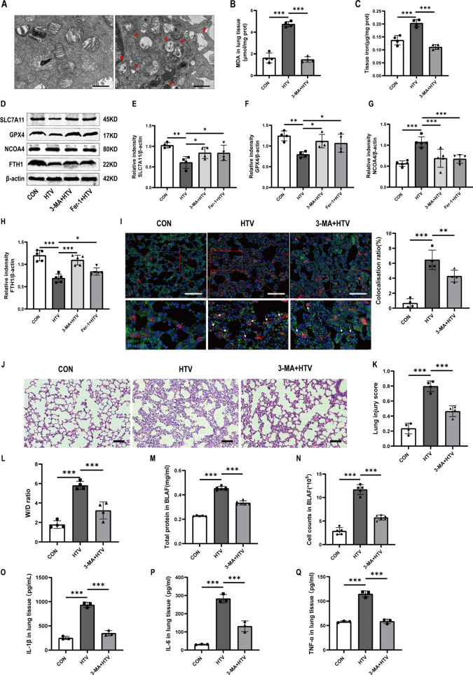
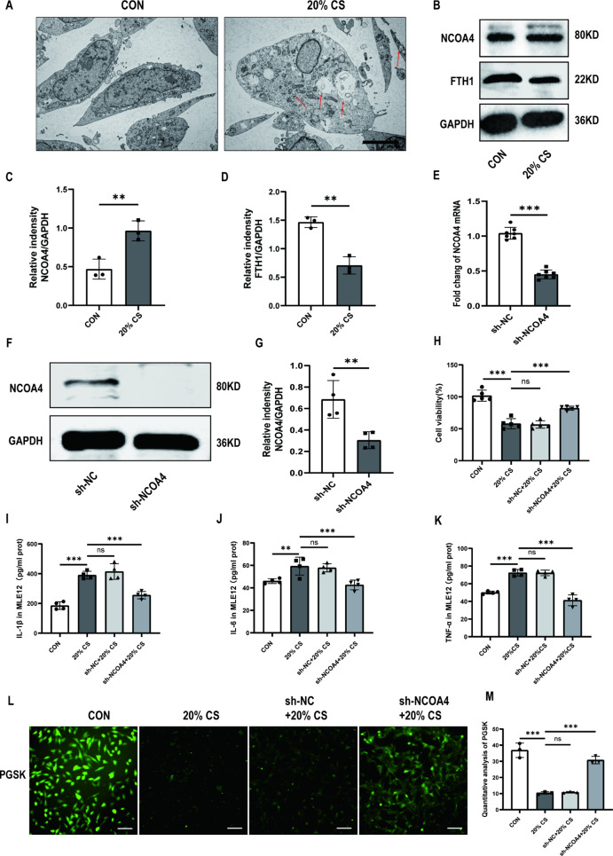
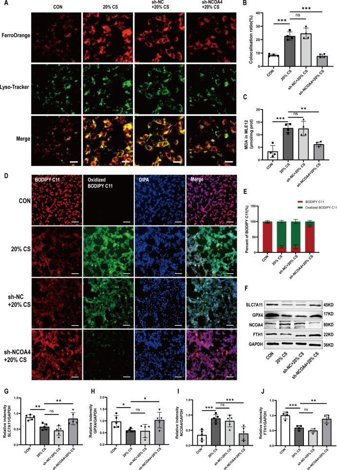
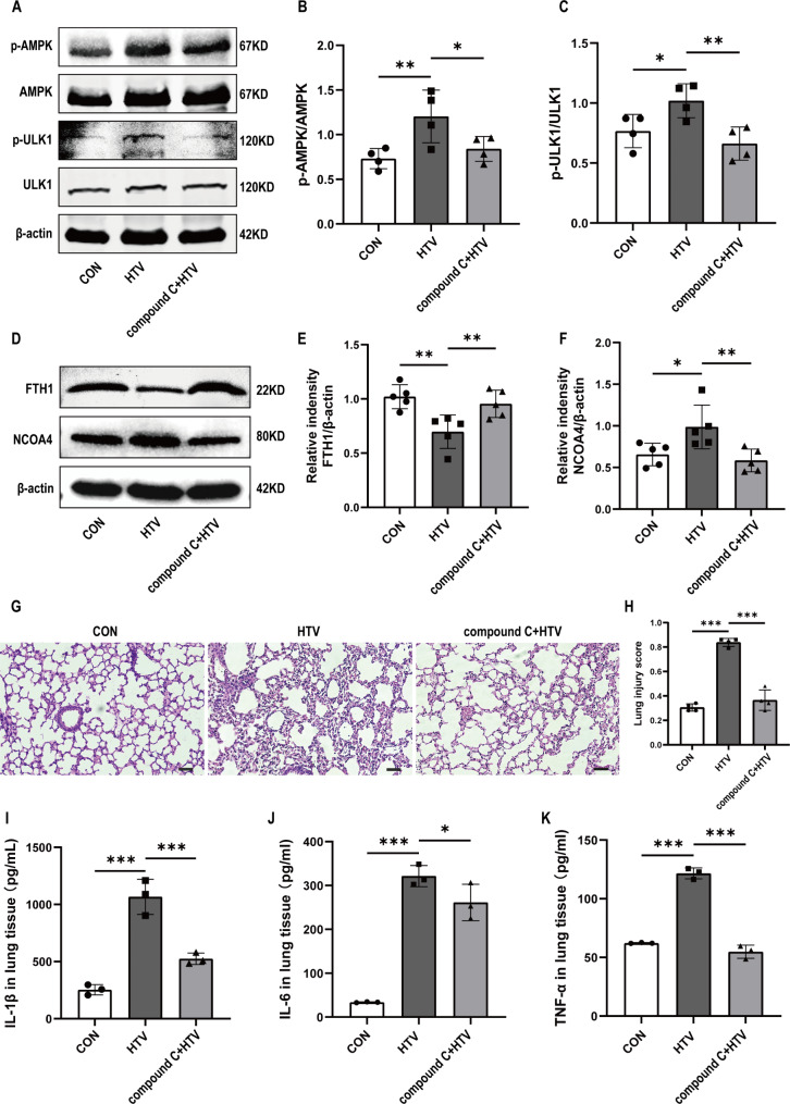
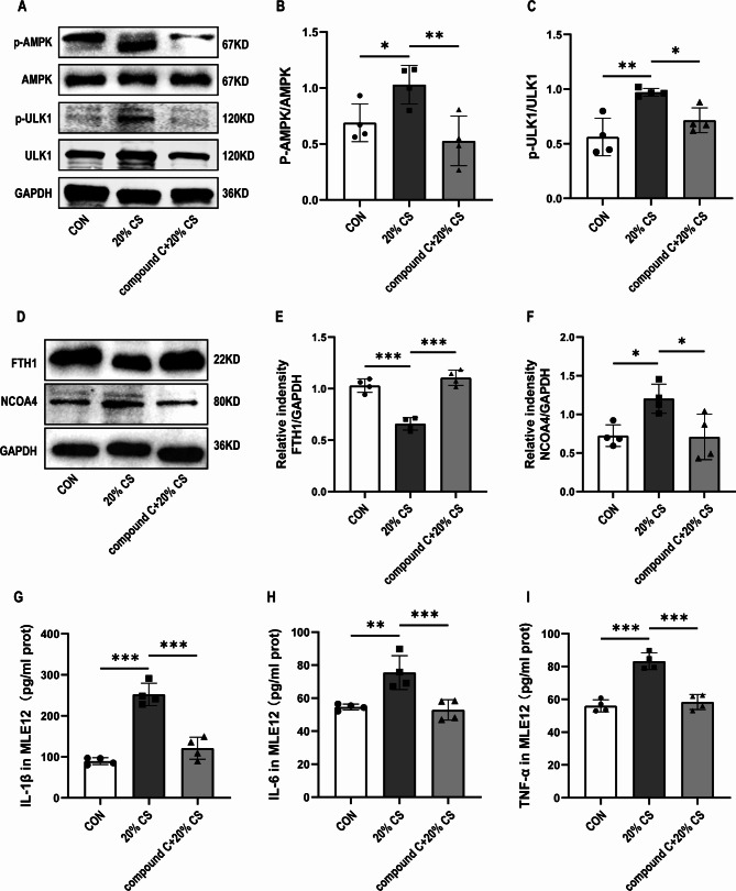
Mechanical ventilation (MV) remains a cornerstone of critical care; however, its prolonged application can exacerbate lung injury, leading to ventilator-induced lung injury (VILI). Although previous studies have implicated ferroptosis in the pathogenesis of VILI, the underlying mechanisms remain unclear. This study investigated the roles of ferritinophagy in ferroptosis subsequent to VILI. Using C57BL/6J mice and MLE-12 cells, we established both in vivo and in vitro models of VILI and cyclic stretching (CS)-induced cellular injury. We assessed lung injury and the biomarkers of ferroptosis and ferritinophagy, after appropriate pretreatments. This study demonstrated that high tidal volumes (HTV) for 4 h enhanced the sensitivity to ferroptosis in both models, evidenced by increased intracellular iron levels, lipid peroxidation and cell death, which can be mitigated by ferrostatin-1 treatment. Notably, nuclear receptor coactivator 4 (NCOA4)-mediated ferritinophagy contributed to ferroptosis in VILI. Inhibition of autophagy with 3-methyladenine or NCOA4 knockdown decreased intracellular Fe2+ levels and inhibited lipid peroxidation, thereby attenuating CS-induced lung injury. Furthermore, it has also been observed that the AMPK/ULK1 axis can trigger ferritinophagy in VILI. Collectively, our study indicated that MV can induce ferroptosis by promoting NCOA4-dependent ferritinophagy, which could be a novel therapeutic target for the prevention and treatment of VILI.

Keywords: AMPK-ULK1 axis; Ferritinophagy; Ferroptosis; Ventilation-induced lung injury.


Conflict of interest statement

Declarations. Ethics approval: Animal studies were reviewed and approved by the Institutional Animal Care and Use Committee of Guangxi Medical University Cancer Hospital. Consent to participate: Not applicable. Competing interests: The authors declare no competing interests.

Figures

Fig. 1
Fig. 1
Ferritinophagy participated in ventilator-induced ferroptosis in vivo. (A) TEM images depict lung tissue sections from both groups. Obvious autophagosomes were found in the HTV group. Red arrows represent autophagosome. Magnifications: 6000 X and acceleration voltage: 80 KV. Scale bar: 1 μm. (B) MDA levels in lung tissues (n = 4). (C) The iron content in the lung tissues of mice (n = 4). (D) Representative Western blotting bands for SLC7A11, GPX4, NCOA4, FTH1, and β-actin in lung tissues. (E-H) Relative protein expression of SLC7A11, GPX4, NCOA4, and FTH1 to β-actin (n = 4). (I) Immunofluorescence assay demonstrated that the co-localization of FTH1 and NCOA4 significantly increased in the HTV group compared to other groups. Scale bar: 100 μm. The percentage of co-localization between FTH1 and NCOA4 was quantified using ImageJ Fiji software (n = 4). (J) H&E staining was performed on tissue samples from both groups, with a scale bar indicating 100 μm. (K) Pathological scoring was conducted based on the H&E staining results (n = 4). (L) Wet/Dry ratios of lung tissue (n = 4). (M) Total protein concentrations in BALF (n = 6). (N) Infiltrating cell counts in BALF (n = 6). (O-Q) The levels of IL-1β, IL-6, and TNF-α in lung tissue (n = 3). Data are expressed as mean ± SD. “*” indicates significant difference between groups (*p < 0.05, ** p < 0.01 or *** p < 0.001)
Fig. 2
Fig. 2
Ferritinophagy promotes ventilator-induced ferroptosis of MLE-12 cells through ferritin degradation mediated by NCOA4. (A) Representative TEM images of MLE12 cells untreated or treated with 20% CS. Red arrows represent autophagosomes. Magnifications: 5000 X, acceleration voltage: 80 kV. Scale bar: 5.0 μm. (B) Representative Western blotting bands of NCOA4, FTH1, and GAPDH in MLE12 cells. (C, D) Relative protein expression of NCOA4 and FTH1 was divided into GAPDH (n = 3). (E) The mRNA levels of NCOA4 knockdown in MLE12 cells (n = 7). (F) Representative Western blotting images of NCOA4 and GAPDH in MLE12 cells. (G) The protein expression of NCOA4 relative to GAPDH (n = 4). (H) Cell viability was detected using a CCK8 assay (n = 5). (I-K) Levels of IL-1β, IL-6, and TNF-α in MLE12 cells (n = 4). (L) PGSK probe staining for Fe2+ in MLE12 cells with different treated groups. Scale bar: 100 μm. (M) Quantification of the fluorescence intensity of PGSK using ImageJ Fiji software (n = 3). Data are expressed as mean ± SD. “*” indicates significant differences between groups (*p < 0.05, ** p < 0.01 or *** p < 0.001)
Fig. 3
Fig. 3
Knockdown NCOA4 contributed to reducing ferroptosis caused by ferritinophagy in MLE-12 cells with cyclic overstretching. (A) Immunofluorescence images of the co-localization of intracellular and lysosomal Fe2+. Scale bar: 20 μm. (B) Quantification of the fluorescence intensity of FerroOrange co-localized with Lyso-Tracker Green by ImageJ Fiji software (n = 4). (C) MDA levels in MLE12 cells (n = 4). (D) Immunofluorescence images of C11-BODIPY in MLE12 cells. Scale bar: 50 μm. (E) Percent of the fluorescence intensity of BODIPY C11 and oxidized BODIPY C11 measured by ImageJ Fiji software. (F) Representative Western blotting images of SLC7A11, GPX4, NCOA4, FTH1, and GAPDH in MLE12 cells. (G-J) Relative protein expression levels of SLC7A11, GPX4, NCOA4 and FTH1 to GAPDH (n = 5). Data are expressed as mean ± SD. “*” indicates significant difference between groups (*p < 0.05, ** p < 0.01 or *** p < 0.001)
Fig. 4
Fig. 4
The AMPK/ULK1 axis was involved in the activation of ferritinophagy activation induced by ventilator-induced lung injury in mice. (A) Representative Western blotting images of p-AMPK, AMPK, p-ULK1, ULK1, and β-actin in lung tissues. (B) The protein expression of P-AMPK relative to AMPK (n = 4). (C) The protein expression of P-ULK1 relative to ULK1 (n = 4). (D) Representative Western blotting images of FTH1, NCOA4, and β-actin in lung tissues. (E, F) The protein expression of FTH1 and NCOA4 relative to β-actin (n = 5). (G) H&E staining was performed on tissue samples from both groups, with a scale bar indicating 100 μm. (H) Pathological scoring was conducted based on the H&E staining results (n = 4). (I) IL-1β levels in lung tissue (n = 3). (J) IL-6 levels in the lung tissue (n = 3). (K) TNF-α levels in the lung tissue (n = 3). Data are expressed as mean ± SD. “*” indicates significant difference between groups (*p < 0.05, ** p < 0.01 or *** p < 0.001)
Fig. 5
Fig. 5
The AMPK-ULK1 axis is involved in ferritinophagy activation induced by cyclic overstretching in vitro. (A) Representative Western blotting images of p-AMPK, AMPK, p-ULK1, ULK1, and GAPDH in MLE12 cells. (B) The protein expression levels of p-AMPK relative to AMPK (n = 4). (C) The protein expression of p-ULK1 relative to ULK1 (n = 4). (D) Representative Western blotting images of FTH1, NCOA4, and GAPDH in MLE12 cells. (E, F) Relative protein expression of FTH1 and NCOA4 relative to GAPDH (n = 4). (G-I) The levels of IL-1β, IL-6, and TNF-α in MLE12 cells (n = 4). Data are expressed as mean ± SD. “*” indicates significant difference between groups (*p < 0.05, ** p < 0.01 or *** p < 0.001)

Similar articles

Cited by

References

    1. Bellani G, Laffey JG, Pham T, Fan E, Brochard L, Esteban A, Gattinoni L, van Haren F, Larsson A, McAuley DF, et al. Epidemiology, patterns of Care, and mortality for patients with Acute Respiratory Distress Syndrome in Intensive Care Units in 50 countries. JAMA. 2016;315:788–800. - PubMed
    1. Mart MF, Ware LB. The long-lasting effects of the acute respiratory distress syndrome. Expert Rev Respir Med. 2020;14:577–86. - PMC - PubMed
    1. Battaglini D, Fazzini B, Silva PL, Cruz FF, Ball L, Robba C, et al. Challenges in ARDS definition, management, and identification of effective personalized therapies. J Clin Med. 2023;12(4):1381. 10.3390/jcm12041381. PMID: 36835919; PMCID: PMC9967510. - PMC - PubMed
    1. Yamamoto R, Okazaki SR, Fujita Y, Seki N, Kokei Y, Sekine S, Wada S, Norisue Y, Narita C. Usefulness of low tidal volume ventilation strategy for patients with acute respiratory distress syndrome: a systematic review and meta-analysis. Sci Rep. 2022;12:9331. - PMC - PubMed
    1. Slutsky AS, Ranieri VM. Ventilator-induced lung injury. N Engl J Med. 2013;369:2126–36. - PubMed

MeSH terms

LinkOut - more resources