

Quantitative analysis of protective Kirschner wire diameters in lateral opening wedge distal femoral osteotomy: A finite element study
- PMID: 39719906
- PMCID: PMC11734846
- DOI: 10.52312/jdrs.2025.1806
Quantitative analysis of protective Kirschner wire diameters in lateral opening wedge distal femoral osteotomy: A finite element study
Abstract
Objectives: This study aims to investigate quantitatively the protective effect of a 1.6-mm or a 2.5-mm Kirschner wire (K-wire) on the medial hinge at different gap distances through finite element analysis (FEA) and to establish whether using a 2.5-mm K-wire can offer benefits compared to a 1.6-mm in preventing medial hinge fractures.
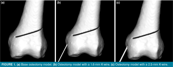
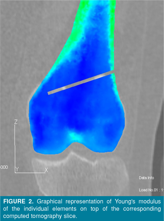
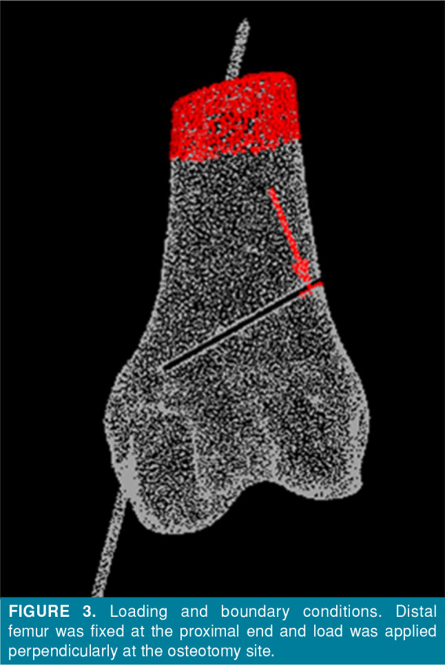
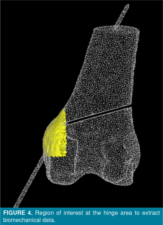
Materials and methods: Between June 2024 and July 2024, three different models simulating a lateral opening wedge (LOW) osteotomy of the distal femur were created from a femoral computed tomography (CT) scan of a 36-year-old male patient: no K-wire (Model I), 1.6-mm K-wire (Model II), and 2.5-mm K-wire (Model III). Finite element analysis was performed to simulate 7- to 13-mm gaps at the osteotomy site. Loads, principal stress, strain, and equivalent stress were analyzed around the medial hinge.
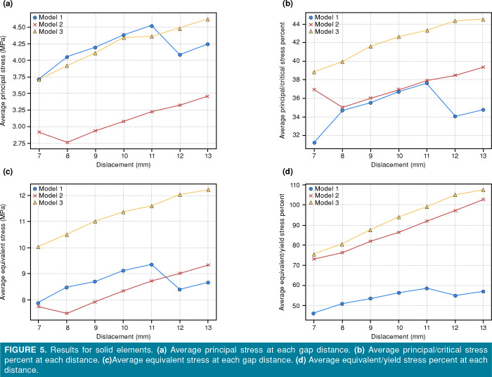
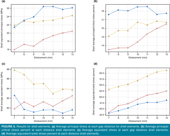
Results: Model I required 123.0±5.2 N, Model II required 181.7±12.2 N, and Model III required 228.7±13.6 N (p<0.001). Cracked shell elements were the lowest in Model II and the highest in Model I. While the average equivalent/yield stress ratio was not significantly lower in Model II compared to Model III (87.0±10.9% vs. 92.7±12.1%), the maximum equivalent/yield stress ratio values in Model II were significantly lower than both Model I and Model III (1206.2±138.3% vs. 1836.2±165.4% and 1689.1±404.0%, respectively), suggesting a superior dispersion of forces.
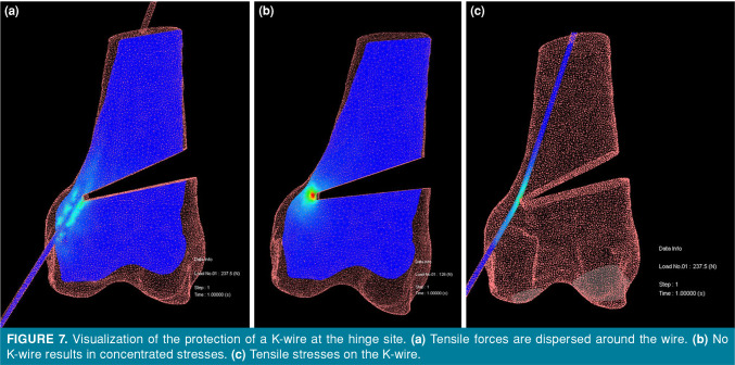
Conclusion: Using a 1.6-mm K-wire during LOW osteotomy of the distal femur provides a balance between structural reinforcement and stress distribution, significantly improving stability and reducing the risk of medial hinge fractures compared to a 2.5-mm K-wire or no K-wire. The 1.6-mm K-wire optimizes stress dispersion, making it the preferred choice for surgical planning in lateral opening wedge distal femoral osteotomy.
Conflict of interest statement
Figures
Similar articles
-
Effects of K-wire diameter and insertion angle on femoral bone medial closing-wedge osteotomies: a finite element study.Sci Rep. 2025 Jun 20;15(1):20116. doi: 10.1038/s41598-025-04260-5. Sci Rep. 2025. PMID: 40541981 Free PMC article.
-
Evaluation of the effectiveness of Kirschner wires in preventing lateral hinge fractures in high tibial osteotomy: A finite element analysis.Jt Dis Relat Surg. 2025 Jul 21;36(3):683-695. doi: 10.52312/jdrs.2025.2252. Epub 2025 Jul 21. Jt Dis Relat Surg. 2025. PMID: 40784001
-
The effect of lateral hinge fracture with hinge hole and protective K-wire for medial opening-wedge high tibial osteotomy by compression testing and finite element analysis.J Orthop Surg Res. 2025 Jun 17;20(1):598. doi: 10.1186/s13018-025-05993-9. J Orthop Surg Res. 2025. PMID: 40528197 Free PMC article.
-
The Ideal Location of the Lateral Hinge in Medial Closing Wedge Osteotomy of the Distal Femur: Analysis of Soft Tissue Coverage and Bone Density.Am J Sports Med. 2019 Oct;47(12):2945-2951. doi: 10.1177/0363546519869325. Epub 2019 Aug 29. Am J Sports Med. 2019. PMID: 31465238
-
Lateral Opening Wedge Distal Femoral Osteotomy for Lateral Compartment Arthrosis/Overload.Clin Sports Med. 2019 Jul;38(3):351-359. doi: 10.1016/j.csm.2019.02.004. Epub 2019 Mar 26. Clin Sports Med. 2019. PMID: 31079767 Review.
References
-
- Liu XY, Yu QP, Chen XM, Zeng WN, Zhou ZK. Effects of preoperative valgus deformity in patients undergoing neutrally aligned total knee arthroplasty: A retrospective cohort study with a minimum five-year follow-up. Jt Dis Relat Surg. 2024;35:529–537. doi: 10.52312/jdrs.2024.1800. - DOI - PMC - PubMed
-
- Matsuoka M, Kondo E, Iwasaki K, Onodera T, Nakamura R, Nakayama H, et al. Simultaneous medial closing wedge distal femoral varus osteotomy and double-bundle anterior cruciate ligament reconstruction in the symptomatic femoral valgus deformity: A case report. Jt Dis Relat Surg. 2024;35:422–432. doi: 10.52312/jdrs.2023.1176. - DOI - PMC - PubMed
-
- Winkler PW, Rupp MC, Lutz PM, Geyer S, Forkel P, Imhoff AB, et al. A hinge position distal to the adductor tubercle minimizes the risk of hinge fractures in lateral open wedge distal femoral osteotomy. Knee Surg Sports Traumatol Arthrosc. 2021;29:3382–3391. doi: 10.1007/s00167-020-06244-6. - DOI - PMC - PubMed
MeSH terms
LinkOut - more resources
Full Text Sources
Miscellaneous
