. 2025 Jan 2;36(1):200-209.
doi: 10.52312/jdrs.2025.2042. Epub 2024 Dec 18.

The effects of gabapentin and pregabalin on fracture healing: A histological, radiological, and biomechanical analysis

Affiliations

The effects of gabapentin and pregabalin on fracture healing: A histological, radiological, and biomechanical analysis

Ahmet Emrah Açan et al. Jt Dis Relat Surg. .

Abstract

Objectives: This study evaluated the impact of different doses of gabapentin and pregabalin on fracture healing in a rat femoral shaft model, with histological, radiological, and biomechanical assessments.

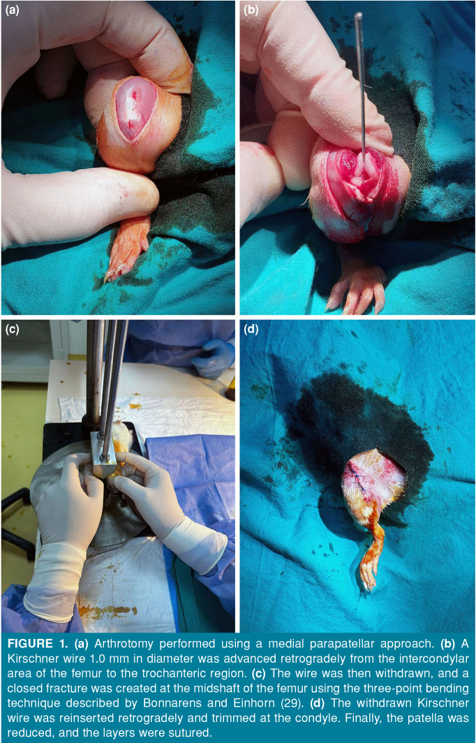
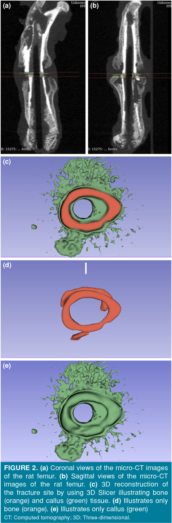
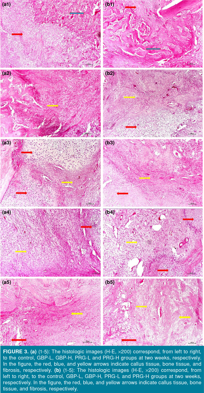
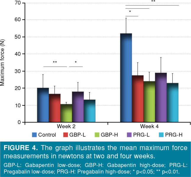
Materials and methods: Seventy male Wistar albino rats were divided into five groups: control, low-dose gabapentin (GBP-L, 300 mg/day), high-dose gabapentin (GBP-H, 3600 mg/day), low-dose pregabalin (PRG-L, 150 mg/day), and high-dose pregabalin (PRG-H, 600 mg/day), based on human equivalent doses. Bilateral femoral fractures were induced; the right femurs were prepared for radiological examination using microtomography, followed by histological analysis, whereas the left femurs were allocated for biomechanical testing. Drug administration began three weeks preoperatively and continued until sacrifice at either two or four weeks. Histological assessments included inflammation and transformation scoring and microtomography-measured callus volume. Biomechanical testing assessed maximum force and stiffness.

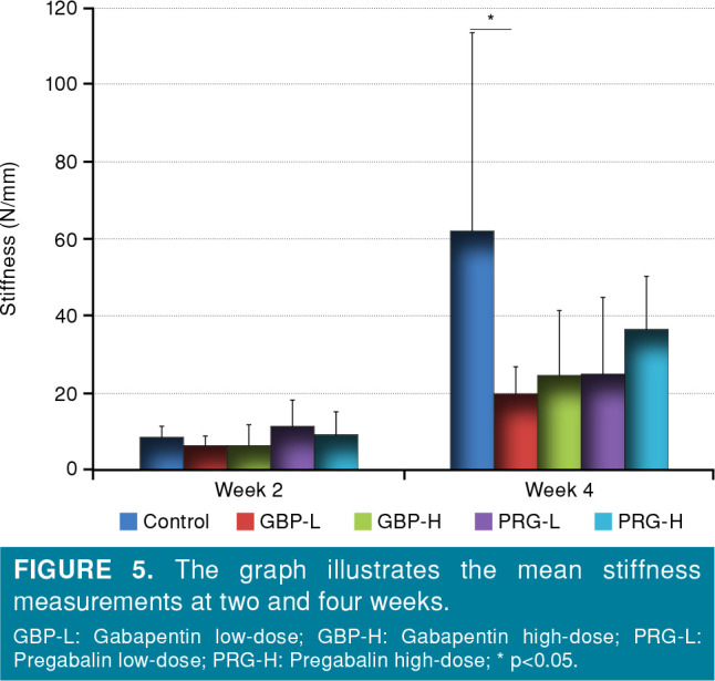
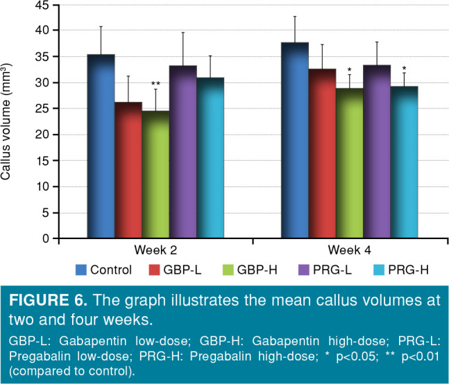
Results: At the fourth week, inflammation levels were significantly higher in the GBP-H, PRG-L, and PRG-H groups compared to control (p<0.01, p<0.05, and p<0.01), while transformation scores were significantly lower in these groups (p<0.01, p<0.05, and p<0.001). Low-dose pregabalin showed a borderline transformation difference (p=0.051). Microtomography analysis showed that the GBP-H group had significantly reduced callus volume versus control by the second week (p<0.01), persisting at a lower significance by week four (p<0.05). By the fourth week, PRG-H also had reduced callus volume (p<0.05). Maximum force values by the fourth week were significantly lower in the GBP-L, GBP-H, and PRG-H groups compared to control (p<0.05 for GBP-L; p<0.01 for GBP-H and PRG-H).

Conclusion: These findings suggest that these drugs, particularly with their high-dose applications, may lead to prolonged inflammation and hinder fracture healing by reducing callus volume and biomechanical integrity, potentially disrupting the transition from the inflammatory to reparative phases of healing.


Conflict of interest statement

Conflict of Interest: The authors declared no conflicts of interest with respect to the authorship and/or publication of this article.

Figures

Figure 1
Figure 1. (a) Arthrotomy performed using a medial parapatellar approach. (b) A Kirschner wire 1.0 mm in diameter was advanced retrogradely from the intercondylar area of the femur to the trochanteric region. (c) The wire was then withdrawn, and a closed fracture was created at the midshaft of the femur using the three-point bending technique described by Bonnarens and Einhorn (29). (d) The withdrawn Kirschner wire was reinserted retrogradely and trimmed at the condyle. Finally, the patella was reduced, and the layers were sutured.
Figure 2
Figure 2. (a) Coronal views of the micro-CT images of the rat femur. (b) Sagittal views of the micro-CT images of the rat femur. (c) 3D reconstruction of the fracture site by using 3D Slicer illustrating bone (orange) and callus (green) tissue. (d) Illustrates only bone (orange). (e) Illustrates only callus (green): CT: Computed tomography; 3D: Three-dimensional.
Figure 3
Figure 3. (a) (1-5): The histologic images (H-E, ×200) correspond, from left to right, to the control, GBP-L, GBP-H, PRG-L and PRG-H groups at two weeks, respectively. In the figure, the red, blue, and yellow arrows indicate callus tissue, bone tissue, and fibrosis, respectively. (b) (1-5): The histologic images (H-E, ×200) correspond, from left to right, to the control, GBP-L, GBP-H, PRG-L and PRG-H groups at two weeks, respectively. In the figure, the red, blue, and yellow arrows indicate callus tissue, bone tissue, and fibrosis, respectively.
Figure 4
Figure 4. The graph illustrates the mean maximum force measurements in newtons at two and four weeks.: GBP-L: Gabapentin low-dose; GBP-H: Gabapentin high-dose; PRG-L: Pregabalin low-dose; PRG-H: Pregabalin high-dose; * p<0.05; ** p<0.01.
Figure 5
Figure 5. The graph illustrates the mean stiffness measurements at two and four weeks.: GBP-L: Gabapentin low-dose; GBP-H: Gabapentin high-dose; PRG-L: Pregabalin low-dose; PRG-H: Pregabalin high-dose; * p<0.05.
Figure 6
Figure 6. The graph illustrates the mean callus volumes at two and four weeks.: GBP-L: Gabapentin low-dose; GBP-H: Gabapentin high-dose; PRG-L: Pregabalin low-dose; PRG-H: Pregabalin high-dose; * p<0.05; ** p<0.01 (compared to control).

Similar articles

Cited by

References

    1. Tzioupis C, Giannoudis PV. Prevalence of long-bone nonunions. S3-9Injury. 2007;38 Suppl 2 doi: 10.1016/s0020-1383(07)80003-9. - DOI - PubMed
    1. Clark D, Nakamura M, Miclau T, Marcucio R. Effects of Aging on fracture healing. Curr Osteoporos Rep. 2017;15:601–608. doi: 10.1007/s11914-017-0413-9. - DOI - PMC - PubMed
    1. Tanios M, Brickman B, Cage E, Abbas K, Smith C, Atallah M, et al. Diabetes and impaired fracture healing: A narrative review of recent literature. Curr Osteoporos Rep. 2022;20:229–239. doi: 10.1007/s11914-022-00740-z. - DOI - PubMed
    1. Stroud S, Katyal T, Gornitzky AL, Swarup I. Effect of nonsteroidal anti-inflammatory drugs on fracture healing in children: A systematic review. World J Orthop. 2022;13:494–502. doi: 10.5312/wjo.v13.i5.494. - DOI - PMC - PubMed
    1. Yurteri A, Yildirim A, Çelik ZE, Vatansev H, Durmaz MS. The effect of quercetin on bone healing in an experimental rat model. Jt Dis Relat Surg. 2023;34:365–373. doi: 10.52312/jdrs.2023.870. - DOI - PMC - PubMed

MeSH terms