. 2024 Nov 14:49:100483.
doi: 10.1016/j.ahjo.2024.100483. eCollection 2025 Jan.

Accurate diagnosis of ischemic heart disease without exposure to radiation using non-stress unshielded magnetocardiography

Affiliations

Accurate diagnosis of ischemic heart disease without exposure to radiation using non-stress unshielded magnetocardiography

Kirsten Tolstrup et al. Am Heart J Plus. .

Abstract

Study objectives: To evaluate the capability and accuracy of magnetocardiography (MCG) to identify patients with ischemic chest pain from those with non-ischemic pain and to verify normalcy in the MCG in healthy subjects.

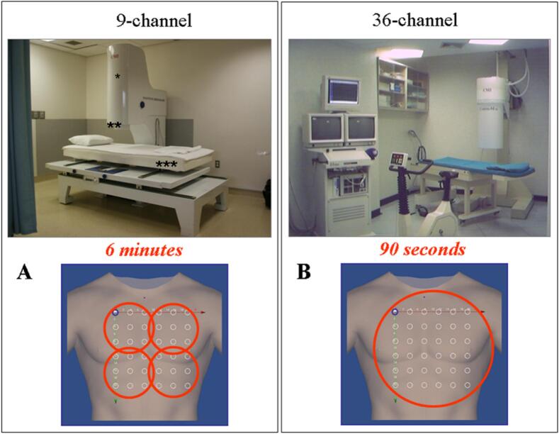
Design: We studied 133 patients (mean age 59 ± 14 years, 69 % male) with chronic or acute chest pain syndrome and 63 healthy subjects (mean age 41.7 ± 12.2 years, 51 % male) using unshielded cryogenically cooled MCG systems (Cardiomag Imaging Inc., 9 and 36 channels) in a general clinical setting. Scan time was 90 s to 6 min. Interventions: The MCG data were processed with the same automated analysis software and results were immediately available. All patients were chest pain free at the time of scanning.

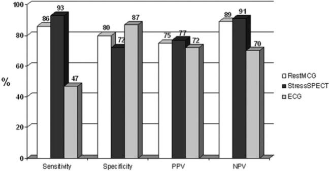
Results: A diagnosis of ischemic chest pain was established in 41 % after non-invasive and invasive testing. Rest MCG was normal in all healthy subjects. An abnormal rest MCG was strongly associated with ischemic chest pain, p < 0.0001 (sensitivity of 86 %, specificity of 80 %, positive (PPV) and negative predictive value (NPV) of 75 % and 89 %, respectively). In comparison, the sensitivity, specificity, PPV and NPV of stress SPECT was 93 %, 72 %, 77 % and 91 %, respectively.

Conclusion: Resting MCG is a rapid risk-free method for the detection of ischemic chest pain without the use of radiation or contrast with results comparable with stress SPECT.

Keywords: Angina; Coronary artery disease; Ischemia; Magnetocardiography.


Conflict of interest statement

The authors declare that they have no known competing financial interests or personal relationships that could have appeared to influence the work reported in this paper.

Figures

Fig. 1
Fig. 1
(A) 9-channel MCG. * Sensor tower containing the cryostat with nine SQUIDs; ** Gantry tower containing the cryostat and SQUID electronics; *** Couch for sequential mapping from four positions over the chest (also shown at the bottom). (B) 36-channel MCG. This system maps the same grid points with a single recording. SQUID = super conducting quantum interference device.
Fig. 2
Fig. 2
A) Magnetic field map imaging of a 41 year old male with known IHD, new class IV angina and non-ischemic 12‑lead ECG. LVEF was 51 % with inferior wall motion abnormality. Coronary angiography of patient 2A demonstrated 2 vessel CAD. MCG obtained same day was abnormal (increased dynamic motion of the effective dipole vector as indicated by the vectors shown in white). B) 50 year old male with body mass index of 36.2 kg/m2, atypical chest pain and normal 12‑lead ECG. Stress nuclear scan showed 15 % reversible defect inferiorly. Cardiac catheterization revealed no CAD. MCG performed on the same day was normal. (Stable motion -no dispersion- of the effective dipole vector as indicated by the vectors shown in white). C) MCG of a 40 year old healthy female demonstrating a very stable dipole vector during repolarization. All images show the MCG time traces of individual channels (*), superposition of MCG traces of the same beat acquired on all channels; red vertical lines show the repolarization epoch between T3 and T4 (**), and the magnetic field topography maps (***). The white vectors demonstrate the magnetic dipole vector trajectory during ventricular repolarization. The vectors (indicated by solid white arrow sequences, each representing a single timepoint within the T3 and T4 interval (repolarization epoch) in the ischemic patient (Fig. 2A) show dispersion in repolarization which is also apparent in the lack of clear dipolar distribution in magnetic field map (there is no clear circular red pole); the non-ischemic patient (Fig. 2B) and the normal healthy subject (Fig. 2C) show no dispersion and clear dipolar distribution in magnetic field map. (For interpretation of the references to colour in this figure legend, the reader is referred to the web version of this article.)
Fig. 3
Fig. 3
Diagnostic value of rest MCG (white bars), stress SPECT (black bars) and 12‑lead ECG (grey bars) for the diagnosis of ischemia. MCG = Magnetocardiography; SPECT = single photon emission tomography; ECG = Electrocardiography; PPV = Positive Predictive Value; NPV = Negative Predictive Value.

Similar articles

Cited by

References

    1. Brisinda D., Fenici P., Fenici R. Clinical magnetocardiography: the unshielded bet-past, present, and future. Front Cardiovasc Med. 2023 Aug;10(10) doi: 10.3389/fcvm.2023.1232882. - DOI - PMC - PubMed
    1. Her A.Y., Dischl D., Kim Y.H., Kim S.W., Shin E.S. Magnetocardiography for the detection of myocardial ischemia. Front Cardiovasc Med. 2023 Jul;7(10) doi: 10.3389/fcvm.2023.1242215. - DOI - PMC - PubMed
    1. Camm A.J., Henderson R., Brisinda D., Body R., Charles R.G., Varcoe B., Fenici R. Clinical utility of magnetocardiography in cardiology for the detection of myocardial ischemia. J. Electrocardiol. 2019 Nov-Dec;57:10–17. doi: 10.1016/j.jelectrocard.2019.07.009. (Epub 2019 Jul 25) - DOI - PubMed
    1. Strand S.A., Strasburger J.F., Wakai R.T. Fetal magnetocardiogram waveform characteristics. Physiol. Meas. 2019 Mar 22;40(3) doi: 10.1088/1361-6579/ab0a2c. - DOI - PMC - PubMed
    1. Wikswo J., Fairbank W. Application of superconducting magnetometers to the measurement of the vector magnetocardiogram. IEEE Trans. Magn. 1977;13(1):354–357. doi: 10.1109/TMAG.1977.1059333. - DOI