. 2024 Dec 17:2024:4811500.
doi: 10.1155/mi/4811500. eCollection 2024.

Hypoxia-Inducible Factor-1α Modulates the Toll-Like Receptor 4/Nuclear Factor Kappa B Signaling Pathway in Experimental Necrotizing Enterocolitis

Affiliations

Hypoxia-Inducible Factor-1α Modulates the Toll-Like Receptor 4/Nuclear Factor Kappa B Signaling Pathway in Experimental Necrotizing Enterocolitis

Yunfei Zhang et al. Mediators Inflamm. .

Abstract

Necrotizing enterocolitis (NEC) is a devastating disease observed in premature infants, characterized by intestinal ischemia and inflammation. Hypoxia-inducible factor-1 alpha (HIF-1α), a master regulator of the cellular response to hypoxia and ischemia, plays a critical role in NEC pathogenesis. However, the precise mechanisms by which HIF-1α influences the intestines in NEC remain poorly understood. Herein, we aimed to explore the role of HIF-1α in NEC using a transgenic mouse model. We induced NEC in neonatal mice from postnatal day 5 to 9, and various parameters, including intestinal injury, oxidative stress, inflammatory responses, intestinal epithelial cell (IEC) proliferation, and apoptosis, were assessed. The results confirmed that the absence of intestinal epithelial HIF-1α increased the susceptibility of mice to NEC-induced intestinal injury, as evidenced by increased oxidative stress, inflammatory responses, apoptosis, and inhibition of proliferation. Additionally, we observed an upregulation of the Toll-like receptor 4 (TLR4)/nuclear factor kappa B (NF-κB) signaling pathway specifically in the intestines of mice lacking HIF-1α in IECs (HIF-1αΔIEC) with NEC. These findings provide crucial insights into the role of HIF-1α in regulating intestinal oxidative stress and inflammation to maintain intestinal homeostasis, highlighting its association with the TLR4-NF-κB signaling pathway. Furthermore, these insights might lead to the identification of novel therapeutic targets for the treatment of NEC.

Keywords: NF-κB; hypoxia-inducible factor-1α; inflammation; necrotizing enterocolitis; oxidative stress.


Conflict of interest statement

The authors declare no conflicts of interest.

Figures

Figure 1
Figure 1
Evaluation of experimental necrotizing enterocolitis (NEC) severity. (A) Hematoxylin and eosin (H&E) staining of terminal ileum specimens. Scale bars: 50 μm. (B) Severity scores based on morphological changes. (C) Body weight changes. (D) Survival rates compared using the Kaplan–Meier method with the log-rank test. (E) Detection of cell death using the Sytox Green necrosis marker. Scale bars: 40 μm. (F) Comparison of necrotic cells between the groups. Two-sided one-way one-way analysis of variance (ANOVA) was utilized for data comparison together with a post hoc Tukey test (n = 8–20, means ± the standard error of the mean [SEM]). p < 0.05, ∗∗p < 0.001. DF, dam feed; HIF-1α, hypoxia-inducible factor-1 alpha; NS, no statistical difference.
Figure 2
Figure 2
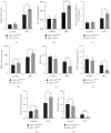
Evaluation of oxidative stress: (A–C) comparison of reactive oxygen species (ROS), superoxide (O2•–), and carbonyl protein levels between the groups, (D, E) measurement of malondialdehyde (MDA) and superoxide dismutase (SOD) levels in intestinal tissues, and (F–H) evaluation of oxidized glutathione (GSSG) and glutathione (GSH) in the intestines. Data represent three separate experiments. Two-sided one-way one-way analysis of variance (ANOVA) was utilized for data comparison together with a post hoc Tukey test (n = 6–8, means ± the standard error of the mean [SEM]). p < 0.05, ∗∗p < 0.01, ∗∗∗p < 0.001. HIF-1α, hypoxia-inducible factor-1 alpha; NEC, necrotizing enterocolitis.
Figure 3
Figure 3
Evaluation of intestinal epithelial cell (IEC) proliferation: (A) BrdU immunostaining in the intestines. Scale bars: 40 µm. (B) Assessment of IEC multiplication (BrdU-positive cells per high-power field). (C–E) Detection of Ccnd2, Vegf, and Bnip3l mRNA levels using quantitative real-time reverse transcription polymerase chain reaction (RT-qPCR). Two-sided one-way one-way analysis of variance (ANOVA) was utilized for data comparison together with a post hoc Tukey test (n = 6–8, means ± the standard error of the mean [SEM]). p < 0.05, ∗∗p < 0.01. HIF-1α, hypoxia-inducible factor-1 alpha; HPF, high power field; NEC, necrotizing enterocolitis.
Figure 4
Figure 4
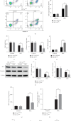
Evaluation of apoptosis and intestinal barrier injury: (A) western blotting analysis of caspase 3 and caspase 8 levels in mouse intestines. β-Actin comprised the loading control. Right panels: densitometry analysis of the immunoreactive protein bands. (B) Flow cytometry analysis of apoptosis in intestinal epithelial cells (IECs). The percentage of annexin-positive and propidium iodide (PI)-negative cells represents the cell apoptosis rate. Right panel: histogram analysis for multiple flow cytometry experiments. (C, D) Estimation of SIgA and β-defensin-2 levels in the terminal ileum. (E) Detection of serum fluorescein isothiocyanate (FITC)–dextran concentrations. (F) Western blotting analysis of ZO-1 and occludin levels in mouse intestines. Right panels: densitometry analysis of the immunoreactive protein bands. (G) Assessment of myeloperoxidase (MPO) activity in the ileum. (H–J) Quantification of bacterial growth in mesenteric lymph nodes, liver, and spleen. Data represent three independent experiments. Two-sided one-way analysis of variance (ANOVA) was utilized for data comparison together with a post hoc Tukey test (n = 6–8, means ± the standard error of the mean [SEM]). ∗∗p < 0.01, ∗∗∗p < 0.001. HIF-1α, hypoxia-inducible factor-1 alpha; NEC, necrotizing enterocolitis.
Figure 5
Figure 5
Evaluation of Toll-like receptor 4 (TLR4)–nuclear factor kappa B (NF-κB) activation and the inflammatory response: (A) western blotting analysis of p-p65, p65, TLR4, and inducible nitric oxide synthase (iNOS) levels in mouse intestines. Right panel: Densitometry analysis was performed. (B, C) Detection of CCL-20 and granulocyte macrophage-colony stimulating factor (GM-CSF) using enzyme-linked immunosorbent assay (ELISA) in the intestines. (D–I) Measurement of inflammatory cytokine concentrations in intestinal tissues using ELISA. Two-sided one-way analysis of variance (ANOVA) was utilized for data comparison together with a post hoc Tukey test (n = 6–8, means ± the standard error of the mean [SEM]). (J) The concentration of the anti-inflammatory cytokine transforming growth factor beta (TGF-β) in the intestines was detected using ELISA. p < 0.05, ∗∗p < 0.01, ∗∗∗p < 0.001. HIF-1α, hypoxia-inducible factor-1 alpha; IFNγ, interferon gamma; IL, interleukin; NEC, necrotizing enterocolitis; TNFα, tumor necrosis factor alpha.

Similar articles

Cited by

References

    1. Bellodas Sanchez J., Kadrofske M. Necrotizing Enterocolitis. Neurogastroenterology & Motility . 2019;31(3) doi: 10.1111/nmo.13569.e13569 - DOI - PubMed
    1. Niemarkt H. J., De Meij T. G., van Ganzewinkel C. J., et al. Necrotizing Enterocolitis, Gut Microbiota, and Brain Development: Role of the Brain-Gut Axis. Neonatology . 2019;115(4):423–431. doi: 10.1159/000497420. - DOI - PMC - PubMed
    1. Yazji I., Sodhi C. P., Lee E. K., et al. Endothelial TLR4 Activation Impairs Intestinal Microcirculatory Perfusion in Necrotizing Enterocolitis via eNOS–NO–Nitrite Signaling. Proceedings of the National Academy of Sciences . 2013;110(23):9451–9456. doi: 10.1073/pnas.1219997110. - DOI - PMC - PubMed
    1. Ballance W. A., Dahms B. B., Shenker N., Kliegman R. M. Pathology of Neonatal Necrotizing Enterocolitis: A Ten-Year Experience. The Journal of Pediatrics . 1990;117(1):S6–S13. doi: 10.1016/S0022-3476(05)81124-2. - DOI - PubMed
    1. Caplan M. S., Hedlund E., Adler L., Hsueh W. Role of Asphyxia and Feeding in a Neonatal Rat Model of Necrotizing Enterocolitis. Pediatric Pathology . 1994;14(6):1017–1028. doi: 10.3109/15513819409037698. - DOI - PubMed

Substances

LinkOut - more resources