. 2024 Dec 18;16(4):1885-1893.
doi: 10.1039/d4sc04951a. eCollection 2025 Jan 22.

Aromatic-aromatic interactions drive fold switch of GA95 and GB95 with three residue difference

Affiliations

Aromatic-aromatic interactions drive fold switch of GA95 and GB95 with three residue difference

Chen Chen et al. Chem Sci. .

Abstract

Proteins typically adopt a single fold to carry out their function, but metamorphic proteins, with multiple folding states, defy this norm. Deciphering the mechanism of conformational interconversion of metamorphic proteins is challenging. Herein, we employed nuclear magnetic resonance (NMR), circular dichroism (CD), and all-atom molecular dynamics (MD) simulations to elucidate the mechanism of fold switching in proteins GA95 and GB95, which share 95% sequence homology. The results reveal that long-range interactions, especially aromatic π-π interactions involving residues F52, Y45, F30, and Y29, are critical for the protein switching from a 3α to a 4β + α fold. This study contributes to understanding how proteins with highly similar sequences fold into distinct conformations and may provide valuable insights into the protein folding code.


Conflict of interest statement

There are no conflicts to declare.

Figures

Fig. 1
Fig. 1. Structures of GA95 and GB95. (A) and (B) The tertiary structures of GA95 (PDB id: 2KDL) and GB95 (PDB id: 2KDM). Aromatic amino acid residues are shown in stick representations with different colors, Y is shown in orange, W is shown in blue, and F is shown in green; (C) sequence comparison and secondary structure of GA95 and GB95. Three residue differences (20, 30, 45) of GA95 and GB95 are highlighted in red. Aromatic amino acid residues are highlighted with colors consistent with the stick representations.
Fig. 2
Fig. 2. Amino acid nodes in GA95 and GB95 folded from secondary to tertiary structures. (A) 1H–15N HSQC spectrum of GA95-38 (red). (B) 1H–15N HSQC spectrum of GA95-45 (green). (C) 1H–15N HSQC spectrum of GA95-51 (orange). (D) 1H–15N HSQC spectrum of GA95-52 (blue). (E) 1H–15N HSQC spectrum of GA95 (black). (F) 1H–15N HSQC spectrum of GB95-38 (red). (G) 1H–15N HSQC spectrum of GB95-45 (green). (H) 1H–15N HSQC spectrum of GB95-54 (orange). (I) 1H–15N HSQC spectrum of GB95-55 (blue). (J) 1H–15N HSQC spectrum of GB95 (black). (K) Histogram of secondary structure content statistics for GA95 and its truncated proteins GA95-38, GA95-45, GA95-51 and GA95-52. (L) Histogram of secondary structure content statistics for GB95 and its truncated proteins GB95-38, GB95-45, GB95-54 and GB95-55. The content of the α-helix is labeled in black, and the content of the β-sheet is labeled in red.
Fig. 3
Fig. 3. Shift in interactions during fold switching of GA95 and GB95. (A) Free energy landscape of folding process of GA95-53. The free energy minima are colored in blue, and the saddle point (state T) between the two minima 3 and 4 is labeled by a dashed circle. (B) The occupancy of important interactions in different states of the folding process of GA95-53. (C) Free energy landscape of folding process of GB95. (D) and (E) The occupancy of important interactions in different states of the folding process of GB95. (F) and (G) The representative structures of important states in the folding process of GA95-53(left) and GB95(right). The native structures of GA95-53 or GB95 are shown in gray. The residues Y29, I30/F30, Y45, and F52, which are important in the folding pathways, are depicted by ball-and-stick. Y29, I30/F30, Y45, and F52 are colored green, yellow, pink, and blue, respectively.
Fig. 4
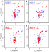
Fig. 4. Validation of F52 interactions. (A) 1H–15N HSQC spectra of GA95-53 and GA95-53 Y29A; (B) 1H–15N HSQC spectra of GA95-53 and GA95-53 F52A; (C)1H–15N HSQC spectra of GB95 and GB95 F30I; (D) 1H–15N HSQC spectra of GB95-55 and GB95-55 Y29A; the wild type is marked in blue and the mutant is marked in red.
Fig. 5
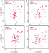
Fig. 5. Validation of the role of Y45 in GB95 to form an aromatic cluster. (A) 1H–15N HSQC spectra of GB95 and GB95 Y45I; (B) 1H–15N HSQC spectra of GB95 and GB95 Y45F; (C)1H–15N HSQC spectra of GA95 and GA95 L45I; (D) 1H–15N HSQC spectra of GB95 and GB95 L32P. The disappearing residues are marked with arrows; the wild type is marked in blue and the mutant is marked in red.
Fig. 6
Fig. 6. The interactions between the N- and C-terminal amino acids stabilize the tertiary structure of GB95. (A) 1H–15N HSQC spectrum of GB95-55 (orange). (B) 1H–15N HSQC spectrum of GB95-55 T55A (red). (C) 1H–15N HSQC spectrum of GB95-55 N8A (blue). (D) 1H–15N HSQC spectrum of GB95 (orange). (E) 1H–15N HSQC spectrum of GB95 E56A (red). (F) 1H–15N HSQC spectrum of GB95 K10A (blue). (G) Hydrogen bonds and bond distance formed in T55–N8 and E56–K10.
Fig. 7
Fig. 7. The mechanism of fold switching in GA95 and GB95. GA95 and GB95 undergo fold switching through mutations at three specific sites, altering their secondary structure preferences and residue interactions. The folded state (left: GA95 and right: GB95) is depicted in gray. In the tertiary structures of GA95 and GB95, the three mutation sites and other crucial amino acids involved in long-range interactions are shown in stick representation: L20A and L45Y in dark grey, I30F in blue, Y29 in orange, F52 in red, T53 in light blue, E56 and T55 in green, N8 and K10 in light pink. The secondary structures of proteins in the unfolded state are related to L20A, I30F, and L45Y, with GA95 preferring α-helices while GB95 prefers β-sheets. The interactions involving F52 during the folding process in GA95 and GB95 are represented as local magnification. In GA95, F52 interacts with Y29, whereas in GB95, the interaction of F52 shifts from Y29 to F30, with Y45 being the key element contributing to the complete shift of this interaction.

Similar articles

References

    1. Anfinsen C. B. Science. 1973;181:223–230. doi: 10.1126/science.181.4096.223. - DOI - PubMed
    1. Anfinsen C. B. Scheraga H. A. Adv. Protein Chem. 1975;29:205–300. doi: 10.1016/S0065-3233(08)60413-1. - DOI - PubMed
    1. Bryan P. N. Orban J. Curr. Opin. Struct. Biol. 2010;20:482–488. doi: 10.1016/j.sbi.2010.06.002. - DOI - PMC - PubMed
    1. Dishman A. F. Volkman B. F. Curr. Opin. Struct. Biol. 2022;74:102380. doi: 10.1016/j.sbi.2022.102380. - DOI - PMC - PubMed
    1. Porte L. L. Looger L. L. Proc. Natl. Acad. Sci. U. S. A. 2018;115:5968–5973. doi: 10.1073/pnas.1800168115. - DOI - PMC - PubMed