. 2024 Dec 10:12:1469238.
doi: 10.3389/fbioe.2024.1469238. eCollection 2024.

The modulating role of uniaxial straining in the IL-1β and TGF-β mediated inflammatory response of human primary ligamentocytes

Affiliations

The modulating role of uniaxial straining in the IL-1β and TGF-β mediated inflammatory response of human primary ligamentocytes

Johannes Heidenberger et al. Front Bioeng Biotechnol. .

Abstract

Biomechanical (over-)stimulation, in addition to inflammatory and fibrotic stimuli, severely impacts the anterior cruciate ligament (ACL) biology, contributing to the overall chronic nature of desmopathy. A major challenge has been the lack of representative two-dimensional (2D) in vitro models mimicking inflammatory processes in the presence of dynamic mechanical strain, both being crucial for ligament homeostasis. Physiological levels of strain exert anti-inflammatory effects, while excessive strain can facilitate inflammatory mechanisms. Adhering to the 3Rs (Replacement, Reduction and Refinement) principles of animal research, this study aims to investigate the role of a dynamic biomechanical in vitro environment on inflammatory mechanisms by combining a Flexcell culture system with primary human ligamentocytes for the study of ligament pathology. Primary ligamentocytes from OA patients were cultured under animal-free conditions with human platelet lysate, and exposed to either IL-1β or TGF-β3 to simulate different inflammatory microenvironments. Cells were subjected to different magnitudes of mechanical strain. Results showed that cells aligned along the force axis under strain. This study highlights the critical role of the mechanical microenvironment in modulating inflammatory and fibrotic cellular responses in ligamentocyte pathology, providing valuable insights into the complex interplay between biomechanical stimuli and cytokine signaling. These findings not only advance our understanding of ligament biology but also can pave the way for the development of more targeted therapeutic strategies for ligament injuries and diseases, potentially improving patient outcomes in orthopedic medicine.

Keywords: fibrosis; in vitro modelling; inflammation; ligamentocytes; mechanical loading.


Conflict of interest statement

The authors declare that the research was conducted in the absence of any commercial or financial relationships that could be construed as a potential conflict of interest.

Figures

FIGURE 1
FIGURE 1
Effect of oxygenation and seeding conditions on ligamentocyte morphology. (A) Mean cell length of 2D ligamentocyte cultures at initial cell seeding of 100 - 300 k cells per well in normoxic and hypoxic conditions over the culturing period. (B) Depiction of the significant differences in cell length between different seeding densities in normoxia and hypoxia. Representative images of ligamentocyte cultures at 300k (top) and 100k (bottom) seeding densities are displayed on the right. Data are expressed as scatter plot with mean for measurements of 43–93 cells of biological duplicates. Significance was determined with a two-way ANOVA. (*P < 0.05, **P < 0.005, ***P < 0.0005, ****P < 0.0001).
FIGURE 2
FIGURE 2
Gene expression of ligamentocytes in hypoxic condition. HIF1A, COL1A1 and COL3A1 gene expression in a hypoxic environment for different cell seeding densities in a BioFlex 2D cell culture including 65 k (n = 8 biological replicates), 100 k (n = 4) and 300 k (n = 2).
FIGURE 3
FIGURE 3
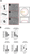
Ligamentocyte cell alignment and gene expression in flex cell culture with 10% and 21% strain and with IL-1β or TGF-β treatment. (A) The polar graph and brightfield images from three different zones (1: Border, 2: Transition, 3: Center) inside a flex cell well show the cell alignment for 10% strain. (B) Ligamentocytes were trained for 48 h in a flexcell system without any treatment. Different amounts of strain were applied to the cells (10%, 21%) and gene expression of COL1A1, COL3A1, MMP1, IL6 and PIEZO1 was analyzed with qPCR. The data is expressed as fold change to the mean of the untreated static control. n = 8 biological replicates. Significance was determined with a one-way ANOVA with Dunnett’s multiple comparisons test. (*P < 0.05, **P < 0.01, ***P < 0.001, ****P < 0.0001).
FIGURE 4
FIGURE 4
Gene expression of ligamentocytes in a static Flexcell culture with and without IL-1 treatment. Ligamentocytes were treated with IL-1β (1 ng/mL) for 48 h in a flexcell system. Gene expression of COL1A1, COL3A1, MMP1, IL6 and PIEZO1 was analyzed with qPCR. The data is expressed as fold change to the mean of the untreated static control. n = 9 biological replicates. Significance was determined with a Wilcoxon test. (*P < 0.05, **P < 0.05).
FIGURE 5
FIGURE 5
Gene expression of ligamentocytes in a static Flexcell culture with and without TGF- β treatment. Ligamentocytes were treated with TGF-β (1 ng/mL) for 48 h in a flexcell system. Gene expression of COL1A1, COL3A1, MMP1, IL6 and PIEZO1 was analyzed with qPCR. The data is expressed as fold change to the mean of the untreated static control. n = 8 biological replicates. Significance was determined with a Wilcoxon test. (*P < 0.05, **P < 0.05).
FIGURE 6
FIGURE 6
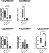
Gene expression of ligamentocytes in a Flexcell culture with 10% and 21% strain and with IL-1β or TGF-β treatment. Ligamentocytes were trained for 48 h in a flexcell system with (A) IL-1β (1 ng/mL) (B) and with TGF-β (1 ng/mL) treatment. Different amounts of strain were applied to the cells (10%, 21%) and gene expression of COL1A1, COL3A1, MMP1, IL6 and PIEZO1 was analyzed with qPCR. The data is expressed as fold change to the mean of the untreated static control. n = 8 biological replicates. Significance was determined with a one-way ANOVA with Dunnett’s multiple comparisons test. (*P < 0.05, **P < 0.01, ***P < 0.001, ****P < 0.0001).
FIGURE 7
FIGURE 7
Gene expression of ligamentocytes in a Flexcell culture with 10% and 21% strain and IL-1β and TGF-β3 treatment. Ligamentocytes were trained and treated together with TGF-β (1 ng/mL) and IL-1β (1 ng/mL) for 48 h in a flexcell system. Different amounts of strain were applied to the cells (10%, 21%) and gene expression of COL1A1, COL3A1, MMP1, IL6 and PIEZO1 was analyzed with qPCR. The data is expressed as fold change to the mean of the untreated static control. n = 8 biological replicates. Significance was determined with a one-way ANOVA with Dunnett’s multiple comparisons test. (*P < 0.05, **P < 0.01, ***P < 0.001, ****P < 0.0001).

Similar articles

References

    1. Agarwal S., Long P., Al S., Piesco N., Shree A., Gassner R. (2003). A central role for nuclear factor-κB pathway in the antiinflammatory and proinflammatory actions of mechanical strain. FASEB J. 17 (8), 1–15. 10.1096/FJ.02-0901FJE - DOI - PMC - PubMed
    1. Altmann N., Bowlby C., Coughlin H., Belacic Z., Sullivan S., Durgam S. (2023). Interleukin-6 upregulates extracellular matrix gene expression and transforming growth factor β1 activity of tendon progenitor cells. BMC Musculoskelet. Disord. 24 (1), 907. 10.1186/S12891-023-07047-9 - DOI - PMC - PubMed
    1. Beaumont R. E., Smith E. J., Zhou L., Marr N., Thorpe C. T., Guest D. J. (2024). Exogenous interleukin-1 beta stimulation regulates equine tenocyte function and gene expression in three-dimensional culture which can be rescued by pharmacological inhibition of interleukin 1 receptor, but not nuclear factor kappa B, signaling. Mol. Cell. Biochem. 479 (5), 1059–1078. 10.1007/S11010-023-04779-Z - DOI - PMC - PubMed
    1. Benhardt H. A., Cosgriff-Hernandez E. M. (2009). The role of Mechanical loading in ligament tissue engineering. Tissue Eng. - Part B Rev. 15 (4), 467–475. 10.1089/ten.teb.2008.0687 - DOI - PubMed
    1. Biela S. A., Su Y., Spatz J. P., Kemkemer R. (2009). Different sensitivity of human endothelial cells, smooth muscle cells and fibroblasts to topography in the nano–micro range. Acta Biomater. 5 (7), 2460–2466. 10.1016/J.ACTBIO.2009.04.003 - DOI - PubMed

LinkOut - more resources