. 2024 Dec 20:18:3881-3892.
doi: 10.2147/OPTH.S491154. eCollection 2024.

The Toronto Tele-Retinal Screening Program for the Elderly in Long-Term Care: A Pilot Project

Affiliations

The Toronto Tele-Retinal Screening Program for the Elderly in Long-Term Care: A Pilot Project

Michelle Lim et al. Clin Ophthalmol. .

Abstract

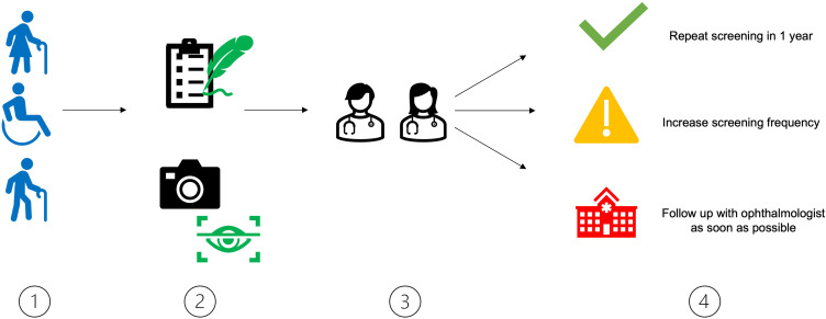
Objective: To report the results and feasibility of a pilot expansion of the Toronto Tele-Retinal Screening Program in an elderly long-term care home.

Methods: Long term care patients with Type II diabetes mellitus (DM) were screened between April 1, 2022, and July 1, 2022. Demographic and health data were collected through surveys.

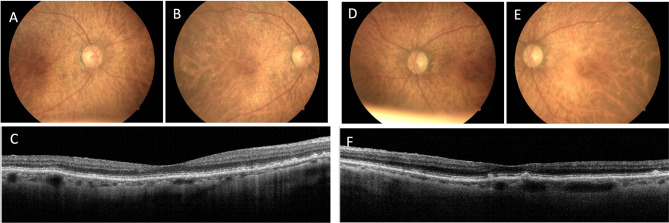
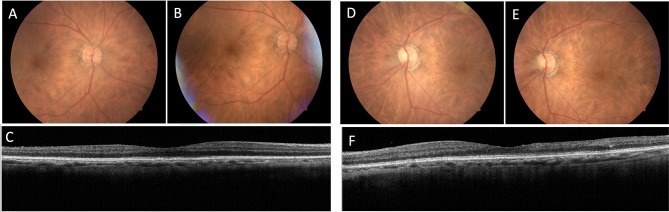
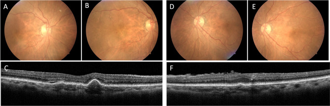
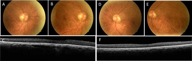
Results: A total of 28 patients were screened, with 85.7% successfully undergoing retinal imaging. Among imaged patients, 8.3% (2/24) required urgent follow-up. Pathologies identified included uncontrolled glaucoma (4.1%, 1/24), non-proliferative diabetic retinopathy (8.3%, 2/24), and age-related macular degeneration (45.8%, 11/24). The handheld camera successfully screened 60% (3/5) of patients with mobility issues. Overall, 90% (17/19) of patients rated their experience as either "brilliant" or "really good".

Discussion: This pilot project demonstrated the necessity for routine eye care in the elderly and the potential for widespread implementation of teleophthalmology in long-term care facilities. With only 14.3% (4/28) of patients unable to be imaged, this program offers a feasible, patient-friendly alternative to in-clinic screening. Future policies and practices in teleophthalmology should consider the unique needs of long-term care residents and the potential for reducing healthcare disparities through such a program.

Keywords: diabetes; elderly care; healthcare accessibility; retinopathy; telemedicine; teleophthalmology.


Conflict of interest statement

The authors disclosed receipt of the following financial support for the research, authorship, and publication of this article: This work was supported by Diabetes Action Canada. This paper has been uploaded to JMIR Publications as a preprint:https://preprints.jmir.org/preprint/49188. The Authors declare that there is no conflict of interest.

Figures

Figure 1
Figure 1
Study design for the screening of residents at Mon Sheong Home for the Aged. Step 1: Patients are identified using the inclusion criteria described in Methods. Step 2: For each patient, demographics, clinical information such as diabetes duration and past ocular history, and survey answers are collected. Visual acuity and intraocular pressures were assessed. Finally, OCT and fundus photos were captured either with a handheld or tabletop camera. Images are uploaded to a secure database. Step 3: Ophthalmologists from a different site review the images for pathology and decide on next steps. Step 4: Patients are categorized into requiring a repeat screening in one year due to lack of pathology; repeat screening in six months due to non-urgent pathology; or immediate follow up with ophthalmologist due to urgent pathology. In some cases, patients are required to repeat screening due to insufficient image quality.
Figure 2
Figure 2
Selected color fundus photographs taken with tabletop camera (A, B, D, and E) and cross-sectional 50-line raster OCT scans (C and F) of right and left eyes of a patient recommended repeated screening in 1 year. This was an 86-year-old female with dementia who could not comment on her ability to see. She was diagnosed with dry age-related macular edema requiring follow-up in one year.
Figure 3
Figure 3
Selected color fundus photographs taken with tabletop camera (A, B, D and E) and cross-sectional 50-line raster OCT scans (C and F) of right and left eyes of a patient recommended repeated screening at an increased frequency of six months. This was an 81-year-old female with dementia found to have moderate (OS) and moderate-severe (OD) non-proliferative diabetic retinopathy as well as dry AMD OS requiring follow-up in six months.
Figure 4
Figure 4
Selected color fundus photographs taken with tabletop camera (A, B, D and E) and cross-sectional 50-line raster OCT scans (C and F) of right and left eyes of a patient recommended urgent follow up with an ophthalmologist within one month. This was an 89-year-old female who had not undergone eye screening in over 10 years. She had some difficulty seeing and was found to have non-proliferative diabetic retinopathy in both eyes, as well as wet AMD OD and epiretinal membrane OS.
Figure 5
Figure 5
Selected color fundus photographs taken with tabletop camera (A, B, D and E) and cross-sectional 50-line raster OCT scans (C and F) of right and left eyes of a patient recommended for urgent follow-up with an ophthalmologist within one month. This was a 77-year-old male who had not undergone eye screening in over three years. He did not express difficulty seeing but was found to have dry AMD OD, epiretinal membranes, and evidence of previous vein occlusion.
Figure 6
Figure 6
The benefit of the tabletop camera’s OCT imaging function. (A and B) While mild NPDR and macular drusen can be identified in the fundus photo, the presence of macular fluid is unclear. (C) OCT supplementation shows pigment epithelial detachment (PED) with a small amount of subretinal fluid (SRF).

Similar articles

References

    1. Saaddine JB, Honeycutt AA, Narayan KMV, Zhang X, Klein R, Boyle JP. Projection of diabetic retinopathy and other major eye diseases among people with diabetes mellitus: United States, 2005-2050. Arch Ophthalmol. 2008;126(12):1740–1747. doi:10.1001/archopht.126.12.1740 - DOI - PubMed
    1. Ting DSW, Cheung GCM, Wong TY. Diabetic retinopathy: global prevalence, major risk factors, screening practices and public health challenges: a review. Clin Exp Ophthalmol. 2016;44(4):260–277. doi:10.1111/ceo.12696 - DOI - PubMed
    1. Flaxman SR, Bourne RRA, Resnikoff S, et al. Global causes of blindness and distance vision impairment 1990–2020: a systematic review and meta-analysis. Lancet Glob Health. 2017;5(12):e1221–e1234. doi:10.1016/S2214-109X(17)30393-5 - DOI - PubMed
    1. Eichenbaum JW. Geriatric vision loss due to cataracts, macular degeneration, and glaucoma. Mount Sinai J Med. 2012;79(2):276–294. doi:10.1002/msj.21303 - DOI - PubMed
    1. Altomare F, Kherani A, Lovshin J. Retinopathy. Can J Diabetes. 2018;42(Suppl 1):S210–S216. doi:10.1016/j.jcjd.2017.10.027 - DOI - PubMed

LinkOut - more resources