Lateralizing Value of Artificial Intelligence-Based Segmentation Software in MRI-Negative Focal Epilepsy
- PMID: 39720198
- PMCID: PMC11664054
- DOI: 10.14581/jer.24011
Lateralizing Value of Artificial Intelligence-Based Segmentation Software in MRI-Negative Focal Epilepsy
Abstract
Background and purpose: The magnetic resonance images (MRIs) ability of lesion detection in epilepsy is crucial for a diagnosis and surgical outcome. Using automated artificial intelligence (AI)-based tools for measuring cortical thickness and brain volume originally developed for dementia, we aimed to identify whether it could lateralize epilepsy with normal MRIs.
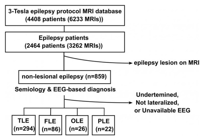
Methods: Non-lesional 3-Tesla MRIs of 428 patients diagnosed with focal epilepsy, based on semiology and electroencephalography findings, were analyzed. AI-based segmentation/volumetry software measured the cortical thickness and the hippocampal volume. The laterality index (LI) was calculated.
Results: We classified into temporal lobe epilepsy (TLE, n=294), frontal lobe epilepsy (FLE, n=86), occipital lobe epilepsy (OLE, n=29), and parietal lobe epilepsy (PLE, n=22). Onset age and MRI age were 24.0±16.6 (0-84) and 35.6±14.8 (16-84) years old. In FLE, the LI of frontal thickness was significantly different between the left and right FLE groups, with LIs of the right FLE group being right-shifted and those of the left FLE group being left-shifted, indicating that the lesion side was thinner than the non-lesion side (p=0.01). The discriminable group, which included the patients with left FLE and a LI lower than minus one standard deviation, as well as the patients with right FLE and a LI higher than one standard deviation, showed a longer duration of epilepsy than the non-discriminable group (12.7±9.9 vs. 8.3±7.7 years; p=0.03). Specifically, the LI of individual regions of interest showed that the rostral middle frontal cortex was significantly different in FLE. However, the TLE, PLE, OLE, and LIs were not significantly different.
Conclusions: AI-based brain segmentation software can be helpful to decide the laterality of non-lesional FLE especially with longer duration of disease.
Keywords: Artificial intelligence; Asymmetry; Frontal lobe epilepsy; MRI-negative epilepsy.
Copyright © 2024 Korean Epilepsy Society.
Conflict of interest statement
Conflict of Interest: None.
Figures


References
-
- Yun CH, Lee SK, Lee SY, Kim KK, Jeong SW, Chung CK. Prognostic factors in neocortical epilepsy surgery: multivariate analysis. Epilepsia. 2006;47:574–9. - PubMed
LinkOut - more resources
Full Text Sources