. 2024 Nov 22;27(12):111452.
doi: 10.1016/j.isci.2024.111452. eCollection 2024 Dec 20.

A negatively charged region within carboxy-terminal domain maintains proper CTCF DNA binding

Affiliations

A negatively charged region within carboxy-terminal domain maintains proper CTCF DNA binding

Lian Liu et al. iScience. .

Abstract

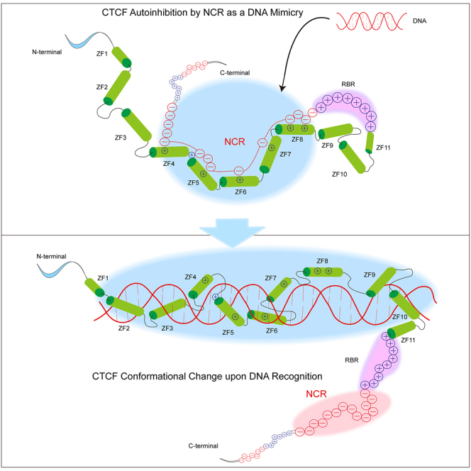
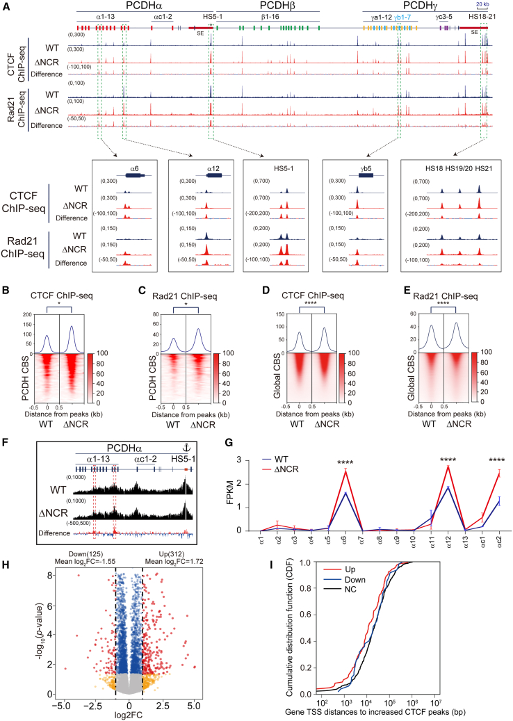
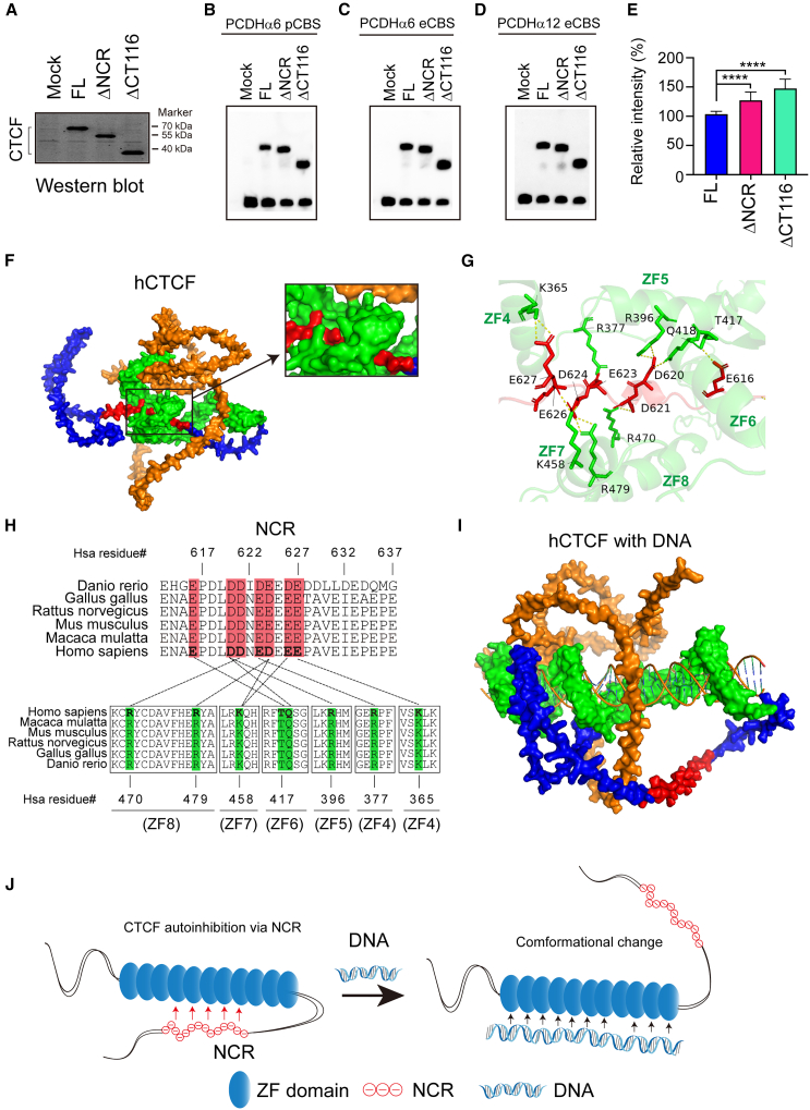
As an essential regulator of higher-order chromatin structures, CCCTC-binding factor (CTCF) is a highly conserved protein with a central DNA-binding domain of 11 tandem zinc fingers (ZFs), which are flanked by amino (N-) and carboxy (C-) terminal domains of intrinsically disordered regions. Here we report that CRISPR deletion of the entire C-terminal domain of alternating charge blocks decreases CTCF DNA binding but deletion of the C-terminal fragment of 116 amino acids results in increased CTCF DNA binding and aberrant gene regulation. Through a series of genetic targeting experiments, in conjunction with electrophoretic mobility shift assay (EMSA), circularized chromosome conformation capture (4C), qPCR, chromatin immunoprecipitation with sequencing (ChIP-seq), and assay for transposase-accessible chromatin with sequencing (ATAC-seq), we uncovered a negatively charged region (NCR) responsible for weakening CTCF DNA binding and chromatin accessibility. AlphaFold prediction suggests an autoinhibitory mechanism of CTCF via NCR as a flexible DNA mimic domain, possibly competing with DNA binding for the positively charged ZF surface area. Thus, the unstructured C-terminal domain plays an intricate role in maintaining proper CTCF-DNA interactions and 3D genome organization.

Keywords: Experimental systems for structural biology; Molecular Structure; Molecular physiology; Properties of biomolecules.


Conflict of interest statement

The authors declare no competing interests.

Figures

None
Graphical abstract
Figure 1
Figure 1
Deletion of CTCF C-terminal domain results in decreased CTCF binding and gene dysregulation (A) Schematic of the CTCF zinc-finger domain (ZFD) and the flanking N-terminal domain (NTD) and C-terminal domain (CTD). (B and C) Amino acid composition of CTCF NTD (B) and CTD (C). (D) Disordered propensity by a computer program indicates that NTD and CTD are intrinsically disorder region (IDR). (E) Multiple sequence alignment of CTD of the vertebrate CTCF proteins. The negatively charged region (NCR) is highlighted in a yellow background. (F) CTCF and Rad21 ChIP-seq peaks at the human cPCDH gene complex. The human cPCDH locus comprises three tandem linked gene clusters: PCDHα, PCDHβ, and PCDHγ. The PCDHα and PCDHγ clusters each consist of a variable region with multiple highly similar and unusually large exons and a constant region with three small exons. Each variable exon is separately cis-spliced to a single set of cluster-specific constant exons. The PCDHβ cluster contains 16 variable exons but with no constant exon. These three clusters form a superTAD with two subTADs: PCDHα and PCDHβγ. Each subTAD has its own downstream super-enhancer. Var, variable; Con, constant; HS, hyper-sensitive site; SE, super-enhancer. (G and H) Heatmaps of CTCF (G) and Rad21 (H) normalized signals at CBS elements in the cPCDH locus. Student’s t test, ∗p < 0.05. (I and J) Heatmaps of CTCF (I) and Rad21 (J) normalized signals at genome-wide CBS elements. Student’s t test, ∗∗∗∗p < 0.0001. (K–M) Three types of CTCF motifs in WT and ΔCTD cells. (N) QHR-4C interaction profiles of the PCDHα gene cluster using HS5-1 as an anchor. (O) RNA-seq shows decreased expression levels of PCDHα6, PCDHα12, and PCDHαc2 upon deletion of CTD. FPKM, fragments per kilobase of exon per million fragments mapped. Data are presented as mean ± SD; Student’s t test, ∗∗∗∗p < 0.0001. See also Figures S1–S3.
Figure 2
Figure 2
Deleting 116 amino acids from the C terminus leads to increased CTCF binding and affects gene expression (A) CTCF and Rad21 ChIP-seq peaks at the cPCDH gene complex. (B and C) Heatmaps of CTCF (B) and Rad21 (C) normalized signals at the cPCDH CBS elements. Student’s t test, ∗p < 0.05. (D and E) Heatmaps of global CTCF (D) and Rad21 (E) ChIP-seq signals. Student’s t test, ∗∗∗∗p < 0.0001. (F) QHR-4C interaction profiles of the PCDHα gene cluster using HS5-1 as an anchor. (G) RNA-seq indicates increased expression levels of PCDHα6, PCDHα12, and PCDHαc2. Data are presented as mean ± SD; Student’s t test, ∗∗∗∗p < 0.0001. See also Figures S1, S3, and S4.
Figure 3
Figure 3
Rescue with a series of C-terminal truncated CTCF proteins reveals an NCR (A) Schematic illustration of different truncated CTCFs used in constructing stable cell lines. All truncated CTCFs are tagged with V5. (B) Western blots of stable cell lines with the V5 antibody indicating truncated CTCF proteins in ΔCTD cells. (C) Binding profiles of different length of CTCFs at the cPCDH locus. (D) Heatmaps of CTCF ChIP-seq signals at the cPCDH locus. Student’s t test, ∗p < 0.05,∗∗p < 0.01,∗∗∗p < 0.001. (E) Heatmaps of different truncated CTCFs, showing global binding profiles. Student’s t test, ∗∗∗∗p < 0.0001. (F–H) CTCF enrichments at the three types of CTCF motifs. Student’s t test, ∗∗∗∗p < 0.0001. See also Figure S5.
Figure 4
Figure 4
NCR deletion increases CTCF binding and affects gene regulation (A) CTCF and Rad21 ChIP-seq signals at the cPCDH locus, showing increased CTCF and cohesin enrichments at CBS elements upon NCR deletion (Δ612-637). (B and C) Heatmaps of CTCF (B) and Rad21 (C) ChIP-seq signals at the cPCDH locus. Student’s t test, ∗p < 0.05. (D and E) Heatmaps of CTCF (D) and Rad21 (E) ChIP-seq signals, indicating genome-wide CTCF and Rad21 enrichments upon NCR deletion. Student’s t test, ∗∗∗∗p < 0.0001. (F) Quantitative high-resolution 4C (QHR-4C) experiments with HS5-1 as an anchor, indicating increased chromatin interactions of the HS5-1 enhancer with PCDHα6 or PCDHα12 promoters. (G) RNA-seq indicates increased expression levels of PCDHα6, PCDHα12, and PCDHαc2 upon NCR deletion. Data are presented as mean ± SD; Student’s t test, ∗∗∗∗p < 0.0001. (H) Volcano plots of differential gene expression analyses for WT and ΔNCR cells. Red dots, fold change of gene expression upon NCR deletion (log2FC > 1 and adjusted p value <0.05). Blue dots, genes only passed adjusted p value <0.05. Yellow dots, gene only passed log2FC > 1. FC, fold change. (I) TSS distances of up-, down-, or none-regulated (NC) genes to the closest increased CTCF peaks in ΔNCR cells (data are shown as a cumulative distribution function, CDF). See also Figures S1, S3, and S6 and Tables S5 and S6.
Figure 5
Figure 5
AlphaFold3 modeling suggests autoregulation of CTCF DNA binding by NCR cis-interactions (A) Western blot of recombinant human CTCF proteins. (B–E) EMSA of full-length human CTCF, ΔNCR, ΔCT116 with three different PCDH promoter or exonic CBS elements (B–D) with quantifications (E). Data are presented as mean ± SD; Student’s t test, ∗∗∗∗p < 0.0001. (F) The surface representation of the AlphaFold3 model of human CTCF in the absence of DNA. NTD, ZF, CTD, and NCR are in orange, green, blue, and red, respectively. (G) Negatively charged residues of NCR form nine pairs of electrostatic interactions with the zinc-finger domain. (H) Pairwise interactions between NCR (top) and ZFs (bottom). (I) The surface representation of the AlphaFold3 model of CTCF with DNA ligand. (J) The model of CTCF autoinhibition via large conformational changes with NCR as a flexible DNA mimicry.

Similar articles

References

    1. Klenova E.M., Nicolas R.H., Paterson H.F., Carne A.F., Heath C.M., Goodwin G.H., Neiman P.E., Lobanenkov V.V. CTCF, a conserved nuclear factor required for optimal transcriptional activity of the chicken c-myc gene, is an 11-Zn-finger protein differentially expressed in multiple forms. Mol. Cell Biol. 1993;13:7612–7624. doi: 10.1128/mcb.13.12.7612-7624.1993. - DOI - PMC - PubMed
    1. Heger P., Marin B., Bartkuhn M., Schierenberg E., Wiehe T. The chromatin insulator CTCF and the emergence of metazoan diversity. Proc. Natl. Acad. Sci. USA. 2012;109:17507–17512. doi: 10.1073/pnas.1111941109. - DOI - PMC - PubMed
    1. Rowley M.J., Corces V.G. Organizational principles of 3D genome architecture. Nat. Rev. Genet. 2018;19:789–800. doi: 10.1038/s41576-018-0060-8. - DOI - PMC - PubMed
    1. Li Y., Haarhuis J.H.I., Sedeño Cacciatore Á., Oldenkamp R., van Ruiten M.S., Willems L., Teunissen H., Muir K.W., de Wit E., Rowland B.D., Panne D. The structural basis for cohesin-CTCF-anchored loops. Nature. 2020;578:472–476. doi: 10.1038/s41586-019-1910-z. - DOI - PMC - PubMed
    1. Wu Q., Liu P., Wang L. Many facades of CTCF unified by its coding for three-dimensional genome architecture. J. Genet. Genom. 2020;47:407–424. doi: 10.1016/j.jgg.2020.06.008. - DOI - PubMed