. 2024 Nov 23;27(12):111448.
doi: 10.1016/j.isci.2024.111448. eCollection 2024 Dec 20.

Accumulation of alpha-synuclein pathology in the liver exhibits post-translational modifications associated with Parkinson's disease

Affiliations

Accumulation of alpha-synuclein pathology in the liver exhibits post-translational modifications associated with Parkinson's disease

Martin Hallbeck et al. iScience. .

Abstract

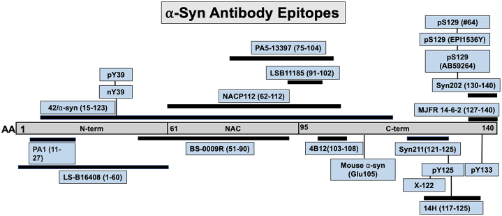
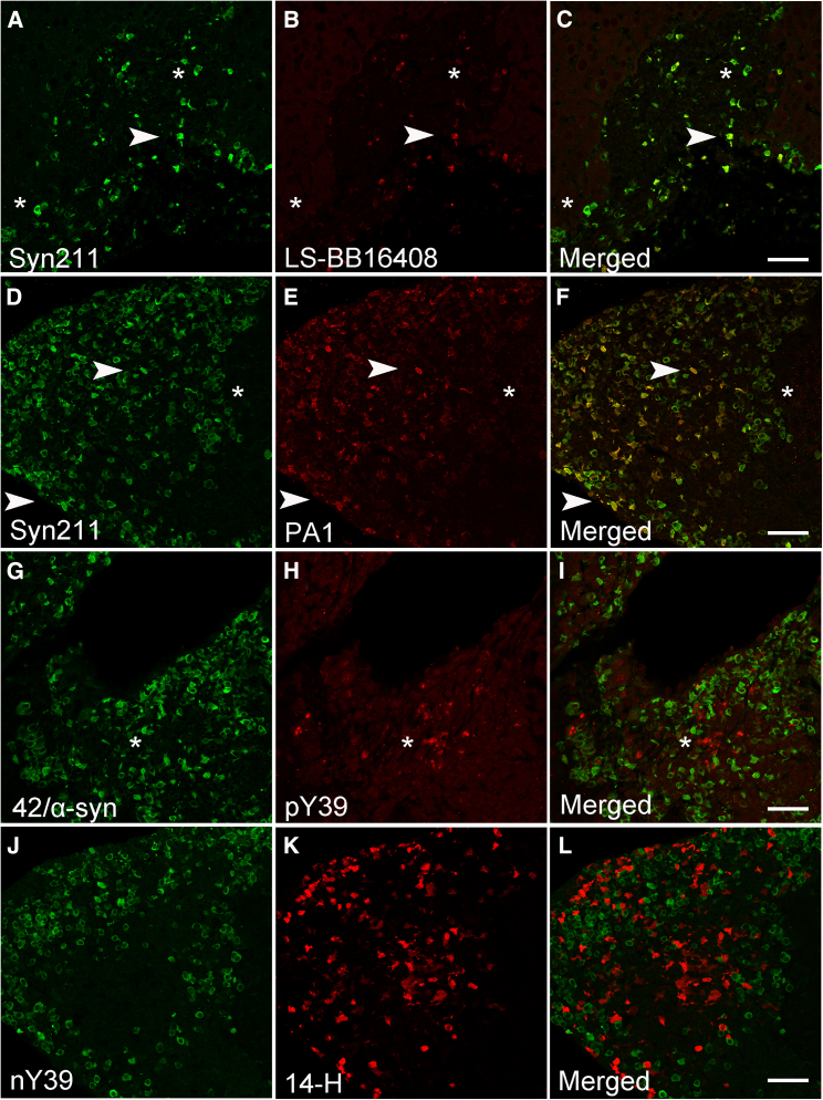
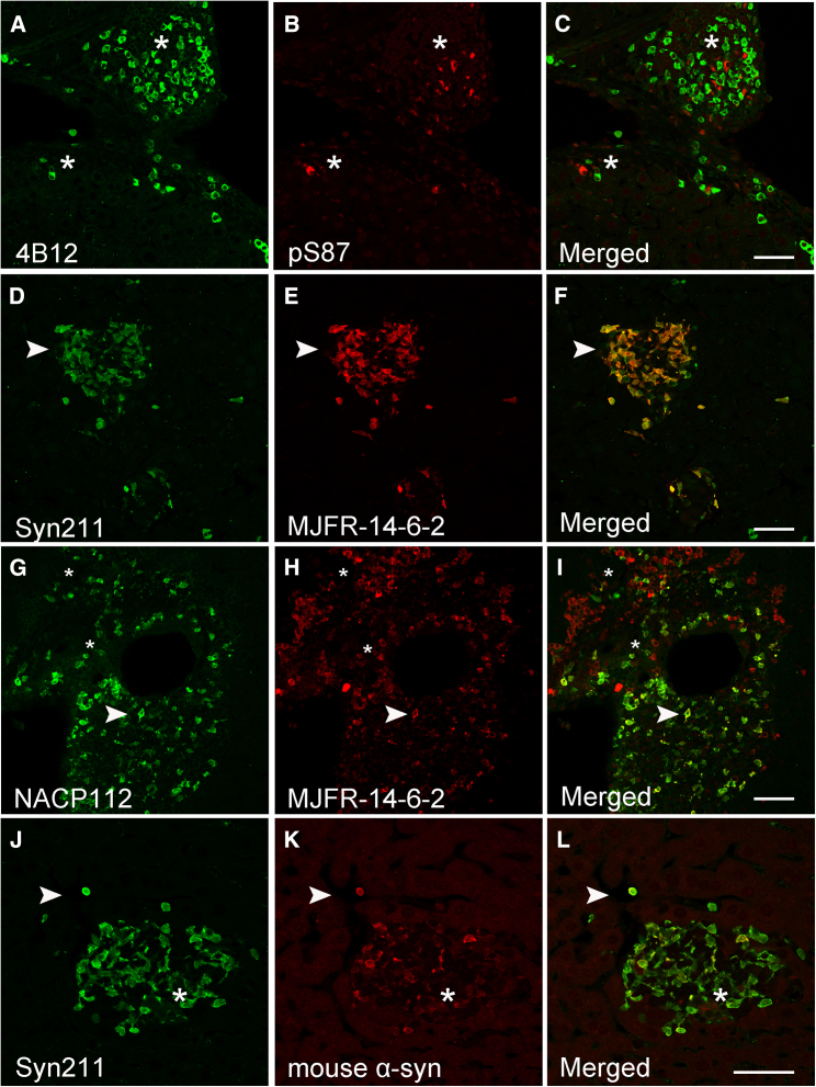
Accumulating evidence demonstrates that alpha-synuclein (α-syn) pathology associated with Parkinson's disease (PD) is not limited to the brain, as it also appears in a select number of peripheral tissues including the liver. In this study, we identified a number of PD-associated α-syn post-translational modifications in the livers of (Thy-1)-h[A30P] mice, a mouse model of familial PD expressing human α-syn harboring the A30P mutation driven by a neuron-specific promoter. Ex vivo, we also demonstrate that human hepatocytes induce post-translational modifications following α-syn fibrillar (PFF) treatment. Moreover, such cells also degrade PFFs over time, whereas oligomeric assemblies are more resistant to degradation, but this process can be enhanced by autophagy stimulators. Collectively, our findings suggest that pathological α-syn is transported to the liver in a modified state or is modified upon arrival, which facilitates its clearance and detoxification, pointing to a role for the liver in the degradation of PD-associated pathology.

Keywords: Biological sciences; Molecular neuroscience; Neuroscience.


Conflict of interest statement

MI is a paid consultant to BioArctic AB.

Figures

None
Graphical abstract
Figure 1
Figure 1
Human α-syn antibody diagram depicting all antibodies used in this study Each antibody epitope is represented schematically by lines under or above the α-syn protein.
Figure 2
Figure 2
Human α-syn inclusions in the A30P liver are partially pS129-positive (A–C) Identification of human α-syn deposits immunolabeled with the Syn202 and 14-H antibodies show extensive co-localization extensively in some regions (∗) whereas in other areas this co-localization is clearly absent (arrowheads). (D–F) Co-localization between Syn211 (green) and 14-H (red) antibodies (∗) and absence (arrowhead) is observed within the same region of the liver. (G–I) The pS129 (#64) antibody clone detects the presence of human α-syn inclusions in the A30P liver which fails to co-localize with the 14-H antibody (arrowhead). (J–L) No pS129 α-syn immunoreactivity (#64) was identified in the liver within α-syn inclusions labeled with the 14-H antibody (arrowhead). Bars = 50 μm.
Figure 3
Figure 3
Differential binding of pS129 antibodies to human α-syn aggregates in the A30P liver (A–C) Partial co-localization (arrowhead) is observed between Syn211 and pS129 clone 59264. (D–F) Abundant Syn211-positve α-syn inclusions are rarely labeled with the pS129 (clone 59264) antibody (arrowheads). (G–I) Rare co-localization is observed (arrowhead) with the pS129 antibodies clone 59264 and pS129 #64 as both antibodies appear to label independent inclusions (∗). (J–L) pS129 (#64) α-syn inclusions in the liver do not co-localize with the conformation specific MJFR-14-6-2 antibody. Bars = 50 μm.
Figure 4
Figure 4
Human α-syn inclusions in the A30P liver are phosphorylated at tyrosine 133 (Y133) or truncated at position X-122 (A–C) Identification of Syn202-positive human α-syn inclusions did not co-localize with a phosphorylation specific antibody at position 133 (pY133, arrowheads) or (D–F) with the 4B12 antibody (arrowheads). (G–I) C-terminal truncation of α-syn at position X-122 is detected within the A30P liver and partially co-localizes with PA5 and (J-L) PA1 pan antibodies (arrowheads). Bars = 50 μm.
Figure 5
Figure 5
Nitration and phosphorylation at position Y39 occur within human α-syn inclusions in the A30P liver (A–C) Abundant Syn211 immunoreactivity human α-syn inclusions which rarely (∗) co-localized with the N-terminal LS-BB16408 (arrowhead). (D–F) Clear Syn211 immunoreactive human α-syn inclusions are present within the A30P liver and are partly labeled (arrowhead) with the N-terminal antibody PA1 but mostly remain unlabeled (∗). (G–I) Phosphorylation of human α-syn inclusions at pY39 fails to co-localize with the 42/α-syn (∗) (J-L) Site-specific nitration at position Y39 (nY39) within α-syn fails to co-localize with the C-terminal antibody 14-H. Bars = 50 μm.
Figure 6
Figure 6
Limited N-terminal α-syn reactivity was observed in the A30P liver (A–C) Rare N-terminal human α-syn immunoreactivity was detected in the liver using the pan-α-syn antibody BS-009R (∗) compared to Syn211. (D–F) Limited co-localization between Syn211 and the NACP112 antibodies (arrowhead). (G–I) Partial co-localization observed with Syn211 and LSB1185 antibodies (arrowhead). (J–L) Rare identification of LSB1185-positive human α-syn inclusions that co-localized with the 4B12 antibody. Bars = 50 μm.
Figure 7
Figure 7
Human α-syn inclusions in the A30P liver seed and co-localize with endogenous mouse α-syn proteins (A–C) Liver α-syn aggregates are phosphorylated at serine 87 (pS87) and fail to co-localize with the pan-α-syn antibody 4B12 (asterisk). (D–F) Identification of Syn211 inclusions which co-localize with the misfolded-specific antibody MJFR-14-6-2 and with the G-I) NACP112 antibody (arrowheads). (J–L) Identification of Syn211-positive human α-syn inclusions which partly co-localize (arrowhead) with endogenous mouse-specific α-syn antibody, while some appear to be completely negative (∗). Bar = 50 μm.
Figure 8
Figure 8
Differential processing of human α-syn assemblies by human Huh-7 hepatocytes in vitro (A) Transmission electron microscopy (TEM) of α-syn monomers, oligomers, and fibrillar assemblies showing the classical donut-like and filamentous ultrastructures, respectively. Bar = 200 nm (B–C) Quantification of human α-syn immuno-reactivity in HuH-7-Cx32 cells at different time periods (24, 48 and 72 h) or in the supernatant (Sup, 72 h) using Western blot analysis with the Syn202, (D-E) Syn211, (F-G) 14-H, and H-I) 4B12 antibodies and analyzed using one-way ANOVA with Tukey’s multiple comparison test. Data are represented as SEM and a number of replicates (n)=4, ∗∗∗p = 0.001, ∗∗∗∗p = 0.0001, respectively.
Figure 9
Figure 9
Human hepatocytes (Huh-7) induce pS129 phosphorylation in vitro (A–B) Western blot analysis of Huh-7 cells treated with different concentrations of PFFs or α-syn monomers (1–5 μM) for 72 h then immunoblotted for pS129 (EP1536Y) and β-actin as loading control. (C–F) Untreated Huh-Cx32 cells show no pS129 immunolabeling whereas (G-J) treated cells with full length PFF show puncta immunoreactive for pS129 and partly co-localize with Cx32-mCherry. (K-N) Untreated control Huh-Cx32-α-syn expressing cells show no pS129 immunoreactivity whereas (O-R) cells treated full length PFF show puncta immunoreactive for pS129 and partly co-localize with Cx32-mCherry. (S–V) Huh-Cx32-α-syn expressing cells treated with truncated PFF (1–120) show puncta immunoreactive for pS129 and show partial co-localization with Cx32-mCherry. Data was analyzed using one-way ANOVA with Tukey’s multiple comparison test. Data are represented as SEM and number of replicates (n)=3,p = 0.05 and ∗∗p = 0.01, and ∗∗∗∗p = 0.0001, respectively). Bars = 20 μm.

References

    1. Varadi C. Clinical Features of Parkinson's Disease: The Evolution of Critical Symptoms. Biology. 2020;9:103. - PMC - PubMed
    1. Braak H., Rüb U., Del Tredici K. Cognitive decline correlates with neuropathological stage in Parkinson's disease. J. Neurol. Sci. 2006;248:255–258. - PubMed
    1. Gasser T. Genetics of Parkinson's disease. Curr. Opin. Neurol. 2005;18:363–369. - PubMed
    1. Konno T., Ross O.A., Puschmann A., Dickson D.W., Wszolek Z.K. Autosomal dominant Parkinson's disease caused by SNCA duplications. Parkinsonism Relat. Disorders. 2016;22:S1–S6. - PMC - PubMed
    1. Petrucci S., Ginevrino M., Valente E.M. Phenotypic spectrum of alpha-synuclein mutations: New insights from patients and cellular models. Parkinsonism Relat. Disorders. 2016;22:S16–S20. - PubMed