. 2024 Dec 10:15:1496169.
doi: 10.3389/fimmu.2024.1496169. eCollection 2024.

Immune regulatory adjuvant approach to mitigate subcutaneous immunogenicity of monoclonal antibodies

Affiliations

Immune regulatory adjuvant approach to mitigate subcutaneous immunogenicity of monoclonal antibodies

Nicole L Jarvi et al. Front Immunol. .

Abstract

Introduction: Immunogenicity continues to be a challenge for development and clinical utility of monoclonal antibodies, and there are gaps in our current ability to prevent anti-drug antibody development in a safe and antigen-specific manner.

Methods: To mitigate immunogenicity of monoclonal antibodies administered subcutaneously, O-phospho-L-serine (OPLS)-the head group of the tolerance-inducing phospholipid, phosphatidylserine-was investigated as an immunoregulatory adjuvant.

Results: Formulations of adalimumab, trastuzumab or rituximab with OPLS showed reduction in relative immunogenicity in mice compared to vehicle formulations, indicated by reduced anti-drug antibody development and significant reductions in CD138+ plasma cell differentiation in bone marrow. Titer development toward recombinant human hyaluronidase, a dispersion enhancer that was co-formulated with monoclonal antibodies, was similarly reduced. Subcutaneous administration of adalimumab with OPLS resulted in a two-fold increase in expression of type 1 regulatory (Tr1) T cell subset in the spleen. This is consistent with in vitro studies where co-culturing of dendritic cells primed with ovalbumin in the presence and absence of OPLS and antigen specific T-cells induced expression of Tr1 phenotype on live CD4+ T cells.

Conclusion: This adjuvant does not impact immune competence of non-human primates and mice, and repeated administration of the adjuvant does not show renal or hepatic toxicity. Formulation of monoclonal antibodies with the immunoregulatory adjuvant, OPLS, was found to be safe and effective at mitigating immunogenicity.

Keywords: anti-drug antibodies; formulation; immune tolerance; immunogenicity; protein therapeutics; subcutaneous administration.


Conflict of interest statement

Author NN was employed by the company Truvai Biosciences, LLC. Author DM was employed by the company Enhanced Pharmacodynamics, LLC. Authors SB, DM, and RS have financial interest in Truvai Biosciences, LLC. SB and KS are inventors on a patent for use of OPLS as an immunomodifier. The remaining authors declare that the research was conducted in the absence of any commercial or financial relationships that could be constructed as a potential conflict of interest. The authors declare that this study received funding from Truvai Biosciences, LLC. The funder had the following involvement in the study: study design.

Figures

Figure 1
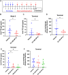
Figure 1
Co-formulation of ADM with OPLS reduces anti-ADM antibody formation. (A) Study timeline. Mice (n=10/group) were administered SC injections of ADM on week 1 (28.5 μg/mouse) and week 3 (14.3 μg/mouse) which were formulated in vehicle containing 0, 25, or 50 mM OPLS. (B) Anti-ADM IgG (μg/mL) in plasma at the terminal endpoint (week 7). Each dot represents an individual mouse and bars are mean ± SD. Likely outliers were removed based on the ROUT test Q=1%. Titers followed a normal distribution (D’Agostino & Pearson test). Statistical significance was tested by Student’s unpaired t-test (one-tailed). *p<0.05.
Figure 2
Figure 2
Co-formulation of OPLS with mAbs and rHuPH20 reduces ADA development. (A) Representative study timeline. (B-D) Mice (n=8/group) were administered TTZ (0.214 mg/mouse) and rHuPH20 (3.57 U/mouse) formulated in vehicle containing 0 or 25 mM OPLS once weekly for four weeks, followed by a ten-week washout period. Anti-TTZ IgG concentration (ng/mL) in plasma at (B) week 8 and (C) week 14 (terminal). (D) Anti-rHuPH20 IgG titers (reciprocal dilution) at the study terminal. (E, F) Mice (n=8/group) were administered RTX (0.5 mg/mouse) and rHuPH20 (8.36 U/mouse) formulated in vehicle containing 0, 25, or 100 mM OPLS subcutaneously once weekly for four weeks, followed by a ten-week washout period (according to (A)). (E) Anti-RTX IgG (ng/mL) in plasma at the terminal endpoint. (F) Anti-rHuPH20 IgG titers (reciprocal dilution) at the study terminal. In all plots, dots represent individual mice and bars are mean ± SD. In all data sets, definitive outliers were removed based on the ROUT test (Q=0.1%). Statistical significance was determined by Student’s unpaired t-test (one-tailed). When titers did not follow a normal distribution (according to D’Agostino & Pearson test), statistical analysis was performed on log-transformed data. ns not significant, *p<0.05.
Figure 3
Figure 3
Reduction of plasma cell development in the bone marrow by OPLS co-administration with mAbs and rHuPH20. Bone marrow cells from the femur were collected at the study terminal—10 weeks past the final dose for RTX + rHuPH20 or TTZ + rHuPH20. Supplementary Figure 2 is the flow cytometry gating strategy. Frequency (%) of CD138+ plasma cells out of CD19midB220mid cells in the bone marrow of mice treated with (A) RTX and rHuPH20 in vehicle, 25 mM OPLS, or 100 mM OPLS or (B) TTZ and rHuPH20 in vehicle or 25 mM OPLS. Dots represent individual mice and bars are mean ± SD. Statistical significance was determined by Student’s unpaired t-test (one-tailed). *p<0.05.
Figure 4
Figure 4
Impact of OPLS on RTX- and ADM-mediated T-cell activation when administered subcutaneously in mice. CD4+ T cells in the draining lymph nodes (DLN) of mice at the study terminal were stained for the activation marker CD44 and classical immune checkpoints CLTA-4 and PD-1. Supplementary Figure 3 is the flow cytometry gating strategy. Frequency (%) of (A) CD44++ and (B) CTLA-4+PD-1+ cells out of live CD3+CD4+ T cells in DLN of mice administered RTX + rHuPH20 in vehicle, 25 mM OPLS, or 100 mM OPLS or (C) in DLN of mice administered ADM in vehicle, 25 mM OPLS, or 50 mM OPLS. Dots represent individual mice and bars are mean ± SD. Statistical significance was determined by Student’s unpaired t-test (one-tailed). *p<0.05, **p<0.01.
Figure 5
Figure 5
Co-administration with OPLS promotes differentiation of regulatory T cells in vivo and in vitro. In the ADM study ( Figure 1 ), mouse splenocytes were collected at the terminal endpoint and analyzed for Tr1 phenotype. Supplementary Figure 4 is the flow cytometry gating strategy. (A) Frequency (%) of LAG-3+CD49b+ FoxP3-CD4+ Tr1 cells in the spleen. Dots represent individual mice and bars are mean ± SD. Statistical significance was determined by Student’s unpaired t-test (one-tailed). *p<0.05. (B, C) Naïve immature SW mouse BMDCs were cultured for 24 h with media, VitD3/Dex, OVA (a model antigen), and OVA + 25 mM or 50 mM OPLS. Splenocytes from SW mice (n=2) subcutaneously immunized with OVA (2 μg/100 μL) once weekly were collected 4 days after the second dose. Isolated CD4+ T cells were co-cultured with treated BMDCs at a ratio of 1:5 BMDC, CD4+ T cell for 72 h. Frequency (%) (B) LAG-3+CD49+ Tr1 and (C) LAP+ of CD4+FoxP3- T cells. Error bars are mean ± SD of triplicate wells.
Figure 6
Figure 6
Proposed mechanism of OPLS immunogenicity reduction. In the effective dosage range (25-100 mM), OPLS signaling to local DCs during antigen uptake/processing induces a tolerogenic DC phenotype, leading to DC migration to the DLN. Antigen presentation by OPLS-primed DCs is more likely to induce tolerogenic or anergic T-cell responses than an immunogenic response. The reduction in ADA development by OPLS likely stems from less T cell help in humoral responses and/or restriction of T cell effector responses by regulatory T cells. This figure was created at Biorender.com.
Figure 7
Figure 7
OPLS does not impact immunocompetence of NHP and mice. CD-1 mice received daily SC doses of vehicle (n=6), 20 mg/kg CYP (n=3), or OPLS (n=6/group) at 18-450 mM for 28 days. NHP (rhesus macaques) (n=3) received 21 daily SC doses of 54 mM OPLS. (A, B) Frequencies of lymphocyte populations—CD19+ B cells, CD3+ T cells, CD3+CD4+ T cells, and CD3+CD8+ T cells—in (A) spleens of mice on day 28 and (B) peripheral blood of NHP on day 0 (baseline), 8, 18, 22, and 35. Supplementary Figure 5 is the gating strategy. (C) Anti-KLH IgM and (D) IgG titers (log2) in plasma of mice on day 28 following a single IV dose of KLH (2 mg) on day 15. (E) Anti-KLH IgM and IgG titers (log2) in plasma of NHP on day 18 and 22, respectively, following a single IM dose of KLH (10 mg) on day 8. Each dot represents an individual animal and all bars are mean ± SD. (F-K) Microscopic images of H&E-stained spleens from mice treated with (F) vehicle or (G) 18 mM, (H) 45 mM, (I) 90 mM, (J) 225 mM, and (K) 450 mM OPLS. Statistical significance was determined by one-way ANOVA with Dunnett’s multiple comparisons test. ****p<0.0001.
Figure 8
Figure 8
OPLS does not induce renal or hepatic toxicity in NHP and mice. (A) Change in body weight (%) from day 1 to day 28 and (B) organ weight (%) of body weight for the liver, spleen, and kidney at day 28 for CD-1 mice (n=6/group) administered daily SC doses of vehicle, 18-450 mM OPLS, or CYP (20 mg/kg). (C) Body weight (kg) of NHP (rhesus macaques) (n=3) administered daily SC doses of 54 mM (25 mg/kg) OPLS. (D, E) Creatinine kinase (CK) activity (U/L) in plasma of (D) CD-1 mice at day 28 and (E) NHP across study days.

Similar articles

Cited by

References

    1. Turner MR, Balu-Iyer SV. Challenges and opportunities for the subcutaneous delivery of therapeutic proteins. J Pharm Sci. (2018) 107:1247–60. doi: 10.1016/j.xphs.2018.01.007 - DOI - PMC - PubMed
    1. Jarvi NL, Balu-Iyer SV. Immunogenicity challenges associated with subcutaneous delivery of therapeutic proteins. BioDrugs. (2021) 35:125–46. doi: 10.1007/s40259-020-00465-4 - DOI - PMC - PubMed
    1. Wynne C, Harvey V, Schwabe C, Waaka D, McIntyre C, Bittner B. Comparison of subcutaneous and intravenous administration of trastuzumab: a phase I/Ib trial in healthy male volunteers and patients with HER2-positive breast cancer. J Clin Pharmacol. (2013) 53:192–201. doi: 10.1177/0091270012436560 - DOI - PubMed
    1. Banugaria SG, Prater SN, McGann JK, Feldman JD, Tannenbaum JA, Bailey C, et al. . Bortezomib in the rapid reduction of high sustained antibody titers in disorders treated with therapeutic protein: lessons learned from Pompe disease. Genet Med. (2013) 15:123–31. doi: 10.1038/gim.2012.110 - DOI - PMC - PubMed
    1. Messinger YH, Mendelsohn NJ, Rhead W, Dimmock D, Hershkovitz E, Champion M, et al. . Successful immune tolerance induction to enzyme replacement therapy in CRIM-negative infantile Pompe disease. Genet Med. (2012) 14:135–42. doi: 10.1038/gim.2011.4 - DOI - PMC - PubMed

MeSH terms

LinkOut - more resources