

Immunoadjuvant therapy in the regulation of cell death in sepsis: recent advances and future directions
- PMID: 39720718
- PMCID: PMC11666431
- DOI: 10.3389/fimmu.2024.1493214
Immunoadjuvant therapy in the regulation of cell death in sepsis: recent advances and future directions
Abstract
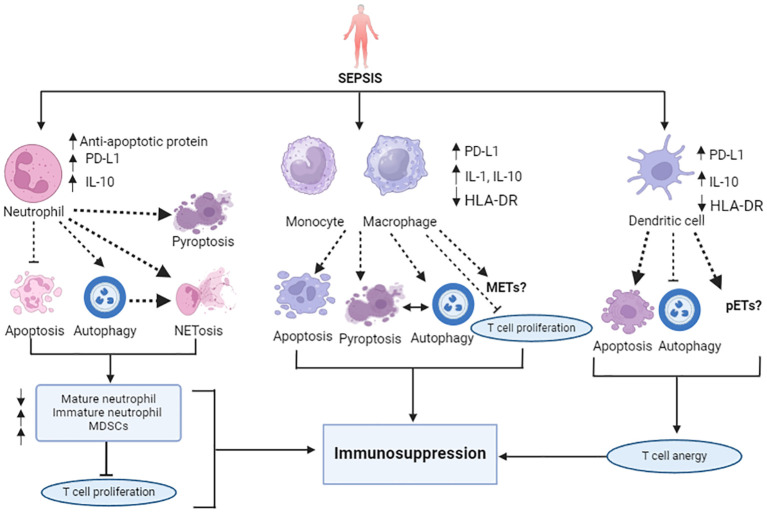
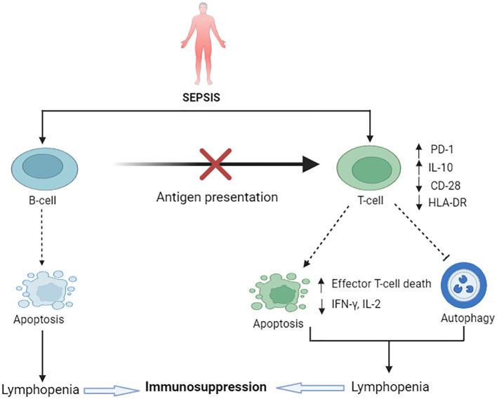
Sepsis is characterized by a concomitant early pro-inflammatory response by immune cells to an infection, and an opposing anti-inflammatory response that results in protracted immunosuppression. The primary pathological event in sepsis is widespread programmed cell death, or cellular self-sacrifice, of innate and adaptive immune cells, leading to profound immunological suppression. This severe immune dysfunction hampers effective primary pathogen clearance, thereby increasing the risk of secondary opportunistic infections, latent viral reactivation, multiple organ dysfunction, and elevated mortality. The types of cell death include apoptosis (type I programmed cell death), autophagy (type II programmed cell death), NETosis (a program for formation of neutrophil extracellular traps (NETs)) and other programmed cell deaths like pyroptosis, ferroptosis, necroptosis, each contributing to immunosuppression in distinct ways during the later phases of sepsis. Extensive apoptosis of lymphocytes, such as CD4+, CD8+ T cells, and B cells, is strongly associated with immunosuppression. Apoptosis of dendritic cells further compromises T and B cell survival and can induce T cell anergy or promote regulatory Treg cell proliferation. Moreover, delayed apoptosis and impaired neutrophil function contribute to nosocomial infections and immune dysfunction in sepsis. Interestingly, aberrant NETosis and the subsequent depletion of mature neutrophils also trigger immunosuppression, and neutrophil pyroptosis can positively regulate NETosis. The interaction between programmed cell death 1 (PD-1) or programmed cell death 1 ligand (PD-L1) plays a key role in T cell modulation and neutrophil apoptosis in sepsis. The dendritic cell growth factor, Fms-like tyrosine kinase (FLTEL), increases DC numbers, enhances CD 28 expression, attenuates PD-L1, and improves survival in sepsis. Recently, immunoadjuvant therapies have attracted attention for their potential to restore host physiological immunity and homeostasis in patients with sepsis. This review focuses on several potential immunotherapeutic agents designed to bolster suppressed innate and adaptive immune responses in the management of sepsis.
Keywords: NETosis; apoptosis; autophagy; ferroptosis; immunosuppression; necroptosis cell death; pyroptosis; sepsis.
Copyright © 2024 Islam, Watanabe, Salma, Ozaki, Irahara, Tanabe, Katsuki, Oishi and Takeyama.
Conflict of interest statement
The authors declare that the research was conducted in the absence of any commercial or financial relationships that could be construed as a potential conflict of interest. The author(s) declared that they were an editorial board member of Frontiers, at the time of submission. This had no impact on the peer review process and the final decision.
Figures
Similar articles
-
Interleukin-7 and anti-programmed cell death 1 antibody have differing effects to reverse sepsis-induced immunosuppression.Shock. 2015 Apr;43(4):334-43. doi: 10.1097/SHK.0000000000000317. Shock. 2015. PMID: 25565644 Free PMC article.
-
The Role of Programmed Cell Death 1/Programmed Death Ligand 1 (PD-1/PD-L1) Axis in Sepsis-Induced Apoptosis.Medicina (Kaunas). 2024 Jul 19;60(7):1174. doi: 10.3390/medicina60071174. Medicina (Kaunas). 2024. PMID: 39064603 Free PMC article.
-
Immunoadjuvant therapy in sepsis: novel strategies for immunosuppressive sepsis coming down the pike.Acute Med Surg. 2018 Aug 6;5(4):309-315. doi: 10.1002/ams2.363. eCollection 2018 Oct. Acute Med Surg. 2018. PMID: 30338075 Free PMC article.
-
Dysregulation of neutrophil death in sepsis.Front Immunol. 2022 Aug 18;13:963955. doi: 10.3389/fimmu.2022.963955. eCollection 2022. Front Immunol. 2022. PMID: 36059483 Free PMC article. Review.
-
Immunotherapy: A promising approach to reverse sepsis-induced immunosuppression.Pharmacol Res. 2016 Sep;111:688-702. doi: 10.1016/j.phrs.2016.07.019. Epub 2016 Jul 25. Pharmacol Res. 2016. PMID: 27468649 Free PMC article. Review.
Cited by
-
Cell death signaling and immune regulation: new perspectives on targeted therapy for sepsis.Cell Mol Biol Lett. 2025 Aug 15;30(1):99. doi: 10.1186/s11658-025-00784-w. Cell Mol Biol Lett. 2025. PMID: 40817040 Free PMC article. Review.
-
Association of PIV value with early mortality in ICU patients with sepsis-associated acute kidney injury from the MIMIC IV database.Sci Rep. 2025 Apr 2;15(1):11212. doi: 10.1038/s41598-025-96320-z. Sci Rep. 2025. PMID: 40175479 Free PMC article.
-
Viral sepsis - pathophysiology and disease manifestation.Infection. 2025 Jun;53(3):775-784. doi: 10.1007/s15010-025-02486-z. Epub 2025 Feb 17. Infection. 2025. PMID: 39961996 Free PMC article. Review.
-
Association between the nutritional inflammation index and mortality among patients with sepsis: insights from traditional methods and machine learning-based mortality prediction.BMC Infect Dis. 2025 Aug 14;25(1):1021. doi: 10.1186/s12879-025-11429-w. BMC Infect Dis. 2025. PMID: 40813626 Free PMC article.
-
The J-shaped association between the ratio of neutrophil counts to prognostic nutritional index and mortality in ICU patients with sepsis: a retrospective study based on the MIMIC database.Front Cell Infect Microbiol. 2025 Jul 22;15:1603104. doi: 10.3389/fcimb.2025.1603104. eCollection 2025. Front Cell Infect Microbiol. 2025. PMID: 40766844 Free PMC article.
References
Publication types
MeSH terms
Substances
LinkOut - more resources
Full Text Sources
Medical
Research Materials
Miscellaneous